Abstract
Objectives:
Early diagnosis of incipient and non-cavitated carious lesions is crucial for performing preventive treatments. The aim of this study was to compare the efficacy of three diagnostic methods of bitewing radiography, DIAGNOdent, and visual examination in diagnosing incipient occlusal caries of permanent first molars.
Materials and Methods:
In this diagnostic cross-sectional study, 109 permanent first molar teeth of 31 patients aged 7–13 years were examined visually, on bitewing radiographs, and using DIAGNOdent. Scoring of visual and radiographic examinations was based on Ekstrand’s classification. Visual examination after pit and fissure opening served as the gold standard. Receiver Operating Characteristic (ROC) curve was used to define the best cutoff point for DIAGNOdent compared with the gold standard. Inter-examiner reproducibility of visual and radiographic examinations was assessed using Kappa test and intraclass correlation coefficient (ICC) was calculated for DIAGNOdent values.
Results:
The sensitivity of detecting caries that had extended into the enamel was 81.4%, 86.3%, and 81.4% for visual examination, DIAGNOdent and radiography, respectively. Moreover, the specificity was 100%, 71.4%, and 100% for visual observation, DIAGNOdent and radiography, respectively in the enamel. The Kappa index for inter-examiner reliability was 0.7 and 0.8 for visual examination and radiography, respectively. The ICC was 0.98 for the values read by DIAGNOdent.
Conclusion:
Visual examination is the first choice for diagnosis of incipient caries. In suspicious cases, radiography and laser DIAGNOdent can be used as adjunct procedures.
Keywords: Dental Caries, Diagnosis, Radiography
INTRODUCTION
Early diagnosis of incipient caries is important. Clinical diagnosis of occlusal caries is challenging due to the complex morphology of pits and fissures and presence of staining [1]. Early diagnosis of incipient and non-cavitated carious lesions is crucial for performing preventive treatments [2]. Both visual and radiographic examinations are conventionally performed. Visual examination is more efficient for diagnosis of cavitated rather than non-cavitated and incipient lesions [3].
Furthermore, this method is subjective and its reproducibility is low, since it involves the clinical experience and scientific knowledge of the clinician. On the other hand, radiographs have high specificity and low sensitivity for the diagnosis of non-cavitated lesions and underestimate the actual depth of caries [2].
Ekstrand index was introduced for visual standardization and improved detection of dental caries [4]. The Ekstrand index scores resemble the clinical situations and are based on signs found on the enamel surface such as opacities, white spots, brown spots, presence of cavities or micro-cavities and a combination of these conditions. This system is expected to increase the sensitivity and reliability of visual examination [4].
New devices using new technologies are applied for quantitative and qualitative diagnosis of incipient demineralized lesions. Fluorescence-based methods are among these modalities, in which the sound and decayed surfaces produce different fluorescence when exposed to a certain light or wavelength [3]. Laser fluorescence DIAGNOdent measures the emitted fluorescent infra-red light and shows the result in whole numbers between 0–99. It seems that bacterial metabolites, especially porphyrins, emit the fluorescent radiation [5]. The results of in vitro studies show the ability of DIAGNOdent in exploring relatively advanced carious lesions. These findings are consistent with histological evidence, but they have no relationship with the depth of the lesions in dentin. This device, with laboratory support, has good reliability and sensitivity [5]. The result of DIAGNOdent is affected by different variables such as dehydration of the lesion, dental plaque and stains in the grooves of the occlusal surface [6].
There are a few in vivo studies in this field. Lussi et al. [7] evaluated the efficacy of visual examination, radiographic assessment and DIAGNOdent for the diagnosis of occlusal caries. They concluded that DIAGNOdent can be useful when visual examination is not efficient. Kouchaji [8] evaluated 156 permanent molars and revealed that the combined use of visual examination and DIAGNOdent laser had the highest sensitivity and specificity. Additionally, Rando-Meirelles and de Sousa [9] applied the fluorescence laser method, radiography and visual examination to assess the extension of occlusal non-cavitated lesions in permanent molars. They concluded that DIAGNOdent cannot replace the radiographic method. The aim of this study was to compare the efficacy of bitewing radiography, fluorescence laser and visual examination in the diagnosis of incipient occlusal caries in permanent first molars.
MATERIALS AND METHODS
This descriptive cross-sectional diagnostic study was conducted on patients aged 7 to 13 years, referred to the Department of Pediatric Dentistry (School of Dentistry, Shahid Sadoughi University of Medical Sciences, Yazd, Iran). Patients’ teeth were visually examined by a dentist and 31 patients who had signs of pit and fissure caries in at least one molar tooth were selected. Informed consent was obtained from all patients before the study. The proposal of this study was approved by the ethics committee of the university (P.17.1.188736). Clinical examination of each tooth was performed under adequate lighting after cleaning the tooth surfaces by two examiners calibrated in a pilot study. The samples used in the pilot study were not included in the main study. One-hundred-fifteen teeth of 31 patients that were intact or had incipient and inconspicuous caries with or without color change were selected. The teeth with occlusal restorations, enamel hypoplasia, hypomineralization or structural defects, cavitated lesions, fissure sealant, orthodontic bands or brackets or pulp necrosis were excluded. Six of the 115 teeth were also excluded due to patient dropout; therefore, the three diagnostic methods were performed for 109 teeth.
Occlusal surfaces of the teeth were cleaned from plaque and debris using water spray and cotton pellets if necessary. Dental explorers were not used for examination.
Occlusal caries were scored (v0–v4) using Ekstrand’s visual scoring system (Table 1) [4]. Then, bitewing radiographs were taken (Planmeca ProStyle, Helsinki, Finland) at 70kV, 8 mA and 0.36s exposure settings using E speed 22×35 mm dental films (Kodak, Rochester, USA) in XCP film holder and processed using a dental film processor (Velopex-Extra X, London, England).
Table 1.
Criteria used in visual examination, radiographic examination and fissure opening
| Visual Examination | |
| V0 | No change or slight change of enamel translucency after air drying |
| V1 | Opacity or discoloration distinctly visible after air drying |
| V2 | Opacity or discoloration visible without air drying |
| V3 | Localized enamel breakdown in opaque or discolored enamel and/or grayish discoloration from the underlying dentin |
| V4 | Cavitation in opaque or discolored enamel exposing dentin |
| Radiographic examination | |
| R0 | No radiolucency |
| R1 | Radiolucency detected in the enamel, but not beyond the dentinoenamel junction |
| R2 | Radiolucency detected and extended into the dentin |
| Fissure opening | |
| B0 | No caries seen |
| B1 | Caries detected and confined to the enamel |
| B2 | Caries detected and extended to the dentin |
Images were initially examined and scored (R0–R2) by a pedodontist on a negatoscope and then confirmed by a radiologist (Table 1) [4].
At this time, the teeth were examined by laser fluorescence pen (DIAGNOdent, Kavo, Biberach, Germany) followed by cleaning with a rubber cup and pumice powder, isolation with cotton rolls, and drying. Dental explorers were used to clean the teeth grooves from powder.
The laser fluorescence pen was placed parallel to the long axis of the tooth on incipient or suspicious dental caries and moved around following calibration of the device on ceramic and intact enamel surfaces.
The peak values were recorded. Cavity preparation, or fissurotomy (Fissurotomy® Miro NTF, SS White, Lakewood, NJ, USA) was performed in cases with obvious or ambiguous dental caries, respectively to assess and score the actual depth of lesions (Table 1) [4]. Then, the cavities were restored by composite or amalgam, and the fissures were sealed.
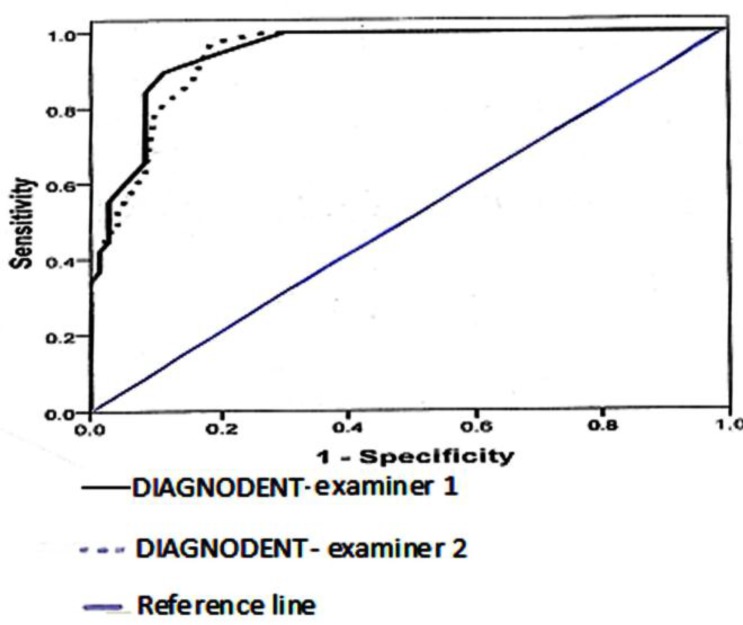
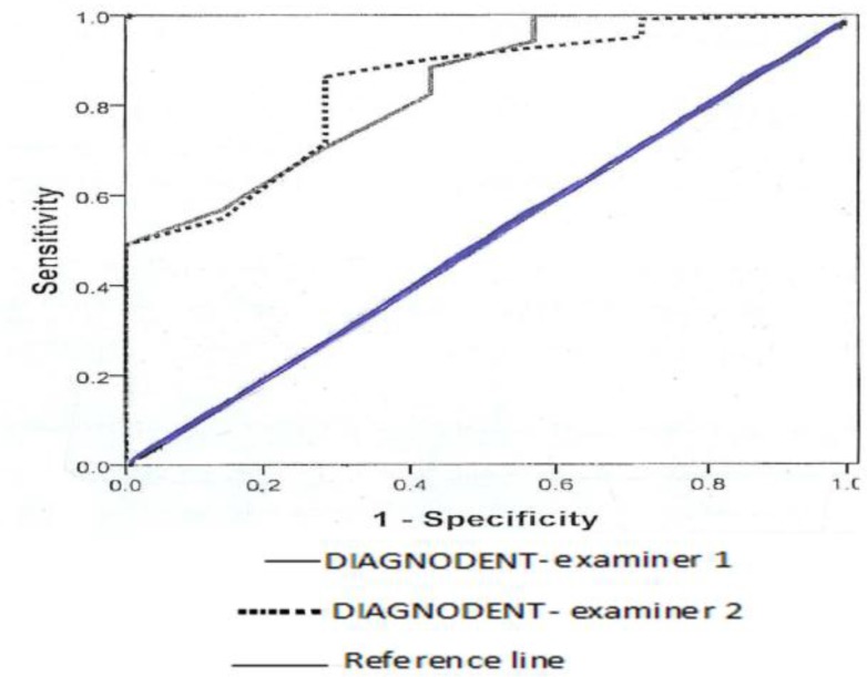
SPSS version 17 software for Windows (SPSS Inc., Chicago, IL, USA) was used to analyze the collected data. ROC curve was used to define the best cut-off point for DIAGNOdent and to compare it with the gold standard (Figs. 1 and 2) and the area under the ROC curve (Az) was calculated; then the values of sensitivity, specificity, and accuracy of this method were determined by calculating the area under the ROC curve.
Fig. 1.
ROC curve for dentin threshold
Fig. 2.
ROC curve for enamel threshold
These values were also calculated for visual and radiographic examinations, and compared with the gold standard (fissure opening). The agreement coefficient for radiography and visual method was measured using a Kappa test and inter-examiner reliability was evaluated using ICC for DIAGNOdent. Altman’s paired samples method for calculating a 95% confidence interval was used to compare the accuracy of the methods.
RESULTS
This study was conducted on 109 teeth of 31 children aged 7 to 13 (mean 11.11±1.34) years. After assessment of information compared to the standard method, seven surfaces were sound, 64 showed enamel caries and 38 showed dentin caries. Table 2 compares the visual method scores with those of the standard method (fissure opening) in enamel and dentin, separately. In Table 3, the radiographic method scores are compared with those of the standard method.
Table 2.
Comparison of Ekstrand’s scoring system and the standard fissure opening method by examiners 1 and 2
| Visual* Scores | Standard Method (Fissure Opening Method) | Total | |||
|---|---|---|---|---|---|
| Sound occlusal surface | Enamel threshold | Dentin threshold | |||
| Examiner 1 | 0 | 4 | 7 | 0 | 11 |
| 1 | 2 | 13 | 0 | 15 | |
| 2 | 1 | 42 | 14 | 57 | |
| 3 | 0 | 2 | 24 | 26 | |
| Total | 7 | 64 | 38 | 109 | |
| Examiner 2 | 0 | 3 | 4 | 0 | 7 |
| 1 | 4 | 15 | 0 | 19 | |
| 2 | 0 | 43 | 17 | 60 | |
| 3 | 0 | 2 | 21 | 23 | |
| Total | 7 | 64 | 38 | 109 | |
We did not find score 4 in any of the teeth.
Table 3.
Frequency of radiographic scores compared to the standard fissure opening scores according to examiners 1 and 2
| Radiographic Scores | Standard Method (Fissure Opening) | Total | |||
|---|---|---|---|---|---|
| Sound occlusal surface | Enamel threshold | Dentin threshold | |||
| Examiner 1 | 0 | 7 | 21 | 0 | 28 |
| 1 | 0 | 43 | 6 | 49 | |
| 2 | 0 | 0 | 32 | 32 | |
| Total | 7 | 64 | 38 | 109 | |
| Examiner 2 | 0 | 7 | 19 | 0 | 26 |
| 1 | 0 | 45 | 6 | 51 | |
| 2 | 0 | 0 | 32 | 32 | |
| Total | 7 | 64 | 38 | 109 | |
All surfaces showing radiolucency in dentine (score 2 based on Table 1) were found to have dentin caries (dentin threshold). The best cutoff point for DIAGNOdent in this study included: sound surfaces: 0–7, enamel decay: 8–10, dentin decay: ≥11.
The Az value for laser method was 0.83 and 0.84 for the first and second examiners, respectively, which shows high efficacy of this method. Inter-examiner kappa coefficients were 0.7 in visual and 0.8 in radiographic methods. The ICC of DIAGNOdent results was 0.98.
The sensitivity of DIAGNOdent method was higher than that of the other two methods, although its specificity in enamel was lower than the other methods (Table 4).
Table 4.
Sensitivity, specificity and accuracy of caries detection by each examiner (1 and 2) for all methods (visual inspection, DIAGNOdent and radiography)
| Diagnostic Methods | Visual inspection | DIAGNOdent | Radiography | ||||
| Examiner1 | Examiner 2 | Examiner 1 | Examiner 2 | Examiner 1 | Examiner 2 | ||
| Enamel Threshold | Sensitivity (%) | 80.4 (74.2–86.6) | 81.4 (75.2–87.6) | 70.6 (64.4–76.8) | 86.3 (80.1–92.5) | 79.4 (73.2–85.6) | 81.4 (75.2–87.6) |
| Specificity (%) | 85.7 (80.1–91) | 100 (94.7–100) | 71.4 (66.1–76.7) | 71.4 (66.1–76.7) | 100 (94.7–100) | 100 (94.7–100) | |
| Accuracy (%) | 80.7 (74.7–86.7) | 82.5 (76.5–88.5) | 70.6 (64.6–76.6) | 85.3 (79.3–91.3) | 81.6 (75.6–87.6) | 82.5 (76.5–88.5) | |
| Dentin Threshold | Sensitivity (%) | 63.2 (56.1–70.3) | 55.3 (48–62.6) | 89.5 (82.2–96.8) | 86.8 (79.5–94.1) | 84.2 (76.9–91.5) | 84.2 (76.9–91.5) |
| Specificity (%) | 97.2 (93.8–100) | 97.2 (93.8–100) | 88.7 (85.3–92.1) | 84.5 (81.1–87.8) | 100 (96.6–100) | 100 (96.6–100) | |
| Accuracy (%) | 85.3 (79.8–90.8) | 82.5 (77–88) | 88.9 (83.4–94.4) | 85.3 (79.8–90.8) | 94.4 (88.9–99.9) | 94.4 (88.9–99.9) | |
The numbers inside the parentheses are calculated with 95% confidence interval.
Totally, all three methods had high sensitivity. The highest accuracy for detection of enamel occlusal caries belonged to DIAGNOdent, but with no significant difference with the other two methods (Table 4).
DISCUSSION
The occlusal surface of the tooth is susceptible to caries. However, making a reliable diagnosis is difficult in some cases. Hence, many researchers have attempted to find techniques to detect occlusal caries [8].
DIAGNOdent for caries detection has been shown in some studies [10,11]; however, controversial results have been reported regarding its efficacy of caries detection [8,12–14]. In this study, surface of the teeth was dried with oil-free air spray before the examination in order to decrease the refractive index between crystals from 1.33 for demineralized wet to 1 for demineralized dried surfaces. This procedure made the caries more visible [7]. Probes and explorers were not used in this study, because they would not increase the diagnostic power [12,15].
Furthermore, use of explorer may damage the dental tissues and impair the remineralization potential [4]. More recent studies have used Ekstrand index for standardization of the stages to detect caries using the visual method [16,17]. Angnes et al. concluded that Ekstrand visual scoring index was the most valuable technique for caries detection [17]. Our results also showed high sensitivity and specificity using this index for detection of enamel occlusal caries.
While in our study the sensitivity of radiographic diagnosis was similar to that of visual inspection for occlusal enamel caries. This difference may be related to the quality of films and the examiner’s effects. In our study, each of the radiographic images was examined by an expert radiologist followed by a pedodontist. Unlike the findings of this study, another study [10] found that DIAGNOdent was not suitable for diagnosing incipient enamel caries. Regarding our results, the specificity of DIAGNOdent in the enamel was lower than that of other methods (Table 4).
Inter-examiner agreement for visual examination was moderately acceptable, which was consistent with the results of earlier studies [8,12]; it may be attributed to the conduction of a pilot study before the main study.
In our study, the sensitivity of radiographic examination was relatively high, which is inconsistent with the results of some studies [2,18].
Souza et al. concluded that bitewing radiography was not suitable for caries detection because of its low sensitivity [2]. In a study conducted by Neuhaus et al, radiographic diagnosis of occlusal caries of deciduous teeth was not as accurate as laser fluorescence pen, laser fluorescence and ICADS methods [18], Bader and Shugars [19] in a review study reported that DIAGNOdent had higher sensitivity, but lower specificity compared to visual examination, and Ricketts [20] found that DIAGNOdent led to a greater possibility of false-positive diagnosis than the visual method. In our study, the diagnostic power of DIAGNOdent was found to be higher in dentin compared to the enamel, which is similar to a study by Hasani-Tabatabaee et al. They found that as the depth of carious lesions increased, DIAGNOdent showed higher values of sensitivity and specificity [21]. DIAGNOdent compared to visual and radiographic methods is more sensitive and accurate for the diagnosis of enamel caries (Table 4).
Costa et al. [22] showed the same results. On the other hand, the specificity of radiography and visual methods for the diagnosis of enamel caries was greater than that of DIAGNOdent. Visual method has a lower cost, is faster and has acceptable sensitivity; therefore, it can still be used as an appropriate method for clinical caries detection. In complicated cases, other methods may be recruited to dispel doubts. This is consistent with the results of Costa et al [22]. They stated that although DIAGNOdent was more accurate, the visual method was preferred because it had no significant difference with DIAGNOdent and it required shorter time.
One limitation in using laser fluorescence pen is receiving fluorescent waves from stains, highly mineralized structures or malformed teeth [23]. This may cause bias, lead to increased sensitivity and cause false-positive results. Exclusion of these teeth in some studies (unlike ours) such as the study by Kouchaji [8], may lead to superior results with regard to the performance of DIAGNOdent compared to studies that included stained teeth. Reproducibility is another important index for assessment of diagnostic methods. Several studies have pointed to the reliability of laser DIAGNOdent for occlusal caries detection in permanent teeth [22,24,25]. In our study, the ICC was used for comparison of the values read by DIAGNOdent and showed a strong agreement between observers. In contrast, Rodrigus et al, [23] in their in vitro study reported a low agreement in values read by DIAGNOdent between observers. Their study was a histological study and the samples were stored in thymol; this antimicrobial agent destroys porphyrins and consequently weakens the signals received by DIAGNOdent. The surface below the ROC curve in our study was 84% at the cut-off point of eight and 94% at the cut-off point of 11. This was similar to the rate (92% at the cut-off point of 12) obtained by Huth et al [26]. Risk factors such as age, history of dental caries, diet, attitude, and topical fluoride application must be considered before treatment of suspicious cases [27]. Considering the recent emphasis on preventive interventions and avoidance of surgical techniques, applying new technologies to detect even the slightest demineralization (incipient caries) seems necessary [22]. One of the limitations of the current study was lack of recording of the depth of lesions, because the calibration and reliability of the measurement of the depth of lesions are difficult to achieve. Additionally, in DIAGNOdent, the qualitative property, i.e., determining the presence or absence of dentinal caries is superior to the quantitative characteristic, i.e., recording the depth of the dentin lesion [12]. In our study, simple radiographic films were used. Future research is needed to compare the validity of digital radiographs (CCD/CMOS) with that of conventional radiography regarding their better contrast and resolution for detection of occlusal caries.
CONCLUSION
Although the visual method was not as accurate as LF, considering the insignificant differences and affordability of visual method, the latter can be the first choice for detection of incipient caries. In suspicious cases, radiography and DIAGNOdent may be used as adjuncts.
REFERENCES
- 1-. Achilleos EE, Rahiotis C, Kakaboura A, Vougiouklakis G. Evaluation of a new fluorescence-based device in the detection of incipient occlusal caries lesions. Lasers Med Sci. 2013. January; 28 (1): 193– 201. [DOI] [PubMed] [Google Scholar]
- 2-. Souza JF, Boldieri T, Diniz MB, Rodrigues JA, Lussi A, Cordeiro RC. Traditional and novel methods for occlusal caries detection: performance on primary teeth. Lasers Med Sci. 2013. January; 28 (1): 287– 95. [DOI] [PubMed] [Google Scholar]
- 3-. Shoaib L, Deery C, Ricketts DN, Nugent ZJ. Validity and reproducibility of ICDAS II in primary teeth. Caries Res. 2009;43 (6): 442– 8. [DOI] [PubMed] [Google Scholar]
- 4-. Ekstrand KR, Ricketts DN, Kidd EA. Reproducibility and accuracy of three methods for assessment of demineralization depth of the occlusal surface: an in vitro examination. Caries Res. 1997; 31 (3): 224– 31. [DOI] [PubMed] [Google Scholar]
- 5-. McDonald RE, Avery DR, Stookey GK, Chin JR, Kowolik JE. Dental Caries in the Child and Adolescent. In: Dean JA, Avery DR, McDonald RE. McDonald and Avery dentistry for the child and adolescent. Missouri, Elsevier Inc.; 2011: 177– 90. [Google Scholar]
- 6-. Novaes TF, Matos R, Braga MM, Imparato JCP, Raggio DP, Mendes FM. Performance of a pen-type laser fluorescence device and conventional methods in detecting approximal caries lesions in primary teeth in vivo study. Caries Res. 2009; 43 (1): 36– 42. [DOI] [PubMed] [Google Scholar]
- 7-. Lussi A, Megert B, Longbottom C, Reich E, Francescut P. Clinical performance of a laser fluorescence device for detection of occlusal caries lesions. Eur J Oral Sci. 2001. February; 109 ( 1): 14– 9. [DOI] [PubMed] [Google Scholar]
- 8-. Kouchaji C. Comparison between a laser fluorescence device and visual examination in the detection of occlusal caries in children. Saudi Dent J. 2012. July; 24 (3–4): 169– 74. [DOI] [PMC free article] [PubMed] [Google Scholar]
- 9-. Rando-Meirelles MP, de Sousa Mda L. Using laser fluorescence (DIAGNOdent) in surveys for the detection of noncavitated occlusal dentine caries. Community Dent Health. 2011. March; 28 (1): 17– 21. [PubMed] [Google Scholar]
- 10-. Lussi A, Imwinkelried S, Pitts N, Longbottom C, Reich E. Performance and reproducibility of a laser fluorescence system for detection of occlusal caries in vitro. Caries Res. 1999. Jul-Aug; 33 (4): 261– 6. [DOI] [PubMed] [Google Scholar]
- 11-. Shi XQ, Welander U, Angmar-Mansson B. Occlusal caries detection with KaVo DIAGNOdent and radiography: an in vitro comparison. Caries Res. 2000. Mar-Apr; 34 (2): 151– 8. [DOI] [PubMed] [Google Scholar]
- 12-. Chu CH, Lo EC, You DS. Clinical diagnosis of fissure caries with conventional and laser-induced fluorescence techniques. Lasers Med Sci. 2010. May; 25 (3): 355– 62. [DOI] [PMC free article] [PubMed] [Google Scholar]
- 13-. Goel A, Chawla HS, Gauba K, Goyal A. Comparison of validity of DIAGNOdent with conventional methods for detection of occlusal caries in primary molars using the histological gold standard: an in vivo study. J Indian Soc Pedod Prev Dent. 2009; 27 (4): 227– 34. [DOI] [PubMed] [Google Scholar]
- 14-. Anttonen V, Seppa L, Hausen H. A follow up study of the use of DIAGNOdent for monitoring fissure caries in children. Community Dent Oral Epidemiol. 2004. August; 32 (4): 312– 8. [DOI] [PubMed] [Google Scholar]
- 15-. Lussi A, Francescut P. Performance of conventional and new methods for the detection of occlusal caries in deciduous teeth. Caries Res. 2003. Jan-Feb; 37 (1): 2– 7. [DOI] [PubMed] [Google Scholar]
- 16-. Landis JR, Koch GG. The measurement of observer agreement for categorical data. Biometrics. 1977. March; 33 (1): 159– 74. [PubMed] [Google Scholar]
- 17-. Angnes V, Angnes G, Batisttella M, Grande RH, Loguercio AD, Reis A. Clinical effectiveness of laser fluorescence, visual inspection and radiography in the detection of occlusal caries. Caries Res. 2005. Nov-Dec; 39 (6): 490– 5. [DOI] [PubMed] [Google Scholar]
- 18-. Neuhaus KW, Rodrigues JA, Hug I, Stich H, Lussi A. Performance of laser fluorescence devices, visual and radiographic examination for the detection of occlusal caries in primary molars. Clin Oral Investig. 2011. October; 15 (5): 635– 41. [DOI] [PubMed] [Google Scholar]
- 19-. Bader JD, Shugars DA. A systematic review of the performance of a laser fluorescence device for detecting caries. J Am Dent Assoc. 2004. October; 135 (10): 1413– 26. [DOI] [PubMed] [Google Scholar]
- 20-. Ricketts D. The eyes have it. How good is DIAGNOdent at detecting caries? Evid Based Dent. 2005; 6 (3): 64– 5. [DOI] [PubMed] [Google Scholar]
- 21-. Hasani-Tabatabaee M, Momeni N, Khorshidian A. The detection of early inter proximal caries: DIAGNOdent, conventional and digital radiography. J Islamic Dent Assoc. 2011; 23 (2): 116– 24. [Google Scholar]
- 22-. Costa AM, Bezzerra AC, Fuks AB. Assessment of the accuracy of visual examination, bite-wing radiographs and DIAGNOdent on the diagnosis of occlusal caries. Eur Arch Paediatr Dent. 2007. June; 8 (2): 118– 22. [DOI] [PubMed] [Google Scholar]
- 23-. Rodrigues JA, Diniz MB, Josgrilberg EB, Cordeiro RC. In vitro comparison of laser fluorescence performance with visual examination for detection of occlusal caries in permanent and primary molars. Laser Med Sci. 2009; 24 (4): 501– 6. [DOI] [PubMed] [Google Scholar]
- 24-. Kuhnisch J, Bucher K, Hickel R. The intra/inter-examiner reproducibility of the new DIAGNOdent pen on occlusal sites. J Dent. 2007. June; 35 (6): 509– 12. [DOI] [PubMed] [Google Scholar]
- 25-. Lussi A, Megert B, Longbottom C, Reich E, Francescut P. Clinical performance of a laser fluorescence device for detection of occlusal caries lesions. Eur J Oral Sci. 2001. February; 109 (1): 14– 9. [DOI] [PubMed] [Google Scholar]
- 26-. Huth KC, Neuhaus KW, Gygax M, Bucher K, Crispin A, Paschos E, et al. Clinical performance of a new laser fluorescence device for detection of occlusal caries lesions in permanent molars. J Dent. 2008. December; 36 (12): 1033– 40. [DOI] [PubMed] [Google Scholar]
- 27-. Akbari M, Ahrari F, Hoseini-Zarch H, Movagharipour F. Assessing the Performance of the Laser Fluorescence Technique in Detecting Proximal Caries Cavities. J Mash Dent Sci. 2013; 37 (3): 195– 204. [Google Scholar]


