

Abstract
The current management of Alzheimer's disease (AD) focuses on acetylcholinesterase inhibitors (AChEIs) and NMDA receptor antagonists, although outcomes are not completely favorable. Hence, novel agents found in herbal plants are gaining attention as possible therapeutic alternatives. The Terminalia chebula (Family: Combretaceae) is a medicinal plant with a wide spectrum of medicinal properties and is reported to contain various biochemicals such as hydrolysable tannins, phenolic compounds, and flavonoids, so it may prove to be a good therapeutic alternative. In this research, we reviewed published scientific literature found in various databases: PubMed, Science Direct, Scopus, Web of Science, Scirus, and Google Scholar, with the keywords: T. chebula, AD, neuroprotection, medicinal plant, antioxidant, ellagitannin, gallotannin, gallic acid, chebulagic acid, and chebulinic acid. This review shows that T. chebula extracts and its constituents have AChEI and antioxidant and anti-inflammatory effects, all of which are currently relevant to the treatment of Alzheimer's disease.
1. Introduction
Alzheimer's disease (AD) is the leading cause of neurodegenerative disease in the geriatric population, accounting for approximately 5 million cases of dementia in the USA according to estimates from the Alzheimer's Association in 2015. Since the disease incidence increases with the progression of age, the risk of developing AD doubles every 5 years, beginning at age 65. Given the growing elderly population in developed countries, projections of future AD prevalence show a fourfold increase through 2050. Consequently, AD has become a major economic health burden because of accrued high healthcare costs, morbidity, and mortality, not to mention the financial burden it has on family members and caregivers due to lost wages and productivity [1].
The pathogenesis of AD is very complicated, but typically cerebral atrophy is clearly evident in postmortem and imaging studies. Combined cortical, limbic, and subcortical pathology leads to dementia typical of AD. Classic microscopic features include neurofibrillary tangles and senile plaques. Also, the clinical features of AD are commonly characterized by psychopathological signs such as language deterioration, memory loss, visuospatial impairment, and poor judgment [2]. Additionally, in AD there is a gradual loss of various neurotransmitters particularly in the basal forebrain. Cholinergic transmission is the earliest and most conspicuously affected in AD. The nucleus basalis in the basal forebrain is affected comparatively early in the process and acetylcholine levels inside the spinal fluid and brain of AD patients quickly reduce with the progression of the disease. This fact supports the cholinergic hypothesis that acetylcholine diminution results in the cognitive decline observed in AD patients finally go to the first symptomatic treatment of AD [3].
Most of the drugs presently available to treat AD are acetylcholinesterase inhibitors (AChEIs): tacrine [4], rivastigmine [5], donepezil [6], and galantamine [7]; but unfortunately all of these drugs have limited effectiveness and side effects [4]. On the other hand, medicinal plants are starting to take an important role in disease treatment, in particular to treat psychiatric and neurological disorders. One reason is because of the discontentment with conventional treatments and another is because patients are seeking greater self-control over their healthcare decisions [8].
It seems that due to the indiscriminate and excessive use of drugs, their costs, side effects, and interactions, herbal medicines can be a suitable alternative to treat diseases because of their low costs, availability, and fewer drug interactions [9]. Therefore, the search for a new pharmacotherapy from medicinal plants to treat neurodegenerative disorders has remarkably advanced and there are several studies and documents that indicate a significant role of herbal medicines in the treatment of AD [10–12]. One particular herbal remedy is Terminalia chebula Retz. (Combretaceae) because of its numerous and different types of phytoconstituents such as polyphenols, terpenes, anthocyanins, flavonoids, alkaloids, and glycosides. In traditional medicine, the fruits of the T. chebula, which hold various chemically active compounds responsible for its medicinal properties, have been used in Unani, Ayurveda, and homeopathic medicine since antiquity to treat geriatric diseases and improve memory and brain function [13, 14]. It is also commonly used to treat numerous diseases such as cancer, cardiovascular diseases, paralysis, leprosy, ulcers, gout, arthritis, epilepsy, cough, fever, diarrhea, gastroenteritis, skin disorders, urinary tract infection, and wound infections [15, 16]. Recent studies show that T. chebula is effective in the treatment of diabetes [17], bacterial and fungal infections [18, 19], immunodeficiency diseases [20, 21], hyperlipidemia [22], liver diseases [15, 23], stomach ulcer [24], and wounds [25]. Other pharmacological properties and beneficial effects of T. chebula are summarized in Table 1.
Table 1.
Structure and pharmacological properties of T. chebula active ingredients.
| Compound | Category | Chemical structure | Pharmacological properties |
|---|---|---|---|
| Gallotannins | Hydrolysable tannin |
|
Antimicrobial [26], antioxidant [27] |
|
| |||
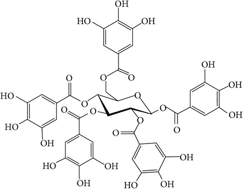
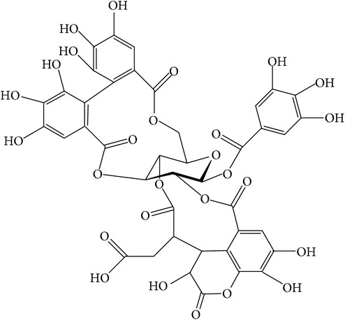
| Ellagitannins | Hydrolysable tannin |
|
Anti-inflammatory, anticancer, cardiovascular protection [28], antioxidant, chemopreventive, antiapoptotic, anti-hepatocellular carcinoma (Anti-HCC) [29] |
|
| |||
| Gallic acid | Phenolic compound |
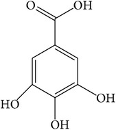
|
Anti-inflammatory [30], antimutagenic [31], cardioprotective [32], antioxidant [33], anticancer [34], antimicrobial [35], neuroprotective [36], immunosuppressive [37], improved cognition [38] |
|
| |||
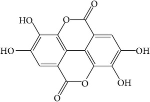
| Chebulic acid | Phenolic compound |
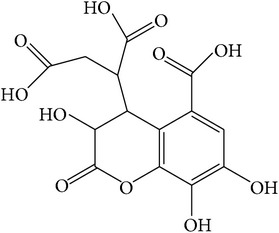
|
Anti-HCV [39], antidiabetic [40], hepatoprotective [41], immunosuppressive [37] |
|
| |||
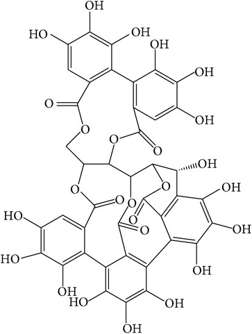
| Chebulagic acid | Hydrolysable tannin |
|
Hepatoprotective [41], antiviral [42], immunosuppressive [43], antidiabetic [44, 45], neuroprotective [46], antiangiogenesis [47], antiproliferative [48], anti-inflammatory [44] |
|
| |||
| Chebulinic acid | Hydrolysable tannin |
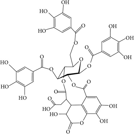
|
Antisecretory, cytoprotective [49], antiangiogenesis [50], antitumor [51] |
|
| |||
| Ellagic acid | Phenolic compound |
|
Antioxidant [52], anti-inflammatory [53], anti-diabetes-induced sexual dysfunction [54], hepatoprotective [55], antiarrhythmic [56], cognitive enhancer [57, 58] |
|
| |||
| Anthraquinone glycosides | Phenolic compound |
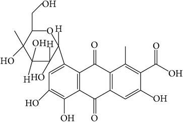
|
Neuroprotective [59], antidiabetic [60] |
Hence, this review article aims to sum up the published literature on the pharmacology and phytochemistry of T. chebula and its effects on the progression and treatment of AD in order to call attention to this plant as a novel alternative in the treatment of Alzheimer's disease.
2. Terminalia chebula Retzius
Terminalia chebula Retzius (Family: Combretaceae), as a shade and ornamental tree with 250 species, is a medicinal plant that grows in the Middle East and tropical regions such as India, China, and Thailand. It can grow to be 25 meters tall and has a variable appearance and spreading branches. The color of the bark is dark brown and is usually cracked. Leaves are thin, elliptic-oblong, cordiform at the base, elliptical, and 7–12 cm long and 4–6.5 cm in width and have a leathery form with entire margins. The upper surface of the leaves is glabrous opposite of the surface beneath. The flowers are futile with a white to yellowish color and unsightly odor. Flowers have 5–7 cm long spikes, simple or branched, about 4 mm across. The ovary is inferior with 10 stamens. Fruits are yellow to orange-brown when ripe and 2.5–5 cm long and unruffled with an ovate-drupe shape [15, 61].
Moreover, it is called by various names by the local people. For example, in the Thai language the plant's common name is “Kot Phung Pla,” and in Indian it is called “Kadukkaai”; and its other names are Black Myrobalan, Ink Tree, or Chebulic Myrobalan (Figure 1) [15].
Figure 1.

T. chebula tree and fruits.
2.1. Scientific Classification
Its classification is Kingdom: Plantae, Division: Magnoliophyta, Class: Magnoliopsida, Order: Myrtales, Family: Combretaceae, Genus: Terminalia, and Species: Chebula [15].
2.2. Phytochemical Compositions of T. chebula
2.2.1. Hydrolysable Tannins
Tannins, as a part of the phenolic compounds, are oligomeric and have multiple structural units with free phenolic groups and their molecular weight ranges from 500 to 3000 D. The fruit pulp and dried pericarp of the seeds contain the highest amount of tannins [62]. Tannins consist of hydrolysable and nonhydrolysable tannins and hydrolysable tannins (i.e., gallotannins and ellagitannins) are the main compounds in T. chebula.
Gallotannins and ellagitannins are polymers found in the fruits of T. chebula [63]. Gallotannins contain gallic acid that has esterified and bonded with the hydroxyl group of a polyol carbohydrate such as glucose [64]. Ellagitannins are formed when oxidative linkage occurs in the galloyl groups in 1,2,3,4,6-pentagalloyl glucose. Ellagitannins differ from gallotannins in that their galloyl groups are linked through C–C bonds, whereas the galloyl groups in gallotannins are linked by depside bonds [65, 66].
Chebulagic acid, a benzopyran tannin, is widely distributed in several plant families: the Combretaceae [67], Euphorbiaceae, Leguminosae, Anacardiaceae, and Fabaceae [31]. Also, in the Combretaceae Family, chebulagic acid is the main constitute of the fruits T. bellerica, T. chebula, and Emblica officinalis [68].
Chebulinic acid, also known as 1,3,6-tri-O-galloyl-2,4-chebuloyl-β-D-glucopyranoside, is an ellagitannin found in the fruits of T. chebula or in the leaves of T. macroptera [31, 68].
2.2.2. Phenolic Compounds
Phenolic compounds include ellagic acid, a natural potent phenolic antioxidant found in the fruits of T. chebula [13]; gallic acid, a trihydroxybenzoic acid found as a part of hydrolysable tannins [69]; tannic acid, a polymer of gallic acid molecules and glucose [70]; and chebulic acid, another phenolic compound in the ripe fruits of T. chebula and a component of transformed ellagitannins such as chebulagic acid or chebulinic acid [41].
Glycosides are other phenolic compounds that are widely present in the Terminalia, rhubarb, Senna, and Aloe species. The pericarp of the T. chebula fruit contains anthraquinone glycosides [71]. Table 1 shows the main ingredients of T. chebula and their chemical structures.
2.2.3. Miscellaneous Compounds
In addition to the above, there are some other compounds that are also present and contribute towards the activity of the plant such as palmitic [72], stearic [73], oleic [74], linoleic [75], and arachidic acids [76], which are present in the fruit kernels.
2.2.4. Methods of Search
This research included articles from 1960 to 2015 that were found in various electronic databases: PubMed, Science Direct, Scopus, Web of Science, Scirus, and Google Scholar by using the search words: T. chebula, AD, neuroprotection, medicinal plant, antioxidant, ellagitannin, gallotannin, gallic acid, chebulagic acid, and chebulinic acid. Only current articles that reported the effects of T. chebula on AD were included in our study. Then, a comparison of the related mechanisms and evaluation of pharmacology effects in the treatment and progression of AD was done.
2.3. T. chebula and Alzheimer's Disease
2.3.1. Acetyl Cholinesterase (AChE)
One of the hypotheses about AD is acetylcholine (ACh) deficiency in the synaptic cleft of the cerebral cortex that causes memory disturbance. Acetyl cholinesterase, also known as AChE or acetyl-hydrolase, is a hydrolase that hydrolyzes the neurotransmitter acetylcholine. AChE is found at mainly neuromuscular junctions and cholinergic brain synapses where its activity serves to terminate synaptic transmission. It is the primary target of inhibition by organophosphorus compounds such as nerve agents and pesticides. AChE has a very high catalytic activity; each molecule of AChE degrades about 25000 molecules of ACh per second, approaching the limit allowed by diffusion of the substrate [77]. During neurotransmission, ACh is released from the nerve into the synaptic cleft and binds to ACh receptors on the postsynaptic membrane, relaying the signal from the nerve. AChE, also located on the postsynaptic membrane, terminates the signal transmission by hydrolyzing Ach [78]. AChE is found in many types of conducting tissue: central and peripheral tissues, motor and sensory fibers, nerve and muscle, and cholinergic and noncholinergic fibers. The activity of AChE is higher in motor neurons than in sensory neurons [79]. AChE is also found on the red blood cell membranes [80]. Traditionally, necrosis is the pathway that had been thought to cause cell death in AD; however, now it is thought that ACh containing neurons are the most important pathway of neuronal death in AD. The cell death mechanisms in AD are deposition of Aβ, reduced energy metabolism and/or mitochondrial dysfunction, excitotoxicity, and oxidative stress and free radical production. These four mechanisms contribute to necrosis in various diseases such as stroke and AD. Inflammatory responses and their effects on trophic factor function have also been proposed as a main factor for cell death in AD. AChE induces an apoptotic and necrotic cell death by inducing membrane depolarization and NMDA receptor activation with consequent Ca2+ influx and modulation of the α7 nicotinic acetylcholine receptor in hippocampal cultures. Therefore, AChE has a main role in neurodegeneration and AChEIs could be effective in neuroprotection [81–83]. Increasing expression of splice variants of AChE (R and S) as neuroprotective forms of AChE, with AChEIs, has been effective in preventing the progression of AD. In a study, expression of AChE-R and AChE-S decreased over a period of 12 months in untreated patients. In contrast, treatment with tacrine causes an upregulation in both variants (up to 117%) [84].
Inhibition of AChE leads to accumulation of ACh in the synaptic cleft and results in impeded neurotransmission [78]. The acetyl cholinesterase inhibitors (AChEIs) attenuate the cholinergic deficit underlying the cognitive and neuropsychiatric dysfunctions in patients with neurodegenerative disorders. So, inhibition of brain AChE has been the major therapeutic target of treatment strategies for AD, Parkinson's disease, senile dementia, ataxia, and myasthenia gravis [85]. There are a few synthetic medicines with this mechanism, for example, tacrine, donepezil, and the natural product-based rivastigmine, to treat cognitive dysfunction and memory loss associated with AD [86]. However, these compounds have been reported to have their adverse effects including gastrointestinal disturbances and problems associated with bioavailability, which necessitates the interest in finding better AChE inhibitors from natural sources [87].
In this section we discuss the effectiveness of T. chebula and its constituents on brain functions.
2.3.2. Anticholinesterase Properties
Anticholinesterase properties of T. chebula have been reported in many studies [3, 88–95]. In Sancheti et al.'s study, the inhibitory effects of T. chebula on AChE and butyrylcholinesterase (BChE) were reported. In the in vitro study, methanolic crude extract of the fruits of T. chebula with a concentration of 5 mg/mL inhibited AChE and BChE about 89% and 95%, respectively. Sancheti et al. extracted 1,2,3,4,6-penta-O-galloyl-β-D-glucose (PGG) with a gallotannin structure from T. chebula by chromatographic methods, and they showed it to be the most potent AChE and BChE inhibitor. Bioassay of PGG exhibited its concentration-dependent inhibitory activity on AChE and BChE with IC50 values of 29.9 ± 0.3 μM and 27.6 ± 0.2 μM, respectively. Then, for deducing the inhibitory activity of PGG, a TLC assay was done with tacrine as a positive control, and the AChE inhibitory effect was visualized clearly in the assay [88]. In addition to the inhibitory effect of methanolic fruit extracts of T. chebula on AChE, the ethyl acetate fraction shows relatively remarkable AChE inhibitory activity. Sulaiman et al. showed that ethyl acetate fraction with doses of 1, 5, 15, and 25 mg/mL inhibited AChE by 29.36%, 32.44%, 45.82%, and 62.32%, respectively. This fraction was more potent than the chloroform and methanolic fraction in AChE inhibition [91]. In a similar in vitro study, Vinutha et al. used the methanolic and aqueous extracts of the fruit of T. chebula and the data showed that IC50 values of the aqueous extract of T. chebula (minimum inhibition: 12.45%) are more potent than the methanolic extract (minimum inhibition: 1.21%) [3].
In addition to the effects of gallotannins on AChE inhibition, tannic acid as an active component of T. chebula has a potential inhibition effect on AChE. Upadhyay and Singh showed the competitive inhibition of AChE by tannic acid in vivo and in vitro. This study focused on the effects of tannic acid from the T. chebula fruit. In vivo treatment with 80% of LC50 of tannic acid during 96 h caused a remarkable inhibition in AChE activity in the nervous tissue of Lymnaea acuminate. Furthermore, maximum inhibition of AChE activity was detected when the tissue was exposed to 80% of LC50 of tannic acid for 96 h, so inhibition of AChE was time- and dose-dependent in the 96 h process. In vitro treatment showed that tannic acid caused dose-dependent AChE inhibition significantly, such that 0.04 mM of tannic acid reduced the AChE activity to 37% of the control in the nervous tissue of L. acuminata [92]. In addition, another in vivo research on AChE activity showed that tannic acid completely neutralized the AChE activity in Naja kaouthia venom [96].
Parle and Vasudevan in their in vivo study showed that Abana (a mixture of medicinal plants containing T. chebula) with doses of 50, 100, and 200 mg/kg administered orally for 15 days can reduce the brain AChE activity in young and aged mice. Passive avoidance apparatus and maze tests were performed on day 16 and the results showed a remarkable dose-dependent reduction in transfer latency and a significant increase in step-down latency tests, indicating significant improvement in memory [93].
Another similar study was done by Dhivya et al. In this in vitro study, AChE inhibitory activity of various plant species containing T. chebula was reported to have AChE inhibition of 41.06 ± 5.6% (0.1 mg/mL) as well [95].
In a similar study, AChE inhibition of the methanolic extract of T. chebula showed a moderate inhibition with an extract with IC50 value of 180 ± 14.6 μg/mL and the percentage of AChE inhibition was 41.06 ± 5.6 at 0.1 mg/mL [97]. In addition, the percentage of AChE inhibition in the extract of T. chebula was 13% (IC50 not detected) [14]. Furthermore, in another research, T. chebula could inhibit 89% of AChE activity (5 mg/mL) and 95% of BChE activity (5 mg/mL) [88].
Gallic acid and ellagic acid, as phenolic compounds in the fruits of T. chebula, could also inhibit AChE. Nag and De in their study showed that the inhibitory effect of the methanolic extract of T. chebula (gallic acid: 0.25 μg, ellagic acid: 0.08 μg) has a linear relationship with increasing dosage (IC50: 10.96) [89].
2.3.3. Anti-Inflammatory Properties
Cyclooxygenase (COX) and 5-lipoxygenase (5-LOX) are the key enzymes that are involved in inflammation. Reddy et al. investigated chebulagic acid, as a COX-LOX dual inhibitor, from the ethanolic extract of fruits of T. chebula. The results showed that chebulagic acid has a potent COX-LOX dual inhibition activity with IC50 values of 15 ± 0.288, 0.92 ± 0.011, and 2.1 ± 0.057 μM for COX-1, COX-2, and 5-LOX, respectively [76].
The water soluble fraction of T. chebula (WFTC) was effective against systemic and local anaphylaxis. Shin et al. showed that the injection of WFTC with doses of 0.01–1.0 g/kg inhibited anaphylactic shock 100%. When WFTC was pretreated at concentrations ranging from 0.005 to 1.0 g/kg, the serum histamine levels were reduced dose-dependently. In addition, this study showed that WFTC increased anti-dinitrophenol and IgE-induced tumor necrosis factor- (TNF-) α production [98].
Lee et al. in their study showed that gallic acid (25 μg) suppressed nuclear factor kappa B (NF-κB) activity and cytokine release. It also remarkably reduced the cyclic AMP response element binding protein/p300 (CBP/p300, a NF-κB coactivator) gene expression, acetylation levels, and CBP/p300 histone acetyltransferase activity. Therefore, gallic acid derived from the extracts of several plants especially T. chebula was shown to exert anti-inflammatory activity via the downregulation of the NF-κB pathway in the development of inflammatory diseases, both in vitro and in vivo [30, 99–101].
Kim et al. showed that the inhibition of NF-κB acetylation with gallic acid resulted in reduced cytokine production in microglia cells and the protection of neuronal cells from amyloid β- (Aβ-) induced neurotoxicity. In addition, this research showed a restorative effect of gallic acid on Aβ-induced cognitive dysfunction in mice in Y-maze and passive avoidance tests. Therefore, gallic acid has several roles in neuroinflammatory diseases including (1) preventing Aβ-induced neuronal cell death by inhibiting RelA (as a gene encoding NF-κB) acetylation and cytokine production; (2) protecting neuronal cells from primary microglia-mediated Aβ neurotoxicity; (3) restoring Aβ-induced memory deficits in mice; and (4) suppressing in vivo cytokine production by inhibiting RelA acetylation in brain [101]. Another similar study showed that polyphenolic extracts (containing gallic acid) of medicinal plants such as T. chebula are able to inhibit Aβ aggregation, reduce Aβ production, and protect against Aβ neurotoxicity, in vitro [102]. Kim et al. reported the inhibitory effects of gallic acid on inflammatory responses via NF- κB and p38 mitogen-activated protein kinase pathways [103].
In another study, chebulagic acid showed a potent anti-inflammatory effect in lipopolysaccharide- (LPS) stimulated RAW 264.7 (mouse macrophage cell line). Reddy and Reddanna showed the effectiveness of chebulagic acid against inflammatory diseases by different mechanisms such as inhibition of nitric oxide (NO) and prostaglandin E2 production, downregulation of iNOS (inducible nitric oxide synthases), COX-2, 5-LOX, TNF-α, interleukin 6, and inhibitory effects on NF-κB, and decreases in nuclear p50 and p65 protein levels. In addition, the generation of reactive oxygen species (ROS) and phosphorylation of p38, ERK 1/2, and JNK in LPS-stimulated RAW 264.7 cells was suppressed by chebulagic acid in a concentration-dependent manner [44]. In another research, chebulagic acid from the immature seeds of T. chebula significantly suppressed the onset and progression of collagen induced arthritis in mice [104]. In a similar study, the aqueous extract of the dried fruit of T. chebula showed an anti-inflammatory effect by inhibiting iNOS [105]. Patil et al. in their study showed that administrating the fruit of T. chebula (500 mg/kg) via inhibition of iNOS expression can be significantly effective in the progression of inflammation [106]. In vivo anti-inflammatory activity of T. chebula fruit extract at different doses (range: 50 to 500 mg/kg) was evaluated against carrageenan-induced inflammation in rats. In this study, Bag et al. reported that 250 mg/kg (administrated orally) caused a 69.96% reduction in carrageenan-induced rat paw edema. This finding suggests that free radical quenching may be one of the mechanisms for its anti-inflammatory activity [107]. Triphala, an Indian Ayurvedic herbal formulation containing T. chebula, at a dosage of 1 g/kg, showed an anti-inflammatory effect. Sabina and Rasool in their research showed that the mechanisms of anti-inflammatory properties of this plant include (a) inhibition of lysosomal enzyme release, (b) a significant decrease of inflammatory mediator TNF-α, and (c) a decrease of beta-glucuronidase and lactate dehydrogenase enzymes release. The anti-inflammatory effect of Triphala might be due to the presence of phenolic components and flavonoids [108]. In another study, Ramani and Pradhan showed that the acetone extract of T. chebula fruits had a significant effect on controlling Complete Freund's Adjuvant-induced arthritis. This effect leads to a good reduction in paw edema and joint thickness (reduction of ESR values and RF values) in comparison with dexamethasone [109].
2.3.4. Antioxidant Properties
Decreases in antioxidant defense or increases in oxidant status in the body tend to lead to the condition called oxidative stress, which plays a major role in neurological degeneration such as AD [110]. Reactive oxygen species and oxidative stress increase the formation of amyloid-β and senile plaques in the brain are the hallmark of AD [111]. High lipid content, deficient antioxidant values, and high oxygen consumption in the brain are the reasons the central nervous system is vulnerable to oxidative stress. Increased lipid peroxidation, decreased glutathione levels, increased dopamine turnover, and elevated iron and aluminum levels in the brain support the role of oxidative stress in Parkinson's, Amyotrophic Lateral Sclerosis, and AD [112].
In Table 2, we have summarized the roles of oxidative stress in the pathogenesis and progression of AD. Thus, reducing oxidative stress by enhancing antioxidant defense or decreasing the production of reactive oxygen species may be effective in treating AD.
Table 2.
Major effects of oxidative stress in the pathogenesis of Alzheimer's disease.
| Effects of ROS | Result | Biological evidence(s) |
|---|---|---|
| Protein oxidation | Increased protein carbonyl content | Increased protein oxidation in frontal pole and occipital pole [113] Decreased ratio of (MAL-6)/(W/S) in AD hippocampus and inferior parietal lobule, decreased the W/S ratio in in vitro models of human synaptosomes oxidation by ROS [114] |
|
| ||
| DNA oxidation | Direct damage to DNA structure | 3-fold increase in mitochondrial DNA oxidation in parietal cortex in AD [115] Increase in oxidative damage to nuclear DNA in AD compared with age-matched control subjects [116] 8-hydroxy-2-deoxyguanosine as a marker of DNA oxidation increases in AD [117] |
|
| ||
| Lipid peroxidation | Brain phospholipid damage | Increased TBARS levels in AD in hippocampus, piriform cortex, and amygdala [118] Increased lipid peroxidation of AD brain homogenates in vitro due to Fe-H2O2 [119] Increased apoptosis in cultured DS and AD neurons inhibited by antioxidant enzymes [112, 120] Decrease in PC, PE, phospholipid precursors, choline, and ethanolamine in hippocampus and inferior parietal lobule in AD [121] Increased aldehydes as a cytotoxic agent in the brain of AD patients [122] |
|
| ||
| Antioxidant enzymes | Changes in enzymes contents | Elevated GSH-Px, GSSG-R, and CAT activity in hippocampus and amygdala in AD [118] Many studies showed no elevation in enzyme activity [123, 124] or decrease in activity [125] |
|
| ||
| AGE formation | Pathological changes in protein structure and action | Accelerates aggregation of soluble nonfibrillar Aβ and tau [126] |
MAL-6: weakly immobilized protein bound spin label; W/S: strongly immobilized protein bound spin label; TBARS: thiobarbituric acid reactive substances; DS: Down syndrome; PC: phosphatidylcholine; PE: phosphatidylethanolamine; GSH-Px: glutathione peroxidase; GSSG-R: glutathione reductase; CAT: catalase; AGE: advanced glycation end products.
Antioxidant effects of T. chebula extract (100 μg/mL) were compared with reference radical scavengers such as quercetin, gallic acid, and t-butylhydroquinone, and T. chebula extract showed 95% activity with IC50 2.2 μg/mL [14]. Terminalia species such as T. chebula and T. arjuna with a high content of phenolic constituents showed strong antioxidant and antiaging properties [88]. Oral administration of aqueous extracts of T. arjuna causes significant elevation in the activities of antioxidant enzymes such as superoxide dismutase, catalase, and glutathione S-transferase. The strong antioxidant action of the aqueous extract of the Terminalia species may play a role in treating age-related diseases such as AD [127]. A similar study done by Mathew and Subramanian showed that the antioxidant activity of the methanolic extract of T. chebula (0.1 mg/mL) and IC50 was 86.3% and 0.5 μg/mL, respectively. In this study the highest amount of antioxidant activity among 20 plants was found in T. chebula due to the presence of gallic acid and ellagic acid. All 20 plants used in this study were traditional neuroprotective plants that have been used for many years and among them T. chebula was the most potent antioxidant and moderate inhibition of AchE [97]. In the evaluation of antioxidant, anti-inflammatory, membrane-stabilizing, and antilipid peroxidative effects of the hydroalcoholic extract of T. chebula fruits in arthritic disorders, Bag et al. showed that the fruit extract of T. chebula (10 to 100 μg/mL) in carrageenan-induced inflammation in rats reduced the formation of thiobarbituric acid reactive substances in the liver with IC50 94.96 mg/kg. The IC50 of the extract in DPPH radical-scavenging activity was 42.14 μg/mL [107]. Antilipid peroxidative capacity of T. chebula was attributed to higher phenolic content, reducing ability, Fe(II) chelating ability, and free radical-scavenging activity. In Khan et al.'s study, the aqueous extracts of yellow T. chebula and black T. chebula showed IC50 values of 20.24 ± 0.9 μg/mL and 17.33 ± 1.1 μg/mL in the scavenging of the DPPH radical in mice brains, respectively [128]. In an in vitro study done to investigate the biological activities of phenolic compounds and triterpenoid constituents of T. chebula, 9 phenolic compounds and 8 triterpenoids were extracted. Radical-scavenging activities of the phenolic compounds were higher than the triterpenoids compounds and had potent inhibitory activities against melanogenesis at a concentration of 10 μM and the IC50 was 1.4–10.9 μM. These results showed that T. chebula with a high content of polyphenolic ingredients was a potent antioxidant [16]. The extract of the dried fruit pulp of T. chebula (600 mg/kg) could diminish acetic acid-induced colitis in rats with antioxidant effects (free radical-scavenging, antioxidant enzymes enhancing activity and decreasing lipid peroxidation) [129].
In the evaluation of neuroprotective effects, the inhibition of H2O2-induced PC12 cell death by T. chebula was investigated. H2O2 at 40 μM for 12 h decreased cell viability to 59%. Also, all of the methanol, water, and 95% ethanol extracts of T. chebula increased cell viability at 0.5–5.0 μg/mL, dose-dependently; and among them the water extract was more potent. Antioxidant effects of the extracts were compared and the results showed that methanol extract had good antioxidant activity based on the luminol-H2O2-horseradish peroxidase assay and the water extract was shown to have good antioxidant activities in cupric sulfate phenanthroline-H2O2 and luminol-H2O2 assays. Also, 95% ethanol extract showed good antioxidant activity in the pyrogallol-luminol assay [130]. Effects of T. chebula seed extracts on brain-derived neurotrophic factor (BDNF), Cu, Zn-superoxide dismutase (SOD1), and Mn-superoxide dismutase (SOD2) were evaluated by Park et al. Extract with a dose of 100 mg/kg once a day was administrated to gerbils for 7 days before induction of transient cerebral ischemia and the neuroprotective effect of the extract was evaluated in the CA1 hippocampal region on the fourth day after ischemia-reperfusion induction. The results showed that astrocytes and microglia remarkably decreased in the ischemic group compared with the vehicle-treated group and neuron nucleus (as the positive neuron) was distinctively abundant (62%) compared to the vehicle-treated ischemia group (62% versus 12.2%). Protein levels and activity of SOD1, SOD2, and BDNF were much higher in the treated group. These results confirmed the neuroprotective effects of T. chebula extracts against neuronal damage in hippocampus [131]. Recently, we have found that the alcoholic extract of T. chebula fruits significantly protects against quinolinic acid-induced neurotoxicity by alleviating the oxidative stress parameters (unpublished data).
Many studies used T. chebula to modulate the oxidative stress adverse effects in various models of oxidation damage and aging [132–134]. T. chebula could enhance the antioxidant defense and modulate oxidative stress due to aging in the liver and kidney of aged rats compared to young albino rats [133] and could protect hepatocytes from damage due to oxidative stress and enhanced total thiol content [41]. Many studies have shown the antioxidant activity of T. chebula in various in vivo and in vitro models [16, 135, 136]. In addition, most of the researchers agree with this hypothesis that “pharmacophore, which contains large numbers of –OH group (such as polyphenols), has greater anti-aging properties than those with less numbers of –OH groups.” Therefore, two constituents that are potent phenolic compounds are ellagic acid and 4-O-xyloside of ellagic acid. These active ingredients were investigated for their activities on free radical-scavenging, elastase inhibition, expression of matrix metalloproteinase-1 (MMP-1), and type I collagen synthesis in normal human fibroblast cells. Thus, ellagic acid may be useful for the geriatric population [137, 138].
According to the points mentioned above regarding the antioxidant activity of T. chebula and the important role of oxidative stress in the pathogenesis of AD, we can conclude that the use of this plant may be effective against the progression of AD.
3. Conclusion
Alzheimer's disease is a debilitating dementia, and only a limited number of therapeutic options are currently available to modify the manifestations of the disease. T. chebula has pharmacological activities relevant to dementia therapy (Figure 2). Different extracts from T. chebula have exhibited concentration-dependent inhibitory activities on AChE and BChE. Such AChE activities are also described for its active ingredients such as PGG, gallic acid, ellagic acid, and tannic acid. Anti-inflammatory properties of T. chebula have been well documented in different experimental systems that could be attributed to chebulagic or gallic acid. T. chebula with a high content of phenolic constituents exhibits strong antioxidant and neuroprotective properties in vitro and in vivo. The efficacy of T. chebula in treating AD should be compared with the current standard pharmacological treatment in animal and clinical testing and researches. Such studies should include the identification of the active principle(s) in order to improve the validation of the clinical trials. Until then, this review provides some evidences on the benefits of T. chebula in the treatment of Alzheimer's disease.
Figure 2.
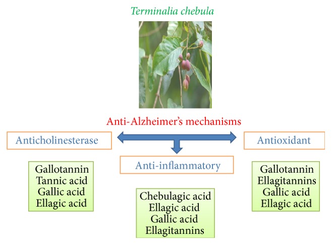
Anticholinesterase, anti-inflammatory, and antioxidant properties of T. chebula relevant to anti-Alzheimer's therapy.
Conflict of Interests
The authors confirm that there is no conflict of interests.
References
- 1.Stern Y. Cognitive reserve and Alzheimer disease. Alzheimer Disease & Associated Disorders. 2006;20(2):112–117. doi: 10.1097/01.wad.0000213815.20177.19. [DOI] [PubMed] [Google Scholar]
- 2.Adhami H.-R., Farsam H., Krenn L. Screening of medicinal plants from Iranian traditional medicine for acetylcholinesterase inhibition. Phytotherapy Research. 2011;25(8):1148–1152. doi: 10.1002/ptr.3409. [DOI] [PubMed] [Google Scholar]
- 3.Vinutha B., Prashanth D., Salma K., et al. Screening of selected Indian medicinal plants for acetylcholinesterase inhibitory activity. Journal of Ethnopharmacology. 2007;109(2):359–363. doi: 10.1016/j.jep.2006.06.014. [DOI] [PubMed] [Google Scholar]
- 4.Chopra K., Misra S., Kuhad A. Current perspectives on pharmacotherapy of Alzheimer's disease. Expert Opinion on Pharmacotherapy. 2011;12(3):335–350. doi: 10.1517/14656566.2011.520702. [DOI] [PubMed] [Google Scholar]
- 5.López S., Bastida J., Viladomat F., Codina C. Acetylcholinesterase inhibitory activity of some Amaryllidaceae alkaloids and Narcissus extracts. Life Sciences. 2002;71(21):2521–2529. doi: 10.1016/s0024-3205(02)02034-9. [DOI] [PubMed] [Google Scholar]
- 6.Ellman G. L., Courtney K. D., Andres V., Jr., Featherstone R. M. A new and rapid colorimetric determination of acetylcholinesterase activity. Biochemical Pharmacology. 1961;7(2):88–95. doi: 10.1016/0006-2952(61)90145-9. [DOI] [PubMed] [Google Scholar]
- 7.Rhee I. K., van de Meent M., Ingkaninan K., Verpoorte R. Screening for acetylcholinesterase inhibitors from Amaryllidaceae using silica gel thin-layer chromatography in combination with bioactivity staining. Journal of Chromatography A. 2001;915(1-2):217–223. doi: 10.1016/s0021-9673(01)00624-0. [DOI] [PubMed] [Google Scholar]
- 8.Anand P., Singh B. A review on cholinesterase inhibitors for Alzheimer's disease. Archives of Pharmacal Research. 2013;36(4):375–399. doi: 10.1007/s12272-013-0036-3. [DOI] [PubMed] [Google Scholar]
- 9.Dirin M. M., Mousavi S., Afshari A. R., Tabrizian K., Ashrafi M. H. Potential drug-drug interactions in prescriptions dispensed in community and hospital pharmacies in East of Iran. Journal of Research in Pharmacy Practice. 2014;3(3):104–107. doi: 10.4103/2279-042x.141118. [DOI] [PMC free article] [PubMed] [Google Scholar]
- 10.May B. H., Lit M., Xue C. C. L., et al. Herbal medicine for dementia: a systematic review. Phytotherapy Research. 2009;23(4):447–459. doi: 10.1002/ptr.2656. [DOI] [PubMed] [Google Scholar]
- 11.Akhondzadeh S., Abbasi S. H. Herbal medicine in the treatment of Alzhelmer's disease. American Journal of Alzheimer's Disease and other Dementias. 2006;21(2):113–118. doi: 10.1177/153331750602100211. [DOI] [PMC free article] [PubMed] [Google Scholar]
- 12.Santos-Neto L. L. D., de Vilhena Toledo M. A., Medeiros-Souza P., de Souza G. A. The use of herbal medicine in Alzheimer's disease—a systematic review. Evidence-Based Complementary and Alternative Medicine. 2006;3(4):441–445. doi: 10.1093/ecam/nel071. [DOI] [PMC free article] [PubMed] [Google Scholar]
- 13.Saleem A., Husheem M., Härkönen P., Pihlaja K. Inhibition of cancer cell growth by crude extract and the phenolics of Terminalia chebula retz. fruit. Journal of Ethnopharmacology. 2002;81(3):327–336. doi: 10.1016/s0378-8741(02)00099-5. [DOI] [PubMed] [Google Scholar]
- 14.Ali S. K., Hamed A. R., Soltan M. M., et al. In-vitro evaluation of selected Egyptian traditional herbal medicines for treatment of Alzheimer disease. BMC Complementary and Alternative Medicine. 2013;13(1):p. 121. doi: 10.1186/1472-6882-13-121. [DOI] [PMC free article] [PubMed] [Google Scholar]
- 15.Rathinamoorthy R., Thilagavathi G. Terminalia chebula—review on pharmacological and biochemical studies. International Journal of PharmTech Research. 2014;6(1):97–116. [Google Scholar]
- 16.Manosroi A., Jantrawut P., Ogihara E., et al. Biological activities of phenolic compounds and triterpenoids from the Galls of Terminalia chebula . Chemistry & Biodiversity. 2013;10(8):1448–1463. doi: 10.1002/cbdv.201300149. [DOI] [PubMed] [Google Scholar]
- 17.Rao N. K., Nammi S. Antidiabetic and renoprotective effects of the chloroform extract of Terminalia chebula Retz. seeds in streptozotocin-induced diabetic rats. BMC Complementary and Alternative Medicine. 2006;6(1, article 17) doi: 10.1186/1472-6882-6-17. [DOI] [PMC free article] [PubMed] [Google Scholar]
- 18.Aneja K., Joshi R. Evaluation of antimicrobial properties of fruit extracts of Terminalia chebula against dental caries pathogens. Jundishapur Journal of Microbiology. 2009;2(3):105–111. [Google Scholar]
- 19.Dutta B., Rahman I., Das T. Antifungal activity of Indian plant extracts: antimyzetische Aktivität indischer Pflanzenextrakte. Mycoses. 1998;41(11-12):535–536. doi: 10.1111/j.1439-0507.1998.tb00718.x. [DOI] [PubMed] [Google Scholar]
- 20.Aher V., Wahi A. Immunomodulatory activity of alcohol extract of Terminalia chebula retz combretaceae. Tropical Journal of Pharmaceutical Research. 2011;10(5):567–575. [Google Scholar]
- 21.Ahn M.-J., Kim C. Y., Lee J. S., et al. Inhibition of HIV-1 integrase by galloyl glucoses from Terminalia chebula and flavonol glycoside gallates from Euphorbia pekinensis . Planta Medica. 2002;68(5):457–459. doi: 10.1055/s-2002-32070. [DOI] [PubMed] [Google Scholar]
- 22.Maruthappan V., Shree K. S. Hypolipidemic activity of Haritaki (Terminalia chebula) in atherogenic diet induced hyperlipidemic rats. Journal of Advanced Pharmaceutical Technology and Research. 2010;1(2):229–235. [PMC free article] [PubMed] [Google Scholar]
- 23.Srigopalram S., Jayraaj I. A. Effect of Terminalia chebula retz on den induced hepatocellular carcinogenesis in experimental rats. International Journal of Pharmacy and Pharmaceutical Sciences. 2012;4(2):440–445. [Google Scholar]
- 24.Sharma P., Prakash T., Kotresha D., et al. Antiulcerogenic activity of Terminalia chebula fruit in experimentally induced ulcer in rats. Pharmaceutical Biology. 2011;49(3):262–268. doi: 10.3109/13880209.2010.503709. [DOI] [PubMed] [Google Scholar]
- 25.Singh M. P., Sharma C. S. Wound healing activity of Terminalia chebula in experimentally induced diabetic rats. International Journal of PharmTech Research. 2009;1(4):1267–1270. [Google Scholar]
- 26.Engels C., Knödler M., Zhao Y.-Y., Carle R., Gänzle M. G., Schieber A. Antimicrobial activity of gallotannins isolated from mango (Mangifera indica L.) kernels. Journal of Agricultural and Food Chemistry. 2009;57(17):7712–7718. doi: 10.1021/jf901621m. [DOI] [PubMed] [Google Scholar]
- 27.Zhao X., Sun H., Hou A., Zhao Q., Wei T., Xin W. Antioxidant properties of two gallotannins isolated from the leaves of Pistacia weinmannifolia . Biochimica et Biophysica Acta (BBA)—General Subjects. 2005;1725(1):103–110. doi: 10.1016/j.bbagen.2005.04.015. [DOI] [PubMed] [Google Scholar]
- 28.Sangiovanni E., Vrhovsek U., Rossoni G., et al. Ellagitannins from Rubus berries for the control of gastric inflammation: in vitro and in vivo studies. PLoS ONE. 2013;8(8) doi: 10.1371/journal.pone.0071762.e71762 [DOI] [PMC free article] [PubMed] [Google Scholar]
- 29.Hussein R. H., Khalifa F. K. The protective role of ellagitannins flavonoids pretreatment against N-nitrosodiethylamine induced-hepatocellular carcinoma. Saudi Journal of Biological Sciences. 2014;21(6):589–596. doi: 10.1016/j.sjbs.2014.03.004. [DOI] [PMC free article] [PubMed] [Google Scholar]
- 30.Lee W., Lee S. Y., Son Y., Yun J. Gallic acid decreases inflammatory cytokine secretion through histone acetyltransferase/histone deacetylase regulation in high glucose-induced human monocytes. Journal of Medicinal Food. 2015;18(7):793–801. doi: 10.1089/jmf.2014.3342. [DOI] [PubMed] [Google Scholar]
- 31.Walia H., Arora S. Terminalia chebula—a pharmacognistic account. Journal of Medicinal Plants Research. 2013;7(20):1351–1361. [Google Scholar]
- 32.Priscilla D. H., Prince P. S. M. Cardioprotective effect of gallic acid on cardiac troponin-T, cardiac marker enzymes, lipid peroxidation products and antioxidants in experimentally induced myocardial infarction in Wistar rats. Chemico-Biological Interactions. 2009;179(2-3):118–124. doi: 10.1016/j.cbi.2008.12.012. [DOI] [PubMed] [Google Scholar]
- 33.Reynolds L. D., Wilson N. G. Scribes and Scholars. Cambridge, UK: Cambridge University Press; 1974. [Google Scholar]
- 34.Ho H.-H., Chang C.-S., Ho W.-C., Liao S.-Y., Wu C.-H., Wang C.-J. Anti-metastasis effects of gallic acid on gastric cancer cells involves inhibition of NF-κB activity and downregulation of PI3K/AKT/small GTPase signals. Food and Chemical Toxicology. 2010;48(8-9):2508–2516. doi: 10.1016/j.fct.2010.06.024. [DOI] [PubMed] [Google Scholar]
- 35.Farag M. A., Al-Mahdy D. A., Salah El Dine R., et al. Structure—activity relationships of antimicrobial gallic acid derivatives from pomegranate and acacia fruit extracts against potato bacterial wilt pathogen. Chemistry & Biodiversity. 2015;12(6):955–962. doi: 10.1002/cbdv.201400194. [DOI] [PubMed] [Google Scholar]
- 36.Ibrahim A., El Kareem R., Sheir M. Elucidation of acrylamide genotoxicity and neurotoxicity and the protective role of gallic acid and green tea. Journal of Forensic Toxicology & Pharmacology. 2015;4(1, article 1) doi: 10.4172/2325-9841.1000135. [DOI] [Google Scholar]
- 37.Hamada S.-I., Kataoka T., Woo J.-T., et al. Immunosuppressive effects of gallic acid and chebulagic acid on CTL-mediated cytotoxicity. Biological and Pharmaceutical Bulletin. 1997;20(9):1017–1019. doi: 10.1248/bpb.20.1017. [DOI] [PubMed] [Google Scholar]
- 38.Sarkaki A., Fathimoghaddam H., Mansouri S. M. T., Korrani M. S., Saki G., Farbood Y. Gallic acid improves cognitive, hippocampal long-term potentiation deficits and brain damage induced by chronic cerebral hypoperfusion in rats. Pakistan Journal of Biological Sciences. 2014;17(8):978–990. doi: 10.3923/pjbs.2014.978.990. [DOI] [PubMed] [Google Scholar]
- 39.Ajala O. S., Jukov A., Ma C.-M. Hepatitis C virus inhibitory hydrolysable tannins from the fruits of Terminalia chebula . Fitoterapia. 2014;99:117–123. doi: 10.1016/j.fitote.2014.09.014. [DOI] [PubMed] [Google Scholar]
- 40.Pham A. T., Malterud K. E., Paulsen B. S., Diallo D., Wangensteen H. α-Glucosidase inhibition, 15-lipoxygenase inhibition, and brine shrimp toxicity of extracts and isolated compounds from Terminalia macroptera leaves. Pharmaceutical Biology. 2014;52(9):1166–1169. doi: 10.3109/13880209.2014.880486. [DOI] [PubMed] [Google Scholar]
- 41.Lee H.-S., Jung S.-H., Yun B.-S., Lee K.-W. Isolation of chebulic acid from Terminalia chebula Retz. and its antioxidant effect in isolated rat hepatocytes. Archives of Toxicology. 2007;81(3):211–218. doi: 10.1007/s00204-006-0139-4. [DOI] [PubMed] [Google Scholar]
- 42.Yang Y., Xiu J., Liu J., et al. Chebulagic acid, a hydrolyzable tannin, exhibited antiviral activity in vitro and in vivo against human enterovirus 71. International Journal of Molecular Sciences. 2013;14(5):9618–9627. doi: 10.3390/ijms14059618. [DOI] [PMC free article] [PubMed] [Google Scholar]
- 43.Banerjee R., Mukherjee G., Patra K. C. Microbial transformation of tannin-rich substrate to gallic acid through co-culture method. Bioresource Technology. 2005;96(8):949–953. doi: 10.1016/j.biortech.2004.08.004. [DOI] [PubMed] [Google Scholar]
- 44.Reddy D. B., Reddanna P. Chebulagic acid (CA) attenuates LPS-induced inflammation by suppressing NF-κB and MAPK activation in RAW 264.7 macrophages. Biochemical and Biophysical Research Communications. 2009;381(1):112–117. doi: 10.1016/j.bbrc.2009.02.022. [DOI] [PubMed] [Google Scholar]
- 45.Huang Y.-N., Zhao D.-D., Gao B., et al. Anti-hyperglycemic effect of chebulagic acid from the fruits of Terminalia chebula Retz. International Journal of Molecular Sciences. 2012;13(5):6320–6333. doi: 10.3390/ijms13056320. [DOI] [PMC free article] [PubMed] [Google Scholar]
- 46.Kim H. J., Kim J., Kang K. S., Lee K. T., Yang H. O. Neuroprotective effect of chebulagic acid via autophagy induction in SH-SY5Y cells. Biomolecules and Therapeutics. 2014;22(4):275–281. doi: 10.4062/biomolther.2014.068. [DOI] [PMC free article] [PubMed] [Google Scholar]
- 47.Athira A. P., Helen A., Saja K., Reddanna P., Sudhakaran P. R. Inhibition of angiogenesis in vitro by chebulagic acid: a COX-LOX dual inhibitor. International Journal of Vascular Medicine. 2013;2013:8. doi: 10.1155/2013/843897.843897 [DOI] [PMC free article] [PubMed] [Google Scholar]
- 48.Kumar N., Gangappa D., Gupta G., Karnati R. Chebulagic acid from Terminalia chebula causes G1 arrest, inhibits NFκB and induces apoptosis in retinoblastoma cells. BMC Complementary and Alternative Medicine. 2014;14, article 319 doi: 10.1186/1472-6882-14-319. [DOI] [PMC free article] [PubMed] [Google Scholar]
- 49.Mishra V., Agrawal M., Onasanwo S. A., et al. Anti-secretory and cyto-protective effects of chebulinic acid isolated from the fruits of Terminalia chebula on gastric ulcers. Phytomedicine. 2013;20(6):506–511. doi: 10.1016/j.phymed.2013.01.002. [DOI] [PubMed] [Google Scholar]
- 50.Lu K., Chakroborty D., Sarkar C., et al. Triphala and its active constituent chebulinic acid are natural inhibitors of vascular endothelial growth factor-a mediated angiogenesis. PLoS ONE. 2012;7(8) doi: 10.1371/journal.pone.0043934.e43934 [DOI] [PMC free article] [PubMed] [Google Scholar]
- 51.Yi Z.-C., Wang Z., Li H.-X., Liu M.-J., Wu R.-C., Wang X.-H. Effects of chebulinic acid on differentiation of human leukemia K562 cells. Acta Pharmacologica Sinica. 2004;25(2):231–238. [PubMed] [Google Scholar]
- 52.Tanaka T., Kouno I., Nonaka G.-I. Glutathione-mediated conversion of the ellagitannin geraniin into chebulagic acid. Chemical and Pharmaceutical Bulletin. 1996;44(1):34–40. doi: 10.1248/cpb.44.34. [DOI] [Google Scholar]
- 53.Mansouri M., Hemmati A., Naghizadeh B., Mard S., Rezaie A., Ghorbanzadeh B. A study of the mechanisms underlying the anti-inflammatory effect of ellagic acid in carrageenan-induced paw edema in rats. Indian Journal of Pharmacology. 2015;47(3):292–298. doi: 10.4103/0253-7613.157127. [DOI] [PMC free article] [PubMed] [Google Scholar]
- 54.Goswami S., Vishwanath M., Gangadarappa S., Razdan R., Inamdar M. Efficacy of ellagic acid and sildenafil in diabetes-induced sexual dysfunction. Pharmacognosy Magazine. 2014;10(supplement 3):581–587. doi: 10.4103/0973-1296.139790. [DOI] [PMC free article] [PubMed] [Google Scholar]
- 55.García-Niño W. R., Zazueta C. Ellagic acid: pharmacological activities and molecular mechanisms involved in liver protection. Pharmacological Research. 2015;97:84–103. doi: 10.1016/j.phrs.2015.04.008. [DOI] [PubMed] [Google Scholar]
- 56.Dianat M., Amini N., Badavi M., Farbood Y. Ellagic acid improved arrhythmias induced by CaCL2 in the rat stress model. Avicenna Journal of Phytomedicine. 2015;5(2):120–127. [PMC free article] [PubMed] [Google Scholar]
- 57.Dolatshahi M., Farbood Y., Sarkaki A., Mansouri S. M. T., Khodadadi A. Ellagic acid improves hyperalgesia and cognitive deficiency in 6-hydroxidopamine induced rat model of Parkinson’s disease. Iranian Journal of Basic Medical Sciences. 2015;18(1):38–46. [PMC free article] [PubMed] [Google Scholar]
- 58.Farbood Y., Sarkaki A., Dianat M., Khodadadi A., Haddad M. K., Mashhadizadeh S. Ellagic acid prevents cognitive and hippocampal long-term potentiation deficits and brain inflammation in rat with traumatic brain injury. Life Sciences. 2015;124:120–127. doi: 10.1016/j.lfs.2015.01.013. [DOI] [PubMed] [Google Scholar]
- 59.Wang C., Zhang D., Ma H., Liu J. Neuroprotective effects of emodin-8-O-β-d-glucoside in vivo and in vitro. European Journal of Pharmacology. 2007;577(1–3):58–63. doi: 10.1016/j.ejphar.2007.08.033. [DOI] [PubMed] [Google Scholar]
- 60.Xu Y.-L., Tang L.-Y., Zhou X.-D., Zhou G.-H., Wang Z.-J. Five new anthraquinones from the seed of Cassia obtusifolia . Archives of Pharmacal Research. 2015;38(6):1054–1058. doi: 10.1007/s12272-014-0462-x. [DOI] [PubMed] [Google Scholar]
- 61.Gupta P. C. Biological and pharmacological properties of Terminalia chebula retz. (haritaki)- an overview. International Journal of Pharmacy and Pharmaceutical Sciences. 2012;4(supplement 3):62–68. [Google Scholar]
- 62.Bag A., Bhattacharyya S. K., Chattopadhyay R. R. The development of Terminalia chebula Retz. (Combretaceae) in clinical research. Asian Pacific Journal of Tropical Biomedicine. 2013;3(3):244–252. doi: 10.1016/s2221-1691(13)60059-3. [DOI] [PMC free article] [PubMed] [Google Scholar]
- 63.Mueller-Harvey I. Analysis of hydrolysable tannins. Animal Feed Science and Technology. 2001;91(1-2):3–20. doi: 10.1016/s0377-8401(01)00227-9. [DOI] [Google Scholar]
- 64.Lokeswari N., Raju K. J. Optimization of gallic acid production from terminalia chebula by Aspergillus niger . E-Journal of Chemistry. 2007;4(2):287–293. doi: 10.1155/2007/649306. [DOI] [Google Scholar]
- 65.Niemetz R., Gross G. G. Enzymology of gallotannin and ellagitannin biosynthesis. Phytochemistry. 2005;66(17):2001–2011. doi: 10.1016/j.phytochem.2005.01.009. [DOI] [PubMed] [Google Scholar]
- 66.Yoshida T., Amakura Y., Yoshimura M. Structural features and biological properties of ellagitannins in some plant families of the order myrtales. International Journal of Molecular Sciences. 2010;11(1):79–106. doi: 10.3390/ijms11010079. [DOI] [PMC free article] [PubMed] [Google Scholar]
- 67.Chen P.-S., Li J.-H. Chemopreventive effect of punicalagin, a novel tannin component isolated from Terminalia catappa, on H-ras-transformed NIH3T3 cells. Toxicology Letters. 2006;163(1):44–53. doi: 10.1016/j.toxlet.2005.09.026. [DOI] [PubMed] [Google Scholar]
- 68.Han Q., Song J., Qiao C., Wong L., Xu H. Preparative isolation of hydrolysable tannins chebulagic acid and chebulinic acid from Terminalia chebula by high-speed counter-current chromatography. Journal of Separation Science. 2006;29(11):1653–1657. doi: 10.1002/jssc.200600089. [DOI] [PubMed] [Google Scholar]
- 69.Sharma H. K., Soni S., Kaushal P., Singh C. Effect of process parameters on the antioxidant activities of bioactive compounds from Harad (Terminalia chebula retz.) Shilpa Soni, H.K. Sharma, Pragati Kaushal and C. Singh Food Engineering & Technology Department, Sant Longowal Institute of Engineeri. Agricultural Engineering International: CIGR Journal. 2015;17(2) [Google Scholar]
- 70.Lopes G. K. B., Schulman H. M., Hermes-Lima M. Polyphenol tannic acid inhibits hydroxyl radical formation from Fenton reaction by complexing ferrous ions. Biochimica et Biophysica Acta (BBA)—General Subjects. 1999;1472(1-2):142–152. doi: 10.1016/s0304-4165(99)00117-8. [DOI] [PubMed] [Google Scholar]
- 71.Surveswaran S., Cai Y.-Z., Corke H., Sun M. Systematic evaluation of natural phenolic antioxidants from 133 Indian medicinal plants. Food Chemistry. 2007;102(3):938–953. doi: 10.1016/j.foodchem.2006.06.033. [DOI] [Google Scholar]
- 72.Onial P., Dayal R., Rawat M., Kumar R. Utilization of Terminalia chebula Retz. fruits pericarp as a source of natural dye for textile applications. Indian Journal of Natural Products and Resources (IJNPR) 2015;6(2):114–121. [Google Scholar]
- 73.Kim H. G., Cho J. H., Jeong E. Y., Lim J. H., Lee S. H., Lee H. S. Growth-inhibiting activity of active component isolated from Terminalia chebula fruits against intestinal bacteria. Journal of Food Protection. 2006;69(9):2205–2209. doi: 10.4315/0362-028x-69.9.2205. [DOI] [PubMed] [Google Scholar]
- 74.Onial P., Rawat M., Dayal R. Chemical studies of fatty oil of Terminalia chebula seeds kernels. Analytical Chemistry Letters. 2014;4(5-6):359–363. doi: 10.1080/22297928.2014.990405. [DOI] [Google Scholar]
- 75.Zhang X., Chen C., He S., Ge F. Supercritical-CO2 fluid extraction of the fatty oil in Terminalia chebula and GC-MS analysis. Journal of Chinese Medicinal Materials. 1997;20(9):463–464. [PubMed] [Google Scholar]
- 76.Reddy D. B., Reddy T. C. M., Jyotsna G., et al. Chebulagic acid, a COX–LOX dual inhibitor isolated from the fruits of Terminalia chebula Retz., induces apoptosis in COLO-205 cell line. Journal of Ethnopharmacology. 2009;124(3):506–512. doi: 10.1016/j.jep.2009.05.022. [DOI] [PubMed] [Google Scholar]
- 77.Anand P., Singh B., Singh N. A review on coumarins as acetylcholinesterase inhibitors for Alzheimer's disease. Bioorganic and Medicinal Chemistry. 2012;20(3):1175–1180. doi: 10.1016/j.bmc.2011.12.042. [DOI] [PubMed] [Google Scholar]
- 78.Pohanka M. Alpha7 nicotinic acetylcholine receptor is a target in pharmacology and toxicology. International Journal of Molecular Sciences. 2012;13(2):2219–2238. doi: 10.3390/ijms13022219. [DOI] [PMC free article] [PubMed] [Google Scholar]
- 79.Suarez-Lopez J. R., Himes J. H., Jacobs D. R., Jr., Alexander B. H., Gunnar M. R. Acetylcholinesterase activity and neurodevelopment in boys and girls. Pediatrics. 2013;132(6):e1649–e1658. doi: 10.1542/peds.2013-0108. [DOI] [PMC free article] [PubMed] [Google Scholar]
- 80.Bartels C. F., Zelinski T., Lockridge O. Mutation at codon 322 in the human acetylcholinesterase (ACHE) gene accounts for YT blood group polymorphism. The American Journal of Human Genetics. 1993;52(5):928–936. [PMC free article] [PubMed] [Google Scholar]
- 81.Greenfield S., Vaux D. J. Parkinson's disease, Alzheimer's disease and motor neurone disease: identifying a common mechanism. Neuroscience. 2002;113(3):485–492. doi: 10.1016/s0306-4522(02)00194-x. [DOI] [PubMed] [Google Scholar]
- 82.Day T., Greenfield S. A. A peptide derived from acetylcholinesterase induces neuronal cell death: characterisation of possible mechanisms. Experimental Brain Research. 2003;153(3):334–342. doi: 10.1007/s00221-003-1567-5. [DOI] [PubMed] [Google Scholar]
- 83.Francis P. T., Nordberg A., Arnold S. E. A preclinical view of cholinesterase inhibitors in neuroprotection: do they provide more than symptomatic benefits in Alzheimer's disease? Trends in Pharmacological Sciences. 2005;26(2):104–111. doi: 10.1016/j.tips.2004.12.010. [DOI] [PubMed] [Google Scholar]
- 84.Darreh-Shori T., Hellström-Lindahl E., Flores-Flores C., Guan Z. Z., Soreq H., Nordberg A. Long-lasting acetylcholinesterase splice variations in anticholinesterase-treated Alzheimer's disease patients. Journal of Neurochemistry. 2004;88(5):1102–1113. doi: 10.1046/j.1471-4159.2003.02230.x. [DOI] [PubMed] [Google Scholar]
- 85.Borisovskaya A., Pascualy M., Borson S. Cognitive and neuropsychiatric impairments in Alzheimer's disease: current treatment strategies. Current Psychiatry Reports. 2014;16(9):1–9. doi: 10.1007/s11920-014-0470-z. [DOI] [PubMed] [Google Scholar]
- 86.Oh M. H., Houghton P. J., Whang W. K., Cho J. H. Screening of Korean herbal medicines used to improve cognitive function for anti-cholinesterase activity. Phytomedicine. 2004;11(6):544–548. doi: 10.1016/j.phymed.2004.03.001. [DOI] [PubMed] [Google Scholar]
- 87.Schulz V. Ginkgo extract or cholinesterase inhibitors in patients with dementia: what clinical trials and guidelines fail to consider. Phytomedicine. 2003;10(4):74–79. doi: 10.1078/1433-187x-00302. [DOI] [PubMed] [Google Scholar]
- 88.Sancheti S., Sancheti S., Um B.-H., Seo S.-Y. 1,2,3,4,6-penta-O-galloyl-β-D-glucose: a cholinesterase inhibitor from Terminalia chebula . South African Journal of Botany. 2010;76(2):285–288. doi: 10.1016/j.sajb.2009.11.006. [DOI] [Google Scholar]
- 89.Nag G., De B. Acetylcholinesterase inhibitory activity of Terminalia chebula, Terminalia bellerica and Emblica officinalis and some phenolic compounds. International Journal of Pharmacy and Pharmaceutical Sciences. 2011;3(3):121–124. [Google Scholar]
- 90.Murray A. P., Faraoni M. B., Castro M. J., Alza N. P., Cavallaro V. Natural AChE inhibitors from plants and their contribution to Alzheimer’s disease therapy. Current Neuropharmacology. 2013;11(4):388–413. doi: 10.2174/1570159x11311040004. [DOI] [PMC free article] [PubMed] [Google Scholar]
- 91.Sulaiman C., Sadashiva C., George S., Balachandran I. Acetylcholinestrase inhibition and antioxidant activity of Terminalia chebula, Retz. Journal of Tropical Medicinal Plants. 2012;13(2):125–127. [Google Scholar]
- 92.Upadhyay A., Singh D. K. Inhibition kinetics of certain enzymes in the nervous tissue of vector snail Lymnaea acuminata by active molluscicidal components of Sapindus mukorossi and Terminalia chebula . Chemosphere. 2011;85(6):1095–1100. doi: 10.1016/j.chemosphere.2011.07.041. [DOI] [PubMed] [Google Scholar]
- 93.Parle M., Vasudevan M. Memory enhancing activity of Abana: an indian ayurvedic poly-herbal formulation. Journal of Health Science. 2007;53(1):43–52. doi: 10.1248/jhs.53.43. [DOI] [Google Scholar]
- 94.Walia H., Kaur J., Arora S. Antioxidant efficacy of fruit extracts of Terminalia chebula prepared by sequential method using TA-102 strain of Salmonella typhimurium . Spatula DD. 2012;2(2):165–171. doi: 10.5455/spatula.20120917112347. [DOI] [Google Scholar]
- 95.Dhivya P., Sobiya M., Selvamani P., Latha S. An approach to Alzheimer's disease treatment with cholinesterase inhibitory activity from various plant species. International Journal of PharmTech Research. 2014;6(5):1450–1467. [Google Scholar]
- 96.Pithayanukul P., Ruenraroengsak P., Bavovada R., Pakmanee N., Suttisri R. In vitro. Investigation of the protective effects of tannic acid against the activities of Naja kaouthia. Venom. Pharmaceutical Biology. 2007;45(2):94–97. doi: 10.1080/13880200601112885. [DOI] [Google Scholar]
- 97.Mathew M., Subramanian S. In vitro screening for anti-cholinesterase and antioxidant activity of methanolic extracts of ayurvedic medicinal plants used for cognitive disorders. PLoS ONE. 2014;9(1) doi: 10.1371/journal.pone.0086804.e86804 [DOI] [PMC free article] [PubMed] [Google Scholar]
- 98.Shin T. Y., Jeong H. J., Kim D. K., et al. Inhibitory action of water soluble fraction of Terminalia chebula on systemic and local anaphylaxis. Journal of Ethnopharmacology. 2001;74(2):133–140. doi: 10.1016/s0378-8741(00)00360-3. [DOI] [PubMed] [Google Scholar]
- 99.Choi K.-C., Lee Y.-H., Jung M. G., et al. Gallic acid suppresses lipopolysaccharide-induced nuclear factor-κB signaling by preventing RelA acetylation in A549 lung cancer cells. Molecular Cancer Research. 2009;7(12):2011–2021. doi: 10.1158/1541-7786.mcr-09-0239. [DOI] [PubMed] [Google Scholar]
- 100.Das N. D., Jung K. H., Park J. H., et al. Terminalia chebula extract acts as a potential NF-κB inhibitor in human lymphoblastic T cells. Phytotherapy Research. 2011;25(6):927–934. doi: 10.1002/ptr.3398. [DOI] [PubMed] [Google Scholar]
- 101.Kim M.-J., Seong A.-R., Yoo J.-Y., et al. Gallic acid, a histone acetyltransferase inhibitor, suppresses β-amyloid neurotoxicity by inhibiting microglial-mediated neuroinflammation. Molecular Nutrition & Food Research. 2011;55(12):1798–1808. doi: 10.1002/mnfr.201100262. [DOI] [PubMed] [Google Scholar]
- 102.Wang Y.-J., Thomas P., Zhong J.-H., et al. Consumption of grape seed extract prevents amyloid-β deposition and attenuates inflammation in brain of an alzheimer's disease mouse. Neurotoxicity Research. 2009;15(1):3–14. doi: 10.1007/s12640-009-9000-x. [DOI] [PubMed] [Google Scholar]
- 103.Kim S.-H., Jun C.-D., Suk K., et al. Gallic acid inhibits histamine release and pro-inflammatory cytokine production in mast cells. Toxicological Sciences. 2006;91(1):123–131. doi: 10.1093/toxsci/kfj063. [DOI] [PubMed] [Google Scholar]
- 104.Nair V., Singh S., Gupta Y. K. Anti-arthritic and disease modifying activity of Terminalia chebula Retz. in experimental models. Journal of Pharmacy and Pharmacology. 2010;62(12):1801–1806. doi: 10.1111/j.2042-7158.2010.01193.x. [DOI] [PubMed] [Google Scholar]
- 105.Moeslinger T., Friedl R., Volf I., Brunner M., Koller E., Spieckermann P. G. Inhibition of inducible nitric oxide synthesis by the herbal preparation Padma 28 in macrophage cell line. Canadian Journal of Physiology and Pharmacology. 2000;78(11):861–866. doi: 10.1139/y00-068. [DOI] [PubMed] [Google Scholar]
- 106.Patil R., Nanjwade B., Manvi F. Evaluation of anti-inflammatory and antiarthritic effect of sesbania grandiflora bark and fruit of Terminalia chebula in rats. International Journal of Pharmacology and Biological Sciences. 2011;5(1):37–46. [Google Scholar]
- 107.Bag A., Kumar Bhattacharyya S., Kumar Pal N., Chattopadhyay R. R. Anti-inflammatory, anti-lipid peroxidative, antioxidant and membrane stabilizing activities of hydroalcoholic extract of Terminalia chebula fruits. Pharmaceutical Biology. 2013;51(12):1515–1520. doi: 10.3109/13880209.2013.799709. [DOI] [PubMed] [Google Scholar]
- 108.Sabina E. P., Rasool M. An in vivo and in vitro potential of Indian ayurvedic herbal formulation Triphala on experimental gouty arthritis in mice. Vascular Pharmacology. 2008;48(1):14–20. doi: 10.1016/j.vph.2007.11.001. [DOI] [PubMed] [Google Scholar]
- 109.Ramani Y. R., Pradhan S. Antiarthritic activity of acetone extract of Terminalia chebula . WebmedCentral Pharmacology. 2012;3(2):1–9. doi: 10.9754/journal.wmc.2012.003057.WMC003057 [DOI] [Google Scholar]
- 110.Srivastava P., Raut H. N., Wagh R. S., Puntambekar H. M., Kulkarni M. J. Purification and characterization of an antioxidant protein (~16 kDa) from Terminalia chebula fruit. Food Chemistry. 2012;131(1):141–148. doi: 10.1016/j.foodchem.2011.08.048. [DOI] [Google Scholar]
- 111.Atwood C. S., Obrenovich M. E., Liu T., et al. Amyloid-β: a chameleon walking in two worlds: a review of the trophic and toxic properties of amyloid-β . Brain Research Reviews. 2003;43(1):1–16. doi: 10.1016/s0165-0173(03)00174-7. [DOI] [PubMed] [Google Scholar]
- 112.Uttara B., Singh A. V., Zamboni P., Mahajan R. T. Oxidative stress and neurodegenerative diseases: a review of upstream and downstream antioxidant therapeutic options. Current Neuropharmacology. 2009;7(1):65–74. doi: 10.2174/157015909787602823. [DOI] [PMC free article] [PubMed] [Google Scholar]
- 113.Hensley K., Hall N., Subramaniam R., et al. Brain regional correspondence between Alzheimer's disease histopathology and biomarkers of protein oxidation. Journal of Neurochemistry. 1995;65(5):2146–2156. doi: 10.1046/j.1471-4159.1995.65052146.x. [DOI] [PubMed] [Google Scholar]
- 114.Hensley K., Carney J., Hall N., Shaw W., Butterfield D. A. Electron paramagnetic resonance investigations of free radical-induced alterations in neocortical synaptosomal membrane protein infrastructure. Free Radical Biology and Medicine. 1994;17(4):321–331. doi: 10.1016/0891-5849(94)90018-3. [DOI] [PubMed] [Google Scholar]
- 115.Santos R. X., Correia S. C., Zhu X., et al. Mitochondrial DNA oxidative damage and repair in aging and Alzheimer's disease. Antioxidants & Redox Signaling. 2013;18(18):2444–2457. doi: 10.1089/ars.2012.5039. [DOI] [PMC free article] [PubMed] [Google Scholar]
- 116.Mecocci P., MacGarvey U., Beal M. F. Oxidative damage to mitochondrial DNA is increased in Alzheimer's disease. Annals of Neurology. 1994;36(5):747–751. doi: 10.1002/ana.410360510. [DOI] [PubMed] [Google Scholar]
- 117.Lee H.-T., Lin C.-S., Lee C.-S., Tsai C.-Y., Wei Y.-H. Increased 8-hydroxy-2′-deoxyguanosine in plasma and decreased mRNA expression of human 8-oxoguanine DNA glycosylase 1, anti-oxidant enzymes, mitochondrial biogenesis-related proteins and glycolytic enzymes in leucocytes in patients with systemic lupus erythematosus. Clinical & Experimental Immunology. 2014;176(1):66–77. doi: 10.1111/cei.12256. [DOI] [PMC free article] [PubMed] [Google Scholar]
- 118.Lovell M. A., Ehmann W. D., Butler S. M., Markesbery W. R. Elevated thiobarbituric acid-reactive substances and antioxidant enzyme activity in the brain in Alzheimer's disease. Neurology. 1995;45(8):1594–1601. doi: 10.1212/WNL.45.8.1594. [DOI] [PubMed] [Google Scholar]
- 119.Subbarao K. V., Richardson J. S., Ang L. C. Autopsy samples of Alzheimer's cortex show increased peroxidation in vitro. Journal of Neurochemistry. 1990;55(1):342–345. doi: 10.1111/j.1471-4159.1990.tb08858.x. [DOI] [PubMed] [Google Scholar]
- 120.Lott I. T. Antioxidants in Down syndrome. Biochimica et Biophysica Acta (BBA)—Molecular Basis of Disease. 2012;1822(5):657–663. doi: 10.1016/j.bbadis.2011.12.010. [DOI] [PMC free article] [PubMed] [Google Scholar]
- 121.Whiley L., Sen A., Heaton J., et al. Evidence of altered phosphatidylcholine metabolism in Alzheimer's disease. Neurobiology of Aging. 2014;35(2):271–278. doi: 10.1016/j.neurobiolaging.2013.08.001. [DOI] [PMC free article] [PubMed] [Google Scholar]
- 122.Mark R. J., Lovell M. A., Markesbery W. R., Uchida K., Mattson M. P. A role for 4-hydroxynonenal, an aldehydic product of lipid peroxidation, in disruption of ion homeostasis and neuronal death induced by amyloid β-peptide. Journal of Neurochemistry. 1997;68(1):255–264. doi: 10.1046/j.1471-4159.1997.68010255.x. [DOI] [PubMed] [Google Scholar]
- 123.Klugman A., Naughton D. P., Isaac M., Shah I., Petroczi A., Tabet N. Antioxidant enzymatic activities in alzheimer's disease: the relationship to acetylcholinesterase inhibitors. Journal of Alzheimer's Disease. 2012;30(3):467–474. doi: 10.3233/jad-2012-120124. [DOI] [PubMed] [Google Scholar]
- 124.Balazs L., Leon M. Evidence of an oxidative challenge in the Alzheimer's brain. Neurochemical Research. 1994;19(9):1131–1137. doi: 10.1007/bf00965146. [DOI] [PubMed] [Google Scholar]
- 125.Omar R. A., Chyan Y.-J., Andorn A. C., Poeggeler B., Robakis N. K., Pappolla M. A. Increased expression but reduced activity of antioxidant enzymes in Alzheimer's disease. Journal of Alzheimer's Disease. 1999;1(3):139–145. doi: 10.3233/jad-1999-1301. [DOI] [PubMed] [Google Scholar]
- 126.Sasaki N., Toki S., Chowei H., et al. Immunohistochemical distribution of the receptor for advanced glycation end products in neurons and astrocytes in Alzheimer's disease. Brain Research. 2001;888(2):256–262. doi: 10.1016/s0006-8993(00)03075-4. [DOI] [PubMed] [Google Scholar]
- 127.Verma N., Vinayak M. Effect of Terminalia arjuna on antioxidant defense system in cancer. Molecular Biology Reports. 2009;36(1):159–164. doi: 10.1007/s11033-008-9279-3. [DOI] [PubMed] [Google Scholar]
- 128.Khan A., Nazar H., Sabir S. M., et al. Antioxidant activity and inhibitory effect of some commonly used medicinal plants against lipid per-oxidation in mice brain. African Journal of Traditional, Complementary and Alternative Medicines. 2014;11(5):83–90. doi: 10.4314/ajtcam.v11i5.14. [DOI] [PMC free article] [PubMed] [Google Scholar]
- 129.Gautam M. K., Goel S., Ghatule R. R., Singh A., Nath G., Goel R. K. Curative effect of Terminalia chebula extract on acetic acid-induced experimental colitis: role of antioxidants, free radicals and acute inflammatory marker. Inflammopharmacology. 2013;21(5):377–383. doi: 10.1007/s10787-012-0147-3. [DOI] [PubMed] [Google Scholar]
- 130.Chang C. L., Lin C. S. Phytochemical composition, antioxidant activity, and neuroprotective effect of Terminalia chebula Retzius extracts. Evidence-Based Complementary and Alternative Medicine. 2012;2012:7. doi: 10.1155/2012/125247.125247 [DOI] [PMC free article] [PubMed] [Google Scholar]
- 131.Park J. H., Joo H. S., Yoo K.-Y., et al. Extract from Terminalia chebula seeds protect against experimental ischemic neuronal damage via maintaining SODs and BDNF levels. Neurochemical Research. 2011;36(11):2043–2050. doi: 10.1007/s11064-011-0528-9. [DOI] [PubMed] [Google Scholar]
- 132.Na M., Bae K., Sik Kang S., et al. Cytoprotective effect on oxidative stress and inhibitory effect on cellular aging of Terminalia chebula fruit. Phytotherapy Research. 2004;18(9):737–741. doi: 10.1002/ptr.1529. [DOI] [PubMed] [Google Scholar]
- 133.Mahesh R., Bhuvana S., Begum V. M. H. Effect of Terminalia chebula aqueous extract on oxidative stress and antioxidant status in the liver and kidney of young and aged rats. Cell Biochemistry and Function. 2009;27(6):358–363. doi: 10.1002/cbf.1581. [DOI] [PubMed] [Google Scholar]
- 134.Cheng H.-Y., Lin T.-C., Yu K.-H., Yang C.-M., Lin C.-C. Antioxidant and free radical scavenging activities of Terminalia chebula . Biological and Pharmaceutical Bulletin. 2003;26(9):1331–1335. doi: 10.1248/bpb.26.1331. [DOI] [PubMed] [Google Scholar]
- 135.Sarkar R., Mandal N. Hydroalcoholic extracts of Indian medicinal plants can help in amelioration from oxidative stress through antioxidant properties. Journal of Complementary and Integrative Medicine. 2012;9(1):1–9. doi: 10.1515/1553-3840.1583. [DOI] [PubMed] [Google Scholar]
- 136.Manosroi A., Jantrawut P., Akihisa T., Manosroi W., Manosroi J. In vitro anti-aging activities of Terminalia chebula gall extract. Pharmaceutical Biology. 2010;48(4):469–481. doi: 10.3109/13880200903586286. [DOI] [PubMed] [Google Scholar]
- 137.Lee K. W., Kim Y. J., Kim D.-O., Lee H. J., Lee C. Y. Major phenolics in apple and their contribution to the total antioxidant capacity. Journal of Agricultural and Food Chemistry. 2003;51(22):6516–6520. doi: 10.1021/jf034475w. [DOI] [PubMed] [Google Scholar]
- 138.Mondal S. C., Singh P., Kumar B., Singh S. K., Gupta S. K., Verma A. Ageing and potential anti-aging phytochemicals: an overview. World Journal of Pharmacy and Pharmaceutical Sciences. 2014;4(1):426–454. [Google Scholar]