
Figure 2.
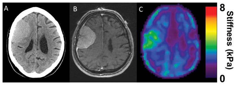
(A) CT head of an isodense right frontal convexity tumor. (B) T1 weighted MRI with contrast shows a homogenously enhancing tumor consistent with meningioma.(C) MRE shows a soft homogenous tumor. Intraoperatively, the tumor was easily removed with ultrasonic aspirator and was consistent throughout.