

Abstract
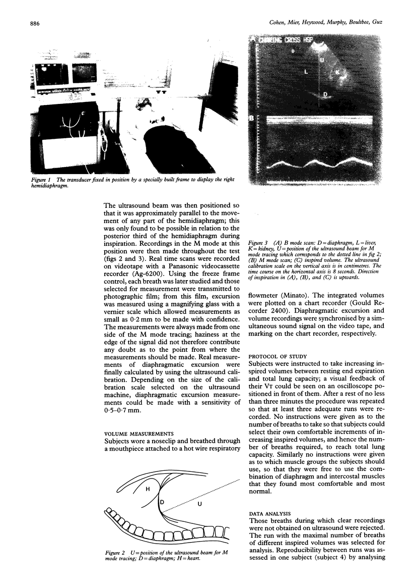
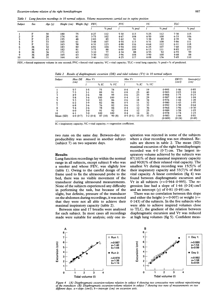
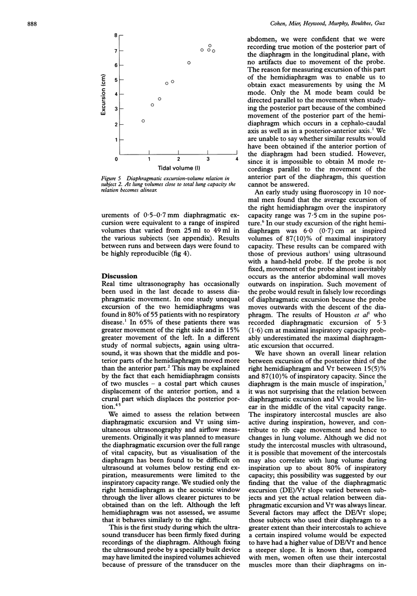
BACKGROUND--Although real time ultrasonography has been used in the last decade to record diaphragmatic motion, the relation between diaphragmatic excursion and different inspired volumes (VT) has not been assessed by ultrasound. METHODS--Ten normal subjects were studied in the supine posture. Diaphragmatic excursion and VT were assessed simultaneously by M mode ultrasonography and respiratory airflow measurements at different inspired volumes. Ultrasound recordings of the movement of the right hemidiaphragm were carried out in the longitudinal plane subcostally. The transducer was held in a fixed position by a frame, built especially to eliminate any artefactual movement caused by outward motion of the anterior abdominal wall on inspiration. RESULTS--Mean (SD) maximal diaphragmatic excursion recorded was 6.0 (0.7) cm. Inspired volumes ranged from 15(5%) to 87(10%) of the subjects' inspiratory capacity. A linear relation between diaphragmatic excursion and VT was found in all subjects (r = 0.976-0.995). The regression line had a slope of 1.66 (0.24) cm/l. This slope had no correlation with either the height (r = 0.007) or weight (r = 0.143) of the subjects. In five subjects in whom diaphragmatic excursion could be recorded at volumes near total lung capacity, the relation between diaphragmatic excursion and VT became alinear at very high lung volumes. CONCLUSIONS--The relation between diaphragmatic excursion and VT was linear between 15(5%) and 87(10%) of inspiratory capacity. Ultrasonography of the diaphragm is a simple technique that could be applied in the clinical investigation of patients with suspected abnormalities of diaphragmatic movement.
Full text
PDF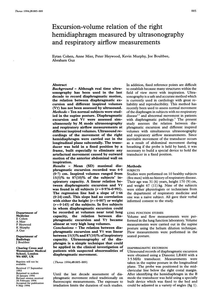
Images in this article
Selected References
These references are in PubMed. This may not be the complete list of references from this article.
- De Troyer A., Sampson M., Sigrist S., Macklem P. T. The diaphragm: two muscles. Science. 1981 Jul 10;213(4504):237–238. doi: 10.1126/science.7244632. [DOI] [PubMed] [Google Scholar]
- Diament M. J., Boechat M. I., Kangarloo H. Real-time sector ultrasound in the evaluation of suspected abnormalities of diaphragmatic motion. J Clin Ultrasound. 1985 Oct;13(8):539–543. doi: 10.1002/1097-0096(199010)13:8<539::aid-jcu1870130805>3.0.co;2-6. [DOI] [PubMed] [Google Scholar]
- Goldman M. D., Mead J. Mechanical interaction between the diaphragm and rib cage. J Appl Physiol. 1973 Aug;35(2):197–204. doi: 10.1152/jappl.1973.35.2.197. [DOI] [PubMed] [Google Scholar]
- Harris R. S., Giovannetti M., Kim B. K. Normal ventilatory movement of the right hemidiaphragm studied by ultrasonography and pneumotachography. Radiology. 1983 Jan;146(1):141–144. doi: 10.1148/radiology.146.1.6849035. [DOI] [PubMed] [Google Scholar]
- Houston J. G., Morris A. D., Howie C. A., Reid J. L., McMillan N. Technical report: quantitative assessment of diaphragmatic movement--a reproducible method using ultrasound. Clin Radiol. 1992 Dec;46(6):405–407. doi: 10.1016/s0009-9260(05)80688-9. [DOI] [PubMed] [Google Scholar]
- Verschakelen J. A., Deschepper K., Demedts M. Relationship between axial motion and volume displacement of the diaphragm during VC maneuvers. J Appl Physiol (1985) 1992 Apr;72(4):1536–1540. doi: 10.1152/jappl.1992.72.4.1536. [DOI] [PubMed] [Google Scholar]
- Verschakelen J. A., Deschepper K., Jiang T. X., Demedts M. Diaphragmatic displacement measured by fluoroscopy and derived by Respitrace. J Appl Physiol (1985) 1989 Aug;67(2):694–698. doi: 10.1152/jappl.1989.67.2.694. [DOI] [PubMed] [Google Scholar]
- WADE O. L. Movements of the thoracic cage and diaphragm in respiration. J Physiol. 1954 May 28;124(2):193–212. doi: 10.1113/jphysiol.1954.sp005099. [DOI] [PMC free article] [PubMed] [Google Scholar]