Abstract
Both enantiomers of lactic acid, l-lactic acid and d-lactic acid, can be produced in a sustainable way by a photosynthetic microbial cell factory and thus from CO2, sunlight, and water. Several properties of polylactic acid (a polyester of polymerized lactic acid) depend on the controlled blend of these two enantiomers. Recently, cyanobacterium Synechocystis sp. strain PCC6803 was genetically modified to allow formation of either of these two enantiomers. This report elaborates on the d-lactic acid production achieved by the introduction of a d-specific lactate dehydrogenase from the lactic acid bacterium Leuconostoc mesenteroides into Synechocystis. A typical batch culture of this recombinant strain initially shows lactic acid production, followed by a phase of lactic acid consumption, until production “outcompetes” consumption at later growth stages. We show that Synechocystis is able to use d-lactic acid, but not l-lactic acid, as a carbon source for growth. Deletion of the organism's putative d-lactate dehydrogenase (encoded by slr1556), however, does not eliminate this ability with respect to d-lactic acid consumption. In contrast, d-lactic acid consumption does depend on the presence of glycolate dehydrogenase GlcD1 (encoded by sll0404). Accordingly, this report highlights the need to match a product of interest of a cyanobacterial cell factory with the metabolic network present in the host used for its synthesis and emphasizes the need to understand the physiology of the production host in detail.
INTRODUCTION
To date, lactic acid has been produced with almost 100% conversion efficiency by several chemotrophic fermentative bacterial and yeast cell factories growing on various sugars (1, 2). Consequently, a large amount of effort is currently exerted to produce lactic acid with lactic acid bacteria (3), in combination with the use of a cheap substrate(s) such as lignocellulosic feedstock, though that feedstock requires an energy-intensive pretreatment of the biomass (1, 4). Lactic acid is used in food preservation, in the chemical and pharmaceutical industries, and as a building block for construction of polymers, the latter for use as an alternative to petroleum-derived plastics. It is compelling that the biodegradability and heat stability of this (bio)plastic depend on the blend of the two optically active, chiral isoforms of lactic acid (5).
Employing a photosynthetic microorganism as the production host has the advantage of enabling the direct conversion of CO2 into (poly)lactic acid (6–9). Such production, which is dependent on cyanobacterial cell factories, allows compound formation without the need to generate complex (plant) biomass first, only to break it down again later for its utilization by a chemotrophic fermentative microorganism (10).
For living organisms, chirality plays an essential role. For example, amino acids are incorporated as l-enantiomers into proteins. Likewise, l-lactic acid seems to be the dominant enantiomeric form of this weak acid in nature. Thus, suitable and effective production hosts for d-lactic acid are more challenging to find and construct. Nonetheless, biosynthetic routes for both enantiomers, and for the corresponding products, exist in various (micro)organisms, facilitated by enantiomer-specific lactate dehydrogenases (LDH) (11), whereas chemical synthesis routinely results in a racemic mixture (12). Earlier, we constructed several l-lactic acid-producing Synechocystis sp. strain PCC6803 (here Synechocystis) variants (7, 13). In the framework of those experiments, we also tested the d-LDH of Escherichia coli (7), but we were not successful in producing d-lactic acid in the engineered Synechocystis strains at that time. However, in another cyanobacterium, Synechococcus elongatus PCC7942, synthesis of the latter enantiomer has been achieved through the expression of the same E. coli enzyme, although its extracellular accumulation required the coexpression of a transporter (6). More recently, d-lactic acid formation in Synechocystis has also been accomplished (9). In the latter study, d-lactic acid formation was achieved via the involvement of a mutated glycerol dehydrogenase, GlyDH*, from Bacillus coagulans (14). Here, we have chosen to use the d-LDH of Leuconostoc mesenteroides (here L. mesenteroides) (15), because of its superior kinetics compared to other characterized d-LDH enzymes (16). As an alternative, we initially decided to also overexpress the native d-LDH (slr1556) of Synechocystis, arguing that a native enzyme might be optimized for the specific conditions present in the cyanobacterial cytosol.
Interestingly, various cyanobacteria have been shown to possess an anaerobiosis-induced fermentation capacity, including the capacity to form lactic acid in the dark (17–19). Further, it has been shown that selected cyanobacterial strains can utilize lactic acid to form biomass aerobically in the dark. For example, suspensions of resting cells of the thermophilic cyanobacterium Synechococcus sp. strain PCC6716 can (aerobically and in the dark) take up lactic acid and use it to form biomass, CO2, and acetate (20). Moreover, the same study showed that cell extracts of various cyanobacteria, including Synechocystis, display lactate dehydrogenase activity. As noted above, slr1556 of Synechocystis is annotated as encoding a putative d-LDH (21) and could thus also represent a d-lactic acid consumption pathway.
In metabolic engineering and strain construction, the choice of an envisioned product has to be matched with a suitable metabolic route and the production host. For an optimized production system employing microorganisms, it is of importance to improve insight into the physiology of these production hosts (22). Indeed, some key intermediates of central metabolism of cyanobacteria are more accessible as a tapping point than metabolites from the periphery of intermediary metabolism and some products consistently allow higher productivities (23). Here, we describe the construction of a d-lactic acid-producing strain of Synechocystis, overexpressing an NADH-dependent d-enantiomer-specific ldh gene of L. mesenteroides. However, we also show that Synechocystis is capable of d-lactic acid consumption which, at least transiently, limits d-lactic acid productivity. Further investigations of the capability of d-lactic acid consumption of Synechocystis showed that not the annotated d-enantiomer-specific ldh gene (carried by slr1556) but the glycolate dehydrogenase glcD1 gene (carried by sll0404) is primarily responsible for the consumption of d-lactic acid. It appears that a significantly higher capacity of production has to be engineered for d-lactic acid production than for l-lactic acid production in order to achieve net production.
MATERIALS AND METHODS
Growth conditions.
For subcloning procedures, plasmid propagation, and isolation, Escherichia coli strains XL-1 Blue (Stratagene) and EPI400 (Epicentre) were cultivated at 37°C in lysogeny broth (LB) liquid medium or on LB agar. Where appropriate, ampicillin was used at a concentration of 100 μg/ml, kanamycin at 50 μg/ml, and spectinomycin at 25 μg/ml, either separately or in combination.
The glucose-tolerant derivative of Synechocystis sp. PCC6803 (wild type; kindly provided by Devaki Bhaya, Stanford, CA, USA) was grown in liquid BG-11 medium (Sigma-Aldrich), supplemented with 10 mM TES-KOH (pH 8). Where appropriate, kanamycin was used at a concentration of 20 μg/ml and spectinomycin at 20 μg/ml, either separately or in combination. Cells were routinely streaked on plates from −80°C dimethyl sulfoxide (DMSO) (5% [vol/vol]) stocks, and colonies were inoculated from the plate to a liquid preculture, which was grown to the early stationary phase for several days. For the replicate cultures, an aliquot of the inoculum was taken to reach a starting optical density at 730 nm (OD730) of 0.05 to 0.1. Cultures were incubated in a shaking incubator (Innova 43; New Brunswick Scientific), equipped with white-light illumination (∼35 μmol photons/m2/s), at 120 rpm and 30°C. BG-11 solid medium was prepared by adding 1.5% (wt/vol) agar and 10 mM TES-KOH (pH 8), 5 mM glucose, and 0.3% (wt/vol) sodium thiosulfate. For the construction and maintenance of the Synechocystis Δslr1556 mutant strain on plates, the addition of glucose was omitted. Growth of Synechocystis wild-type and mutant strains (including ΔglcD1 and ΔglcD2 mutants [24]) was followed by OD730 determinations (Lightwave II spectrophotometer; Biochrom) at selected time points. Roughly, every 24 h approximately 3% of the culture volume was removed for the analysis of growth and subsequent determination of the lactic acid concentration, when applicable. The water evaporation rate under these conditions was determined and found to be 1% of the culture volume per day (see Fig. S1 in the supplemental material); hence, the lactic acid concentration was increased only marginally. The method for rate calculations of product formation has been reported earlier (7, 13).
Strain and plasmid construction.
Synechocystis mutant strains were constructed essentially as described before (7). Briefly, we constructed plasmids to be used for natural transformation with subsequent integration of the gene cassette of interest and a kanamycin resistance cassette at the slr0168 locus of the Synechocystis genome. Additionally, plasmid pGemT_slr1556_Sp122 targets slr1556 with a streptomycin/spectinomycin resistance cassette to create the particular knockout strain. Before selection of one clone for further investigation, three individual colonies originating from the transformation were routinely checked for full segregation and correct sequence alteration. Moreover, cultures started from them were compared with respect to growth and, if applicable, lactic acid production. Segregation was evaluated by colony PCR with primers flanking the integration site (Table 1) employing Taq DNA polymerase (Thermo Scientific). For verification of correct insertions, the sequence was evaluated by capillary electrophoresis sequencing (Macrogen Netherlands) of relevant PCR fragments (for information concerning the flanking primers, see Table 1) amplified in colony PCRs employing the proofreading Pfu DNA polymerase (Thermo Scientific).
TABLE 1.
Primers used in this study
| Name | Sequence | Additional information |
|---|---|---|
| H1_fw | AATGGTCCCAAAATTGTC | Colony PCR for segregation at slr0168 insertion and sequencing |
| H2_rv | CTGTGGGTAGTAAACTGGC | Colony PCR for segregation at slr0168 insertion and sequencing |
| KanF_back_Seq | TCCCGTTGAATATGGCTC | Colony PCR for insertion at slr0168 and sequencing |
| 1556_Nde_F | AATCATATGAAAATCGCTTTTTTTAGCAG | slr1556 ORF amplification |
| 1556_Bam_R | AATGGATCCTTAATGGGGACAGATTACTTGGT | slr1556 ORF amplification |
| 1556_out_fw | AATTTGGGGTGAAGCTGGGT | Colony PCR for slr1556 deletion |
| 1556_out_rv | CCGTTAATACTATCGTTGCTTCATTT | Colony PCR for slr1556 deletion |
| slr1556_800bp_fw | AAGGTTGACCAAGGAATGGTTGGTT | slr1556 knockout plasmid |
| slr1556_800bp_rv | GTTTACGAATGGACTGATTCCACCA | slr1556 knockout plasmid |
| slr1556_EcorI_fw | GAATTCATGAAAATCGCTTTTTTTAGC | Slr1556 overexpression for purification |
| slr1556_XhoI_rev | CTCGAGTTAATGGGGACAGATTACTTGG | Slr1556 overexpression for purification |
The plasmid used to make Synechocystis strain DLEU is basically a derivative of the previously published pHKH001 integration vector (7). The (protein) sequence of the d-LDH of L. mesenteroides was taken from UniProt database entry Q2ABS1_LEUME based on data in reference 25. The (gene) sequence was codon optimized using a codon usage table generated from a subset of highly transcribed Synechocystis genes (for the exact procedure, see reference 26). Relevant restriction sites needed for subcloning procedures have been avoided in the design. The gene was further flanked on the 5′ end with Ptrc and a ribosome binding site (RBS) and at the 3′ end with a transcriptional terminator (BBa_B0014; http://parts.igem.org/). The gene cassette was synthesized and directly cloned into pHKH001 at the SacII/EcoRV site by Genscript (NJ, USA). The plasmid used to make Synechocystis strain ODSYN was constructed from a derivative of pHKHDLEU which carries, in addition to the NdeI site of the synthesized construct at the 5′ end, a BamHI site at the 3′ end for exact open reading frame (ORF) replacement. First, Synechocystis slr1556 was amplified from pIBA43plus_slr1556 with primers introducing the flanking NdeI and BamHI restriction sites employing Pfu DNA polymerase (Thermo Scientific). The gene was then subcloned into the pHKHDLEU derivative, replacing the L. mesenteroides ldh gene with slr1556 and creating pHKHDSYN. pIBA43plus_slr1556 was constructed as follows: to generate an Slr1556-overexpressing E. coli strain, the slr1556 gene was amplified by PCR using genomic DNA from Synechocystis sp. PCC6803, anchored gene-specific primers (Table 1), and proofreading Elongase enzyme (Invitrogen). The resulting fragments were first cloned in pGemT vector (Promega). After sequence confirmation, the coding sequence of slr1556 was transferred to pIBA43plus using EcoRI/XhoI. The verified recombinant pIBA43plus vector was transformed into E. coli strain BL21(DE3). The plasmid insertions were verified by sequencing of the relevant part of the plasmid. We used pGemT_slr1556_Sp122 to make the ΔDSYN strain, disrupting slr1556. pGemT_slr1556_Sp122 inserts a streptomycin/spectinomycin resistance cassette (for strain construction details, compare also reference 27), replacing a 585-nucleotide-long region (nucleotides 3359843 to 3360428) on the Synechocystis genome and thereby disrupting the ORF of slr1556. The slr1556 gene is the putative ddh gene (encoding the d-isomer-specific lactate dehydrogenase) of Synechocystis, as annotated in the genome and essentially inferred by automated homology determination (21). All restriction enzymes and the ligase used were from Thermo Scientific.
Protein purification.
A 5-ml overnight culture of E. coli BL21 harboring pIBA43plus_slr1556 grown for the heterologous production of Slr1556 was diluted in 200 ml LB medium. After incubation for 6 h at 30°C with shaking at 170 rpm, expression of the recombinant protein was induced overnight with the addition of 200 ng/ml anhydrotetracyclin (Clontech). Cells were harvested by centrifugation and were resuspended in 5 ml homogenization buffer consisting of 20 mM sodium phosphate buffer (pH 7.4) and 500 mM NaCl and lysed by ultrasonic treatment (Labsonic U; B. Braun). Cell debris was removed by centrifugation (30 min at 10,000 rpm at 4°C). Subsequently, the soluble lysates were loaded on a nickel-nitrilotriacetic acid (Ni-NTA) Sepharose column primed with ProBond slurry (Invitrogen), washed with sterile water, and equilibrated with homogenization buffer. After careful addition of soluble fraction of the cell extract, the column was washed with the same buffer but supplemented with 20 mM imidazole and next with a buffer supplemented with 40 mM imidazole. The protein bound to the column was eluted with elution buffer consisting of 20 mM sodium phosphate (pH 7.4), 500 mM NaCl, and 300 mM imidazol and collected in 500-μl fractions. The fraction containing the His-tagged protein was collected on a washed PD-10-desalting column (GE Healthcare), and the buffer was exchanged by applying approximately two volumes of 20 mM sodium phosphate buffer (pH 7.4) in a sequential manner. For protein storage, 20% (vol/vol) glycerol was added. The purity of the protein was verified by visual inspection of a Coomassie brilliant blue (CBB)-stained sodium dodecyl sulfate-polyacrylamide gel electrophoresis (SDS-PAGE) gel. The concentration of Slr1556 protein was determined by the method of Bradford (28) with bovine albumin as the standard protein.
Determination of lactic acid.
The lactic acid concentration in supernatant samples of Synechocystis cultures was determined essentially as described earlier (7). Briefly, aliquots of supernatant were assayed with the d-/l-lactic acid (Rapid) assay (Megazyme), adapted to a 96-well plate format. Occasionally, the supernatant was subjected to high-performance liquid chromatography (HPLC) analysis. Samples were separated on a Rezex ROA-Organic Acid H+ (8%) column (Phenomenex) at 45°C at a flow rate of 0.5 ml/min. Signals were obtained by the use of a refractive index detector (RI-1530; Jasco) and analyzed with the Azur 4.5 software package (Datalys). For both detection methods, the sample signal was compared to a set of known standard concentrations of d-/l-lactic acid (Megazyme) for quantification.
Cell extracts and lactate dehydrogenase enzymatic activity assays.
Crude cell extracts of Synechocystis cells were obtained essentially as described earlier (13). Briefly, 10 ml of Synechocystis cultures in the late exponential phase of growth (OD730 of ∼1.0) was harvested and pelleted at 4°C. Aliquots of the supernatant were kept for lactic acid determinations, and the excess supernatant was discarded. The cell pellets were resuspended in the respective prechilled buffers (see below) and disrupted with glass beads (Sigma) (diameter, 100 μm) in a Precellys24 bead beater (Bertin Technologies). Cell debris was removed by centrifugation at 4°C, and supernatant was collected and either used on the same day for the LDH enzymatic activity assay (stored at 4°C) or flash-frozen in liquid nitrogen and stored at −20°C for later use. The protein concentration was determined with the standard bicinchoninic acid (BCA) protein assay (Pierce). Absorption at 570 nm was determined in a 96-well plate reader (Fluostar Optima; BMG LabTech) and compared to results from a set of known standard concentrations of bovine serum albumin for quantification.
LDH enzymatic activity assays were performed essentially as described earlier (13). Briefly, the consumption (or formation) of NADH at 340 nm was followed in a 96-well plate reader (Multiscan FC microplate reader; Thermo Scientific) set to 30°C. For cell extracts containing the LDH of L. mesenteroides, the reaction mixture contained 100 mM Tris-HCl buffer (pH 7.5), 5 mM MgCl2, and 300 μM NAD(P)H. For cell extracts of the wild-type, ODSYN, and ΔDSYN strains, the reaction was performed using 100 mM TEA (Tris base, acetic acid, EDTA) buffer at pH 7.0. The reaction was done in 100 mM potassium phosphate buffer at pH 7.5 for cell extracts containing the LDH of E. coli (7), in 100 mM Tris-HCl at pH 7.2 for the LDH of L. lactis (13), and in 50 mM sodium phosphate buffer at pH 6.5 for the LDH of B. subtilis (7). After a background reading, allowing time for equilibration, the reaction was started by the addition of 30 mM sodium pyruvate (Sigma). For the d-lactic acid consumption assay, the same reaction mixture was used but with NAD(P)+ instead of NAD(P)H. The reaction was started by the addition of 100 mM lithium d-lactic acid (Roche). For the reactions destined for HPLC analysis, 50 μg/ml cell extract was incubated in 100 mM TEA buffer containing 5 mM MgCl2, 1 mM NAD(P)H, and 30 mM sodium pyruvate for 3 h with gentle agitation at 30°C and pH 7.0.
The LDH enzymatic activity assays and the half-maximal activity constant (KH) determination for the purified enzyme were performed as follows. A 50-μl volume of purified protein was added to 900 μl of 100 mM MES (morpholineethanesulfonic acid) buffer (pH 6.5) containing (freshly added) dithiothreitol (DTT) at 1 mg/ml and NADH at 187 μM. After 2 min of equilibration, 50 μl of pyruvate (20 mM) was added to start the reaction (29). The decrease of absorption at 340 nm was observed with a Cary-50 spectrophotometer (Varian) at 30°C, and the resulting activity was expressed in micromoles per minute per milligram, considering the equimolar conversion of NADH and pyruvate into NAD and lactic acid. For kinetic parameter determinations, concentrations of 0.5 to 50 mM for glyoxylate or 0.1 to 2.5 mM for pyruvate were used, whereby the NADH concentration was kept constant. For d-lactic acid, concentrations of 1 to 30 mM were used, and the NAD+ concentration was kept constant. The kinetic parameters were calculated using Sigma Plot 11.0.
Sample preparation for SDS-PAGE analysis.
Aliquots (18 μl) of the same protein concentration (2 μg/μl) of the crude cell extract samples were supplemented with 2 μl protein solubilization buffer, consisting of 50 mM Tris-HCl (pH 6.8), 100 mM dithiothreitol, 50 mM EDTA, 2% (wt/vol) sodium dodecyl sulfate, and 10% (vol/vol) glycerol. Samples were boiled at 95°C and mixed thoroughly before application on a 12% (wt/vol) SDS-polyacrylamide (PA) gel. Samples were electrophoresed on these SDS-PAGE gels, fixed with an isopropanol/acetic acid solution, and subsequently stained with CBB G-250 (PageBlue staining solution; Thermo Scientific) according to the manufacturer's protocol.
RESULTS
Construction of d-lactic acid-producing Synechocystis strains.
Motivated by previous work (7), we attempted to construct a Synechocystis strain that is capable of d-lactic acid production in order to have an enantiomeric complement to our l-lactic acid-producing strains. On the basis of data available in BRENDA (16), we chose the d-isomer-specific lactate dehydrogenase (LDH) of L. mesenteroides for the experiment. This enzyme is characterized by having the most optimal kinetic properties, such as a low KM value for pyruvate and a high molecular turnover number (15), of those available in this database. To avoid potential competition between related pathways, we additionally deleted the putative endogenous LDH (encoded by slr1556 [21]) of Synechocystis. Moreover, we constructed an slr1556 overexpression strain. The resulting Synechocystis strains are listed in Table 2. For the overexpression strains, slr0168 served as the neutral integration site on the Synechocystis chromosome. The slr1556 gene was disrupted by the deletion of a part of its open reading frame, achieved by the insertion of a spectinomycin resistance cassette (see Fig. S2A in the supplemental material). Colony PCR with flanking primers confirmed full segregation of the genomes with the respective insertions (see Fig. S2B).
TABLE 2.
Strains and plasmids used and constructed in this study
| Strain or plasmid | Genotype or relevant feature(s)a | Source or reference(s) |
|---|---|---|
| E. coli XL-1 Blue | Cloning host | Stratagene |
| E. coli EPI400 | Cloning host | Epicentre |
| Synechocystis sp. PCC6803 | Wild type, glucose-tolerant derivative | D. Bhaya (Stanford, CA, USA) |
| pHKH001 | Δslr0168::Knr in sp. strains; in E. coli, additionally Apr | 7 |
| pIBA43plus_slr1556 | slr1556; Apr | This study |
| pGemT_slr1556_Sp122 | Δslr1556::Srr; in E. coli, additionally Apr | This study |
| pHKHDLEU | Δslr0168::Ptrc::Lmldhco::tt::Knr | This study |
| pHKHDSYN | Δslr0168::Ptrc::Synslr1556na::tt::Knr | This study |
| Synechocystis strain DLEU | Ptrc::Lmldhco::tt::Kanr | This study |
| Synechocystis strain ODSYN | Ptrc::Synslr1556na::tt::Kanr | This study |
| Synechocystis strain ΔDSYN | Δslr1556::Spcr | This study |
| Synechocystis strain DLEU/ΔDSYN | Ptrc::Lmldhco::tt::Kanr; Δslr1556::Spcr | This study |
| Synechocystis strain ΔGlcD1Ro | sll0404::Kanr | 24, 30 |
| Synechocystis strain ΔGlcD2Ro | slr0806::Kanr | 24 |
| Synechocystis strain ΔDSYNRo | Δslr1556::Spcr in the same wild-type background as the glycolate dehydrogenase mutants | This study |
| Synechocystis strain SAA013 | Δslr0168::Ptrc::Ecldhna::tt::Kanr | 7 |
| Synechocystis strain SAA015 | Δslr0168::Ptrc::Bsldhna::tt::Kanr | 7 |
| Synechocystis strain SAA023 | Δslr0168::Ptrc::Llldhco::tt::Kanr | 13 |
Kanr or Knr, kanamycin resistance cassette; Spcr or Srr, streptomycin/spectinomycin resistance cassette; Ampr or Apr, ampicillin resistance cassette; Ptrc1, trc promoter; tt, transcriptional terminator (BBa_B0014); co, codon optimized sequence; na, native sequence; Lm, Leuconostoc mesenteroides; Syn, Synechocystis sp. PCC6803; Ec, Escherichia coli; Bs, Bacillus subtilis; Ll, Lactococcus lactis.
d-Lactic acid production with a genetically engineered Synechocystis strain.
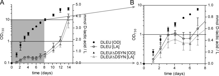
Synechocystis strain DLEU, expressing the d-LDH of L. mesenteroides, indeed showed d-lactic acid production (Fig. 1A). It accumulated d-lactic acid to extracellular concentrations of 0.60 ± 0.19 mmol/liter within 1 week and 3.93 ± 0.27 mmol/liter within 2 weeks. Strikingly, upon exit of the initial phase of exponential growth, d-lactic acid is transiently consumed, before the production resumes after the “transition phase” (Fig. 1B). The Synechocystis genome harbors the slr1556 gene, annotated as the d-LDH gene, which could be held responsible for the observed d-lactic acid consumption. Therefore, we modified the DLEU strain with a disruption of slr1556, resulting in the DLEU/ΔDSYN strain. Significantly, this double mutant displays the same d-lactic acid production characteristics as the single-mutant DLEU strain (Fig. 1). Consistent with previous observations (7), we could not detect any d-lactic acid formation in wild-type cultures. Also, as expected, the slr1556 deletion ΔDSYN mutant did not show lactic acid production (see Fig. S3A and B in the supplemental material).
FIG 1.
(A) Time course of d-lactic acid (LA) production by a Synechocystis strain overexpressing d-LDH of L. mesenteroides (DLEU) solely or in combination with the slr1556 disruption strain (DLEU/ΔDSYN). (B) Closeup of data in panel A, allowing a detailed view of the changes in the d-lactic acid concentration during the early growth phase. Error bars indicate the standard deviations (SD) (n = 3) of results from biological replicates; where error bars are not visible, they are smaller than the data point symbol.
In vitro characterization of the putative d-LDH Slr1556.
To verify the specificity of Slr1556, we purified recombinant Slr1556 and tested it for activity without cleaving the His label. The enzyme can utilize pyruvate but also d-lactic acid, glyoxylate, and hydroxypyruvate as a substrate in vitro. Interestingly, the recombinant enzyme showed sigmoidal kinetics with increasing d-lactate concentrations (see Fig. S4 in the supplemental material). Thus, we applied the Hill equation to determine substrate affinities. This evaluation revealed that pyruvate was used with the highest affinity (lowest KH value) using NADH as the redox cofactor (Table 3). The in vitro activity detected with d-lactic acid as the substrate and NAD+ as the cofactor was more than 20 times lower than the activity detected with pyruvate. Therefore, the high KH for d-lactic acid suggests that in vivo utilization of this substrate with NAD+ as an electron acceptor under photoautotrophic conditions is unlikely. Consequently, the in vitro data indicate that the preferred activity of Slr1556 is the conversion of pyruvate into d-lactic acid and not the reverse reaction. In agreement with this notion, we observed that overexpression of slr1556 under the control of Ptrc, resulting in Synechocystis slr1556 overexpression strain ODSYN, did not result in any measurable extracellular d-lactic acid formation (see Fig. S3C).
TABLE 3.
Characterization of the kinetic properties of purified, recombinant Slr1556a
| Compound | Measured directionality | KH (mM) | Vmax (μmol/mg/min) | Hill coefficient |
|---|---|---|---|---|
| Pyruvate | Pyruvate reductase | 0.39 ± 0.17 | 236.12 ± 13.12 | 1.97 ± 0.47 |
| Hydroxypyruvate | Hydroxypyruvate reductase | 6.7 ± 1.4 | 78.14 ± 8.42 | 1.38 ± 0.24 |
| Glyoxylate | Glyoxylate reductase | 2.43 ± 0.01 | 175.52 ± 12.08 | 2.34 ± 0.02 |
| d-Lactic acid | Lactate dehydrogenase | 7.93 ± 0.22 | 10.65 ± 0.82 | 2.52 ± 0.29 |
The kinetic parameters for the Hill equation were calculated in SigmaPlot 11.0 as described by Goutelle et al. (31), assuming a baseline response. Data are mean values of results (n = 2).
Native d-LDH activity of Synechocystis.
It was found earlier that a cell extract of Synechocystis, at that time also named Aphanocapsa PCC6803, has specific activity toward d-lactic acid but not l-lactic acid (20). Similarly, here we show that the soluble fraction of the crude cell extract of wild-type Synechocystis is capable of reduction of NAD(P)+ when d-lactic acid is provided as a substrate (see Fig. S5A in the supplemental material). Interestingly, the slr1556 deletion ΔDSYN mutant lost that property (see Fig. S5B). Further investigation of the wild-type extracts shows that the conversion of pyruvate into lactic acid proceeds in an NAD(P)H-dependent manner (see Fig. S6A). The wild-type cell extract thus has oxidoreductase activity with respect to pyruvate and lactic acid, utilizing NAD(P)+ and NAD(P)H as redox cofactors, respectively. The soluble lysate of the ΔDSYN strain lacks the ability to reduce pyruvate with NADH (see Fig. S6A). Additionally, an NADPH-dependent LDH activity assay shows activity in extracts of the ODSYN slr1556 overexpression strain, suggesting that Slr1556 can utilize both NADH and NADPH (see Fig. S6B). However, NADPH-dependent activity of the wild-type extract was not detected, probably because of the lower expression level of Slr1556 in the wild-type strain than in the ODSYN overexpression strain.
Enzyme activity in cell extracts shows a high capacity for d-LDHs.
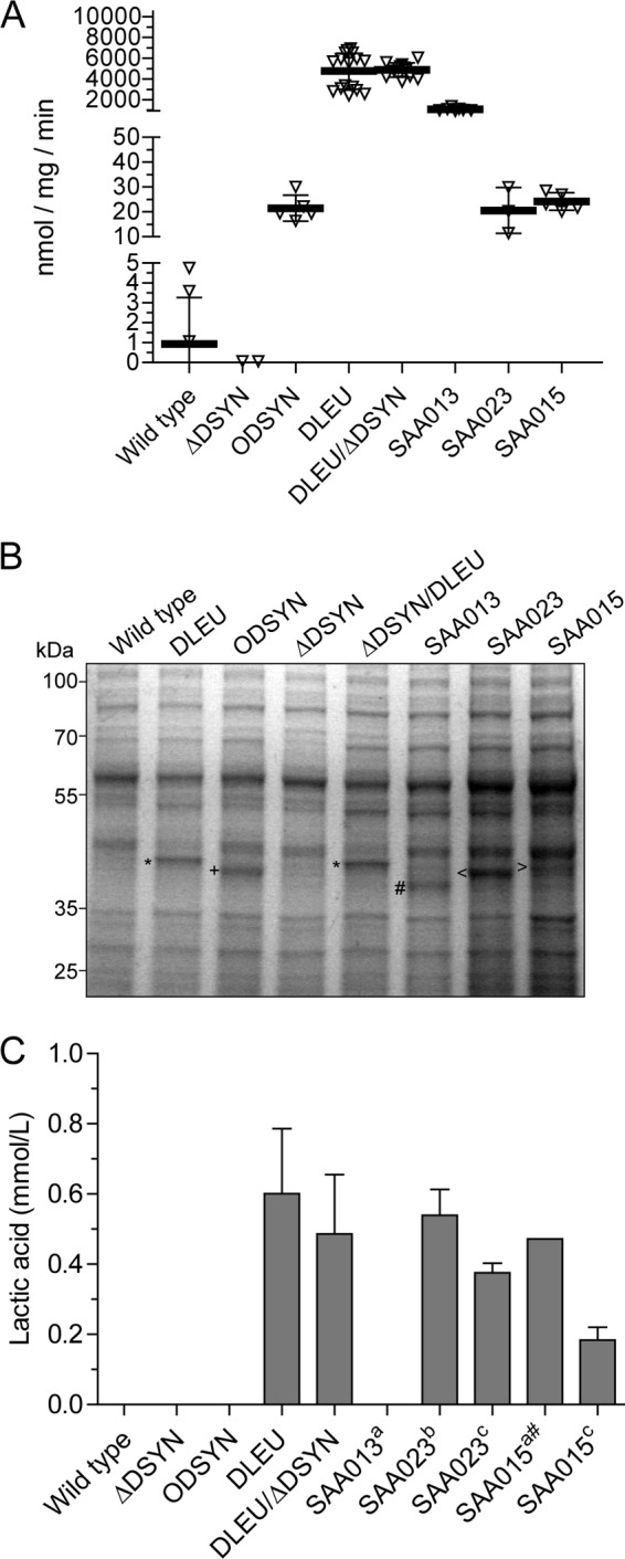
Next, we compared the activity of the different LDHs in the lysates of the overexpression strains and included some previously constructed d-- and l-LDH-gene-carrying strains into this analysis (7, 13). Crude cell extracts of the slr1556 overexpression strain, ODSYN, which does not produce d-lactic acid, shows NADH-dependent oxidoreductase activity with pyruvate as the starting substrate that is significantly higher than in wild-type-soluble lysate (Fig. 2A). Although the direction that is measured in such an assay actually verifies the respective pyruvate reduction activities, it is common to refer to this activity as LDH activity. Unless noted otherwise, or where the distinction is necessary, we follow this convention throughout the rest of the manuscript. Strikingly, the measured LDH activities for the DLEU and DLEU/ΔDSYN strains were much higher than anticipated, in the range of 4,700 nmol/mg/min (Fig. 2A). To verify protein levels, we analyzed the soluble lysates with SDS-PAGE, followed by staining with Coomassie brilliant blue. Comparison with the wild-type results showed that all of our overexpressed LDH enzymes were expressed to similar levels (Fig. 2B), as judged by visual inspection.
FIG 2.

(A and B) Soluble lysates of Synechocystis wild-type, ΔDSYN, ODSYN, DLEU, ΔDSYN/DLEU, SAA013, SAA023, and SAA015 strains assayed for pyruvate-dependent oxidoreductase activity with NADH as the redox cofactor (note the three segments of the left y axis) (A) and analyzed using CBB staining and SDS-PAGE (B). Different LDHs are indicated as follows: *, d-LDH from L. mesenteroides (36.3 kDa); +, Slr1556 (36.9 kDa); #, d-LDH from E. coli (36.5 kDa); <, LDH from L. lactis (35.1 kDa); >, d-LDH from B. subtilis (34.9 kDa). (C) Production titers after 7 days of culturing of Synechocystis wild-type, ΔDSYN, ODSYN, DLEU, ΔDSYN/DLEU, SAA013, SAA023, and SAA015 strains. Data indicated with a superscript “a” were derived from reference 7; data indicated with a superscript “b” were derived from reference 13; data indicated with a superscript “c” were derived from reference 39. #, extrapolated value.
However, the enzymatic activities observed for the overexpressed d-LDHs are in general much higher than those observed for overexpressed l-LDHs under their respective optimal in vitro conditions (Fig. 2A). We even observed high LDH activity levels for SAA013, a strain expressing the d-LDH of E. coli, for which no lactic acid production could be observed (7). Since the production titers after 7 days for d-lactic acid in DLEU and DLEU/ΔDSYN strains are similar to those observed for l-lactic acid (7, 13) (Fig. 2C), while the enzymes involved showed much higher LDH activity (an approximately 200-fold difference; compare Fig. 2A), this result is another indication that production of d-lactic acid in Synechocystis is inherently more difficult than production of l-lactic acid. The lack of a high d-lactic acid titer could be partially explained by the transient consumption of d-lactic acid.
d-Lactic acid, but not l-lactic acid, allows mixotrophic growth of Synechocystis.
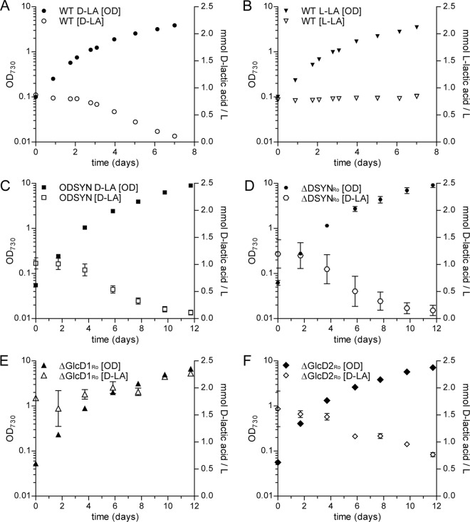
When growing mixotrophically in the light, Synechocystis is able to assimilate glucose to form biomass. It can also utilize glucose heterotrophically in the dark when provided daily with short periods of illumination with blue light (32). Acetate was also previously reported to be a suitable organic carbon source for mixotrophic growth for selected cyanobacteria (17). Here, we tested the ability of Synechocystis to use lactic acid as a potential carbon source. Remarkably, Synechocystis wild-type cultures, growing under continuous illumination, consume and grow on d-lactic acid but not on l-lactic acid (Fig. 3A and B). The growth rate for cultures supplemented with d-lactic acid is significantly higher during the main growth phase than for those without addition of an organic carbon source (see Fig. S7 in the supplemental material). Interestingly, the slr1556 deletion ΔDSYN mutant shows the same lactic acid consumption pattern as the wild type (Fig. 3D). These results suggest that the ability to consume (and grow on) d-lactic acid is not dependent on the presence of slr1556 in the wild-type strain. Furthermore, the slr1556 overexpression strain ODSYN also showed the same lactic acid consumption pattern (i.e., no increase in the rate of assimilation; Fig. 3C), suggesting again that Slr1556 does not function as a d-lactic acid-consuming enzyme under the conditions tested.
FIG 3.
Mixotrophic growth and consumption of d-lactic acid (D-LA) for the wild-type (A), ODSYN (C), ΔDSYNRo (D), ΔGlcD1Ro (E), and ΔGlcD2Ro (F) Synechocystis strains. (B) Growth of the wild-type strain on l-lactic acid (L-LA) is also shown. Error bars indicate the SD (n = 2 [except for the wild type, where n = 3]) of results of biological replicates; where error bars are not visible, they are smaller than the data point symbol.
As evident from the data shown in Fig. 1, the production and consumption rates for lactic acid are not constant over the course of a typical batch culture experiment, which suggests a correlation between growth phase and lactic acid consumption.
d-Lactic acid consumption by glycolate dehydrogenase.
Our investigation of the crude soluble lysates showed that Slr1556 is able to interconvert pyruvate and d-lactic acid, with NAD(P)(H) as the redox cofactor. However, we have also shown that Slr1556 is not the enzyme responsible for the consumption of d-lactic acid during batch culturing. Thus, we hypothesized that a different enzyme catalyzes the d-lactic acid channeling into the metabolism. As no obvious, or annotated, candidates for such an enzyme are available, we performed a BLASTp analysis against the Synechocystis proteome with several different known d-LDH enzymes from the literature. While we mostly found similarity with Slr1556, a search using the membrane-bound d-lactic acid dehydrogenase of E. coli (33) indicated minor (i.e., 23%) identity with the glycolate dehydrogenase GlcD1 of Synechocystis, which is encoded by sll0404. sll0404 also shows 46% similarity to the E. coli glycolate dehydrogenase, an enzyme that shows specific d-LDH activity (34, 35). Since many glycolate dehydrogenase enzymes can have a specific affinity for d-lactic acid (36), we decided to follow this lead. Synechocystis actually has two glycolate dehydrogenase enzymes, but GlcD1 is the predominant one (24). To test the involvement of these enzymes in d-lactate assimilation, we repeated the d-lactic acid consumption assay with the ΔglcD1 and ΔglcD2 mutants (24) and compared the results obtained with those derived from a Δslr1556 mutant, the ΔDSYNRo mutant, with the same wild-type background (Fig. 3D). We observed diminished consumption of d-lactic acid in the ΔglcD2 mutant (Fig. 3E) compared to wild-type results. However, strikingly, this activity was completely abolished in the ΔglcD1 mutant (Fig. 3F). This result implies that the enzyme involved in the consumption of d-lactic acid in Synechocystis is in fact the GlcD1 glycolate dehydrogenase.
DISCUSSION
In this report, we show that considering the chiral nature of lactic acid is of importance for production of this weak acid in Synechocystis. While production of the l-enantiomer of lactic acid was established some time ago, the attempted production of d-lactic acid by the use of Synechocystis initially failed (7); however, others have succeeded more recently (9, 37). Here, we have successfully engineered a production strain for d-lactic acid by overexpression of the d-LDH of L. mesenteroides. However, we noticed a transient decrease in d-lactic acid formation during a transitional growth phase. Additionally, the activity of a cell extract of the DLEU production strain is 2 orders of magnitudes higher than the LDH activities observed in related studies aiming at l-lactic acid production (compare Fig. 2A to data in references 7 and 13). Recently, comparably high LDH activity (i.e., 51.92 ± 2.31 μmol/mg/min) in a Synechocystis strain expressing the gene encoding d-LdhE of E. coli K-12 has been reported (38), but lactic acid production data were not included in that manuscript. The LDH activity in our Synechocystis strain expressing the d-LDH of E. coli (Synechocystis strain SAA013 [7]) was also much higher than the activity typically measured for the l-LDHs (Fig. 2A). However, SAA013 does not produce d-lactic acid (7). The protein level of the heterologously expressed d-LDH of E. coli is similar to the amount of heterologous LDH of the mutant strains presented here (Fig. 2B). Arguably, the protein expression levels in the strains expressing the l-LDH of B. subtilis (SAA015) or of L. lactis (SAA023) are also similar to those in the strains expressing a d-LDH, but the measured activities are much lower. Nonetheless, they are achieving l-lactic acid formation (7, 13, 39). We argue that a huge overcapacity of the biosynthetic enzyme is necessary for significant d-lactic acid formation. Additionally, the LDH activity measured in the soluble lysate of the DLEU production strain is more than 2,000-fold higher than the actual maximal production rate achieved. We conclude that the d-LDH of L. mesenteroides possibly does not function under Vmax conditions in the cyanobacterial cytoplasm and/or that competing pathways in the cell interfere with d-lactic acid production. The observed transient decrease in product formation in the DLEU mutant indicates specific interference with d-lactic acid production by other pathways in the cell. d-Lactic acid is consumed by wild-type Synechocystis cultured under mixotrophic conditions, while l-lactic acid is not used. In a recent computational metabolic network analysis (40), based on a genome-scale model of wild-type Synechocystis, the only annotated route involving lactic acid [from dihydroxyacetone-phosphate via methylglyoxal, (R)-S-lactoylglutathione, and lactic acid to pyruvate] did not show any flux under the conditions tested (continuous-light and day/night cycles) and thus appears nonessential for (optimal) growth. According to this scheme, the metabolite d-lactic acid resides between pyruvate and lactoylglutathione, linked by the reversible putative Slr1556 lactate dehydrogenase, and the unidirectional (R)-S-lactoylglutathione hydrolase that is exclusively active in the direction toward lactic acid. The latter is a metabolic route that, in many organisms, is functioning as a glutathione-dependent detoxifying mechanism of methylglyoxal toward lactic and pyruvic acid and thus does not offer the option of d-lactic acid uptake and utilization. In Synechocystis, glyoxalases I (slr0381) and II (sll1019) are annotated in the genome (21) and have recently been studied, resulting in confirmation of their respective activities (41). Thus, this pathway could be considered a stress response pathway, employed only under specific conditions, and possibly not a pathway that could carry significant amounts of carbon flux toward d-lactic acid under physiological conditions. Interestingly, Slr1556 has been suspected before to function in a competing pathway, i.e., when aiming at the production of alka(e)nes. However, the deletion of slr1556 failed to show a clear benefit. Although an increase of alka(e)ne production was observed, this was primarily achieved by gene duplication of the production pathway employed; the respective contributions to the production increase of the modifications have not been disentangled (42).
Here, we have shown that Slr1556 can indeed function as an NAD(P)H-dependent pyruvate reductase under physiological conditions. But we have also shown that it has NAD(P)+-dependent d-lactate dehydrogenase activity. Although the presence of the His tag on the recombinant protein might have influenced the enzymatic properties of the protein, these observations are consistent with the activity in Synechocystis. While specific oxidoreductase/dehydrogenase activity was seen in the extracts of the wild-type strain and the slr1556 overexpression strain ODSYN, it was abolished in the extract of the slr1556 deletion ΔDSYN strain. However, this deletion strain still consumed d-lactic acid. The d-lactic acid-consuming reaction during mixotrophic growth is thus not encoded by slr1556. This is in line with the proposed preferred direction of pyruvate reduction for Slr1556 activity. Interestingly, the GlcD1 glycolate dehydrogenase seems to be responsible for d-lactic acid consumption. It further seems likely that an intact GlcD2 is needed for wild-type-like consumption. These glycolate dehydrogenases mainly function in the photorespiratory C2 pathway and are involved in the recycling of glycolate back into metabolism. Since we grow Synechocystis under conditions with relatively low carbon, it seems likely that this GlcD1 is expressed to significant levels (43). This might also be the reason for the glycolate dehydrogenase-dependent d-lactic acid consumption seen in our experiments. The expression levels of GlcD1 also seem to be higher in exponential phase than in stationary phase (44), which may explain the transient d-lactate consumption that we observed in our DLEU mutant. Clearly, the consumption activity of GlcD1 is limited to the d-enantiomer of lactic acid. Recombinant GlcD1 has been studied in vitro but only for its activity toward glycolate (30). However, it seems likely that its activity is restricted to the d-enantiomer of lactic acid, as this has also been observed for the GlcD of several green algae (36) and E. coli (34). Interestingly, we did not detect any d-lactic acid consumption activity using NAD(P)+ as the acceptor in the soluble lysates of the ΔDSYN deletion strain (see Fig. S4 in the supplemental material), although one could expect that GlcD1 has a role in this consumption, provided that it has strong LDH activity under those in vitro conditions. A possible explanation could be that GlcD1 has weak activity or is present in only small amounts and is hence present in amounts lower than the detection limit of the assay. Alternatively, the enzyme might be unstable in our soluble lysate preparation or it might fractionate with the insoluble material, such as the membrane fraction. Nonetheless, we suspect that d-lactic acid, in vivo, can enter metabolism through GlcD1. Speculating that this happens through the conversion to pyruvate, this would also give GlcD1 the physiological role of an LDH. In vivo, however, such an activity may require an acceptor other than NAD(P)+.
Summarizing, we assume that a part of the d-lactic acid (produced by the heterologously expressed d-LDH in the DLEU- and the DLEU/ΔDSYN strains) is utilized by the cellular metabolism to support growth, which can also diminish the d-lactic acid excretion rate. The intracellular d-lactic acid is possibly metabolized via a route similar to that seen with added extracellular d-lactic acid (note that import is necessary in the latter case). As a result, it seems likely that production titers for the d-enantiomer of lactic acid are lower than they potentially could be or than they would be for the l-enantiomer. Without consumption of d-lactic acid, i.e., in an engineered strain with the ΔglcD1 background, production levels of this enantiomer would likely be higher, an issue that which will be the focus of further studies. In short, for lactic acid production in Synechocystis, chirality matters.
Supplementary Material
ACKNOWLEDGMENTS
We are thankful to Chris Olberts for his help with initial enzymatic activity assays and to the members of the Molecular Microbial Physiology group for inspiring and helpful discussions. We thank Johan H. van Heerden, Lucas J. Stal, and Hans C. P. Matthijs for helpful comments.
Funding Statement
The research program of BioSolar Cells, cofinanced by the Dutch Ministry of Economic Affairs, Agriculture and Innovation, provided funding to S. Andreas Angermayr and Klaas J. Hellingwerf. Deutsche Forschungsgemeinschaft (DFG) provided funding to Ramona Kern and Martin Hagemann under grant number Forschergruppe FOR 1186 - Promics. The funders had no role in study design, data collection and interpretation, or the decision to submit the work for publication.
Footnotes
Supplemental material for this article may be found at http://dx.doi.org/10.1128/AEM.03379-15.
REFERENCES
- 1.Yang X, Lai Z, Lai C, Zhu M, Li S, Wang J, Wang X. 2013. Efficient production of l-lactic acid by an engineered Thermoanaerobacterium aotearoense with broad substrate specificity. Biotechnol Biofuels 6:124. doi: 10.1186/1754-6834-6-124. [DOI] [PMC free article] [PubMed] [Google Scholar]
- 2.Ilmén M, Koivuranta K, Ruohonen L, Rajgarhia V, Suominen P, Penttilä M. 2013. Production of l-lactic acid by the yeast Candida sonorensis expressing heterologous bacterial and fungal lactate dehydrogenases. Microb Cell Fact 12:53. doi: 10.1186/1475-2859-12-53. [DOI] [PMC free article] [PubMed] [Google Scholar]
- 3.John RP, Nampoothiri KM, Pandey A. 2007. Fermentative production of lactic acid from biomass: an overview on process developments and future perspectives. Appl Microbiol Biotechnol 74:524–534. doi: 10.1007/s00253-006-0779-6. [DOI] [PubMed] [Google Scholar]
- 4.Abdel-Rahman MA, Tashiro Y, Sonomoto K. 2010. Lactic acid production from lignocellulose-derived sugars using lactic acid bacteria: overview and limits. J Biotechnol 156:286–301. [DOI] [PubMed] [Google Scholar]
- 5.Datta R, Tsai S-P, Bonsignore P, Moon S-H, Frank JR. 1995. Technological and economic potential of poly(lactic acid) and lactic acid derivatives. FEMS Microbiol Rev 16:221–231. doi: 10.1111/j.1574-6976.1995.tb00168.x. [DOI] [Google Scholar]
- 6.Niederholtmeyer H, Wolfstädter BT, Savage DF, Silver PA, Way JC. 2010. Engineering cyanobacteria to synthesize and export hydrophilic products. Appl Environ Microbiol 76:3462–3466. doi: 10.1128/AEM.00202-10. [DOI] [PMC free article] [PubMed] [Google Scholar]
- 7.Angermayr SA, Paszota M, Hellingwerf KJ. 2012. Engineering a cyanobacterial cell factory for production of lactic acid. Appl Environ Microbiol 78:7098–7106. doi: 10.1128/AEM.01587-12. [DOI] [PMC free article] [PubMed] [Google Scholar]
- 8.Joseph A, Aikawa S, Sasaki K, Tsuge Y, Matsuda F, Tanaka T, Kondo A. 2013. Utilization of lactic acid bacterial genes in Synechocystis sp. PCC 6803 in the production of lactic acid. Biosci Biotechnol Biochem 77:966–970. [DOI] [PubMed] [Google Scholar]
- 9.Varman AM, Yu Y, You L, Tang YJ. 2013. Photoautotrophic production of d-lactic acid in an engineered cyanobacterium. Microb Cell Fact 12:117. doi: 10.1186/1475-2859-12-117. [DOI] [PMC free article] [PubMed] [Google Scholar]
- 10.Angermayr SA, Hellingwerf KJ, Lindblad P, de Mattos MJT. 2009. Energy biotechnology with cyanobacteria. Curr Opin Biotechnol 20:257–263. doi: 10.1016/j.copbio.2009.05.011. [DOI] [PubMed] [Google Scholar]
- 11.Garvie EI. 1980. Bacterial lactate dehydrogenases. Microbiol Rev 44:106–139. [DOI] [PMC free article] [PubMed] [Google Scholar]
- 12.Hofvendahl K, Hahn-Hägerdal B. 2000. Factors affecting the fermentative lactic acid production from renewable resources. Enzyme Microb Technol 26:87–107. doi: 10.1016/S0141-0229(99)00155-6. [DOI] [PubMed] [Google Scholar]
- 13.Angermayr SA, Hellingwerf KJ. 2013. On the use of metabolic control analysis in the optimization of cyanobacterial biosolar cell factories. J Phys Chem B 117:11169–11175. doi: 10.1021/jp4013152. [DOI] [PubMed] [Google Scholar]
- 14.Wang Q, Ingram LO, Shanmugam KT. 2011. Evolution of d-lactate dehydrogenase activity from glycerol dehydrogenase and its utility for d-lactate production from lignocellulose. Proc Natl Acad Sci U S A 108:18920–18925. doi: 10.1073/pnas.1111085108. [DOI] [PMC free article] [PubMed] [Google Scholar]
- 15.Stoll VS, Manohar AV, Gillon W, Macfarlane ELA, Hynes RC, Pai EF. 1998. A thioredoxin fusion protein of VanH, a d-lactate dehydrogenase from Enterococcus faecium: cloning, expression, purification, kinetic analysis, and crystallization. Protein Sci 7:1147–1155. doi: 10.1002/pro.5560070508. [DOI] [PMC free article] [PubMed] [Google Scholar]
- 16.Scheer M, Grote A, Chang A, Schomburg I, Munaretto C, Rother M, Sohngen C, Stelzer M, Thiele J, Schomburg D. 2011. BRENDA, the enzyme information system in 2011. Nucleic Acids Res 39:D670–D676. [DOI] [PMC free article] [PubMed] [Google Scholar]
- 17.Stal LJ, Moezelaar R. 1997. Fermentation in cyanobacteria. FEMS Microbiol Rev 21:179–211. doi: 10.1111/j.1574-6976.1997.tb00350.x. [DOI] [Google Scholar]
- 18.Heyer H, Krumbein WE. 1991. Excretion of fermentation products in dark and anaerobically incubated cyanobacteria. Arch Microbiol 155:284–287. doi: 10.1007/BF00252213. [DOI] [Google Scholar]
- 19.McNeely K, Xu Y, Bennette N, Bryant DA, Dismukes GC. 11 June 2010. Redirecting reductant flux into hydrogen production via metabolic engineering of fermentative carbon metabolism in a cyanobacterium. Appl Environ Microbiol doi: 10.1128/AEM.00862-10. [DOI] [PMC free article] [PubMed] [Google Scholar]
- 20.Sanchez JJ, Palleroni NJ, Doudoroff M. 1975. Lactate dehydrogenases in cyanobacteria. Arch Microbiol 104:57–65. doi: 10.1007/BF00447300. [DOI] [PubMed] [Google Scholar]
- 21.Kaneko T, Sato S, Kotani H, Tanaka A, Asamizu E, Nakamura Y, Miyajima N, Hirosawa M, Sugiura M, Sasamoto S, Kimura T, Hosouchi T, Matsuno A, Muraki A, Nakazaki N, Naruo K, Okumura S, Shimpo S, Takeuchi C, Wada T, Watanabe A, Yamada M, Yasuda M, Tabata S. 1996. Sequence analysis of the genome of the unicellular cyanobacterium Synechocystis sp. strain PCC6803. II. Sequence determination of the entire genome and assignment of potential protein-coding regions. DNA Res 3:109–136. [DOI] [PubMed] [Google Scholar]
- 22.Keasling JD. 2010. Manufacturing molecules through metabolic engineering. Science 330:1355–1358. doi: 10.1126/science.1193990. [DOI] [PubMed] [Google Scholar]
- 23.Angermayr SA, Gorchs Rovira A, Hellingwerf KJ. 21 April 2015, posting date Metabolic engineering of cyanobacteria for the synthesis, of commodity products. Trends Biotechnol doi: 10.1016/j.tibtech.2015.03.009. [DOI] [PubMed] [Google Scholar]
- 24.Eisenhut M, Ruth W, Haimovich M, Bauwe H, Kaplan A, Hagemann M. 2008. The photorespiratory glycolate metabolism is essential for cyanobacteria and might have been conveyed endosymbiontically to plants. Proc Natl Acad Sci U S A 105:17199–17204. doi: 10.1073/pnas.0807043105. [DOI] [PMC free article] [PubMed] [Google Scholar]
- 25.Ishida N, Suzuki T, Tokuhiro K, Nagamori E, Onishi T, Saitoh S, Kitamoto K, Takahashi H. 2006. d-Lactic acid production by metabolically engineered Saccharomyces cerevisiae. J Biosci Bioeng 101:172–177. doi: 10.1263/jbb.101.172. [DOI] [PubMed] [Google Scholar]
- 26.Savakis PE, Angermayr SA, Hellingwerf KJ. 2013. Synthesis of 2,3-butanediol by Synechocystis sp. PCC6803 via heterologous expression of a catabolic pathway from lactic acid- and enterobacteria. Metab Eng 20:121–130. [DOI] [PubMed] [Google Scholar]
- 27.Hackenberg C, Kern R, Huge J, Stal LJ, Tsuji Y, Kopka J, Shiraiwa Y, Bauwe H, Hagemann M. 2011. Cyanobacterial lactate oxidases serve as essential partners in N2 fixation and evolved into photorespiratory glycolate oxidases in plants. Plant Cell 23:2978–2990. doi: 10.1105/tpc.111.088070. [DOI] [PMC free article] [PubMed] [Google Scholar]
- 28.Bradford MM. 1976. A rapid and sensitive method for the quantitation of microgram quantities of protein utilizing the principle of protein-dye binding. Anal Biochem 72:248–254. doi: 10.1016/0003-2697(76)90527-3. [DOI] [PubMed] [Google Scholar]
- 29.Timm S, Nunes-Nesi A, Pärnik T, Morgenthal K, Wienkoop S, Keerberg O, Weckwerth W, Kleczkowski LA, Fernie AR, Bauwe H. 2008. A cytosolic pathway for the conversion of hydroxypyruvate to glycerate during photorespiration in Arabidopsis. Plant Cell 20:2848–2859. doi: 10.1105/tpc.108.062265. [DOI] [PMC free article] [PubMed] [Google Scholar]
- 30.Eisenhut M, Kahlon S, Hasse D, Ewald R, Lieman-Hurwitz J, Ogawa T, Ruth W, Bauwe H, Kaplan A, Hagemann M. 2006. The plant-like C2 glycolate cycle and the bacterial-like glycerate pathway cooperate in phosphoglycolate metabolism in cyanobacteria. Plant Physiol 142:333–342. doi: 10.1104/pp.106.082982. [DOI] [PMC free article] [PubMed] [Google Scholar]
- 31.Goutelle S, Maurin M, Rougier F, Barbaut X, Bourguignon L, Ducher M, Maire P. 2008. The Hill equation: a review of its capabilities in pharmacological modelling. Fundam Clin Pharmacol 22:633–648. doi: 10.1111/j.1472-8206.2008.00633.x. [DOI] [PubMed] [Google Scholar]
- 32.Anderson SL, McIntosh L. 1991. Light-activated heterotrophic growth of the cyanobacterium Synechocystis sp. strain PCC 6803: a blue-light-requiring process. J Bacteriol 173:2761–2767. [DOI] [PMC free article] [PubMed] [Google Scholar]
- 33.Kohn LD, Kaback HR. 1973. Mechanisms of active transport in isolated bacterial membrane vesicles. XV. Purification and properties of the membrane-bound d-lactate dehydrogenase from Escherichia coli. J Biol Chem 248:7012–7017. [PubMed] [Google Scholar]
- 34.Lord JM. 1972. Glycolate oxidoreductase in Escherichia coli. Biochim Biophys Acta 267:227–237. doi: 10.1016/0005-2728(72)90111-9. [DOI] [PubMed] [Google Scholar]
- 35.Pellicer MT, Badía J, Aguilar J, Baldomà L. 1996. glc locus of Escherichia coli: characterization of genes encoding the subunits of glycolate oxidase and the glc regulator protein. J Bacteriol 178:2051–2059. [DOI] [PMC free article] [PubMed] [Google Scholar]
- 36.Frederick SE, Gruber PJ, Tolbert NE. 1973. The occurrence of glycolate dehydrogenase and glycolate oxidase in green plants: an evolutionary survey. Plant Physiol 52:318–323. doi: 10.1104/pp.52.4.318. [DOI] [PMC free article] [PubMed] [Google Scholar]
- 37.Li C, Tao F, Ni J, Wang Y, Yao F, Xu P. 2015. Enhancing the light-driven production of d-lactate by engineering cyanobacterium using a combinational strategy. Sci Rep 5:9777. doi: 10.1038/srep09777. [DOI] [PMC free article] [PubMed] [Google Scholar]
- 38.Zhou J, Zhang H, Meng H, Zhu Y, Bao G, Zhang Y, Li Y, Ma Y. 28 March 2014, posting date Discovery of a super-strong promoter enables efficient production of heterologous proteins in cyanobacteria. Sci Rep doi: 10.1038/srep04500. [DOI] [PMC free article] [PubMed] [Google Scholar]
- 39.Angermayr SA, van der Woude AD, Correddu D, Vreugdenhil A, Verrone V, Hellingwerf KJ. 2014. Exploring metabolic engineering design principles for the photosynthetic production of lactic acid by Synechocystis sp. PCC6803. Biotechnol Biofuels 7:99. doi: 10.1186/1754-6834-7-99. [DOI] [PMC free article] [PubMed] [Google Scholar]
- 40.Maarleveld TR, Boele J, Bruggeman FJ, Teusink B. 2014. A data integration and visualization resource for the metabolic network of Synechocystis sp. PCC 6803. Plant Physiol 164:1111–1121. [DOI] [PMC free article] [PubMed] [Google Scholar]
- 41.Shimakawa G, Suzuki M, Yamamoto E, Nishi A, Saito R, Sakamoto K, Yamamoto H, Makino A, Miyake C. 2013. Scavenging systems for reactive carbonyls in the cyanobacterium Synechocystis sp. PCC 6803. Biosci Biotechnol Biochem 77:2441–2448. [DOI] [PubMed] [Google Scholar]
- 42.Wang W, Liu X, Lu X. 2013. Engineering cyanobacteria to improve photosynthetic production of alka(e)nes. Biotechnol Biofuels 6:69. doi: 10.1186/1754-6834-6-69. [DOI] [PMC free article] [PubMed] [Google Scholar]
- 43.Eisenhut M, Huege J, Schwarz D, Bauwe H, Kopka J, Hagemann M. 2008. Metabolome phenotyping of inorganic carbon limitation in cells of the wild type and photorespiratory mutants of the cyanobacterium Synechocystis sp. strain PCC 6803. Plant Physiol 148:2109–2120. doi: 10.1104/pp.108.129403. [DOI] [PMC free article] [PubMed] [Google Scholar]
- 44.Kopf M, Klähn S, Scholz I, Matthiessen JKF, Hess WR, Voß B. 2014. Comparative analysis of the primary transcriptome of Synechocystis sp. PCC 6803. DNA Res 21:527–539. doi: 10.1093/dnares/dsu018. [DOI] [PMC free article] [PubMed] [Google Scholar]
Associated Data
This section collects any data citations, data availability statements, or supplementary materials included in this article.


