

Abstract
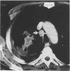
BACKGROUND--The aim of preoperative computed tomographic (CT) assessment of patients with carcinoma of the bronchus is to stage the tumour accurately, and forewarn the surgeon of any possible local extrapulmonary extension of tumour in patients considered to have potentially resectable disease. The ability of CT scanning to differentiate between conventionally resectable lung cancer (TNM stages I and II), locally advanced but resectable lung cancer (TNM stage IIIa), and locally advanced but unresectable lung cancer (TNM stage IIIb) was determined in a group of patients accepted for surgery. METHODS--Computed tomographic scans of 110 patients who underwent thoracotomy for intended resection of carcinoma of the bronchus, including 52 cases with stage III and 58 cases with stage I or II disease, were reviewed and the CT features and radiological interpretations correlated with the surgical and pathological findings. RESULTS--Thirteen CT scans were judged not to have been of diagnostic quality: of the remaining 97 cases 45 had stage III lung cancer, of whom 30 had successful resections, and 52 had stage I or stage II tumours. There was no difference in the frequencies of CT observations--including contiguity of tumour and mediastinum or chest wall, apparent mediastinal or chest wall invasion, proximity of tumour to the carina, mediastinal nodal enlargement, pulmonary collapse or consolidation and pleural effusion--in patients with stage I/II disease and patients with stage III disease. Similar results were found when the same observations were compared in all patients with resected disease and those with unresectable tumour. Sensitivity and specificity of CT was 27% and 96% respectively for tumour unresectability, 50% and 89% for mediastinal invasion, 14% and 99% for chest wall invasion, and 61% and 76% for mediastinal nodal metastases. Only 19 of 45 stage III tumours were correctly identified as being stage III and resectable or unresectable. CONCLUSIONS--In patients being considered for thoracotomy for resection of lung cancer, CT scanning used as the sole method of staging is of limited value for differentiating between stage I/II and stage III tumours. Patients should not be denied the opportunity for curative surgery on the basis of equivocal CT signs.
Full text
PDF

Images in this article

Selected References
These references are in PubMed. This may not be the complete list of references from this article.
- Armstrong P., Vincent J. M. Staging non-small cell lung cancer. Clin Radiol. 1993 Jul;48(1):1–10. doi: 10.1016/s0009-9260(05)80099-6. [DOI] [PubMed] [Google Scholar]
- Friedman P. J. Lung cancer staging: efficacy of CT. Radiology. 1992 Feb;182(2):307–309. doi: 10.1148/radiology.182.2.1732940. [DOI] [PubMed] [Google Scholar]
- Glazer G. M., Orringer M. B., Gross B. H., Quint L. E. The mediastinum in non-small cell lung cancer: CT-surgical correlation. AJR Am J Roentgenol. 1984 Jun;142(6):1101–1105. doi: 10.2214/ajr.142.6.1101. [DOI] [PubMed] [Google Scholar]
- Glazer H. S., Duncan-Meyer J., Aronberg D. J., Moran J. F., Levitt R. G., Sagel S. S. Pleural and chest wall invasion in bronchogenic carcinoma: CT evaluation. Radiology. 1985 Oct;157(1):191–194. doi: 10.1148/radiology.157.1.4034965. [DOI] [PubMed] [Google Scholar]
- Glazer H. S., Kaiser L. R., Anderson D. J., Molina P. L., Emami B., Roper C. L., Sagel S. S. Indeterminate mediastinal invasion in bronchogenic carcinoma: CT evaluation. Radiology. 1989 Oct;173(1):37–42. doi: 10.1148/radiology.173.1.2781028. [DOI] [PubMed] [Google Scholar]
- Lewis J. W., Jr, Pearlberg J. L., Beute G. H., Alpern M., Kvale P. A., Gross B. H., Magilligan D. J., Jr Can computed tomography of the chest stage lung cancer? Yes and no. Ann Thorac Surg. 1990 Apr;49(4):591–596. doi: 10.1016/0003-4975(90)90306-q. [DOI] [PubMed] [Google Scholar]
- McLoud T. C., Bourgouin P. M., Greenberg R. W., Kosiuk J. P., Templeton P. A., Shepard J. A., Moore E. H., Wain J. C., Mathisen D. J., Grillo H. C. Bronchogenic carcinoma: analysis of staging in the mediastinum with CT by correlative lymph node mapping and sampling. Radiology. 1992 Feb;182(2):319–323. doi: 10.1148/radiology.182.2.1732943. [DOI] [PubMed] [Google Scholar]
- Patterson G. A., Ilves R., Ginsberg R. J., Cooper J. D., Todd T. R., Pearson F. G. The value of adjuvant radiotherapy in pulmonary and chest wall resection for bronchogenic carcinoma. Ann Thorac Surg. 1982 Dec;34(6):692–697. doi: 10.1016/s0003-4975(10)60911-3. [DOI] [PubMed] [Google Scholar]
- Pearlberg J. L., Sandler M. A., Beute G. H., Lewis J. W., Jr, Madrazo B. L. Limitations of CT in evaluation of neoplasms involving chest wall. J Comput Assist Tomogr. 1987 Mar-Apr;11(2):290–293. doi: 10.1097/00004728-198703000-00019. [DOI] [PubMed] [Google Scholar]
- Pennes D. R., Glazer G. M., Wimbish K. J., Gross B. H., Long R. W., Orringer M. B. Chest wall invasion by lung cancer: limitations of CT evaluation. AJR Am J Roentgenol. 1985 Mar;144(3):507–511. doi: 10.2214/ajr.144.3.507. [DOI] [PubMed] [Google Scholar]
- Piehler J. M., Pairolero P. C., Weiland L. H., Offord K. P., Payne W. S., Bernatz P. E. Bronchogenic carcinoma with chest wall invasion: factors affecting survival following en bloc resection. Ann Thorac Surg. 1982 Dec;34(6):684–691. doi: 10.1016/s0003-4975(10)60909-5. [DOI] [PubMed] [Google Scholar]
- Rendina E. A., Bognolo D. A., Mineo T. C., Gualdi G. F., Caterino M., Di Biasi C., Facciolo F., Ricci C. Computed tomography for the evaluation of intrathoracic invasion by lung cancer. J Thorac Cardiovasc Surg. 1987 Jul;94(1):57–63. [PubMed] [Google Scholar]
- Scott I. R., Müller N. L., Miller R. R., Evans K. G., Nelems B. Resectable stage III lung cancer: CT, surgical, and pathologic correlation. Radiology. 1988 Jan;166(1 Pt 1):75–79. doi: 10.1148/radiology.166.1.3336705. [DOI] [PubMed] [Google Scholar]
- Stitik F. P. Staging of lung cancer. Radiol Clin North Am. 1990 May;28(3):619–630. [PubMed] [Google Scholar]
- Templeton P. A., Caskey C. I., Zerhouni E. A. Current uses of CT and MR imaging in the staging of lung cancer. Radiol Clin North Am. 1990 May;28(3):631–646. [PubMed] [Google Scholar]