Abstract
A method is described for examining viruses in faeces by direct electron microscopy using negative staining. The particles found in a group of patients with gastroenteritis and a group with other conditions are compared. Small particles in the range of sizes covering parvoviruses and enteroviruses were found about as frequently in each group.
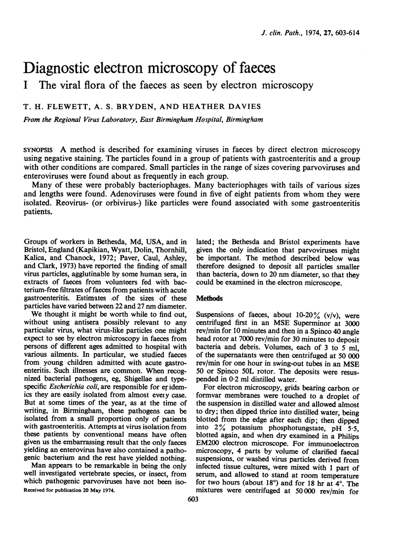
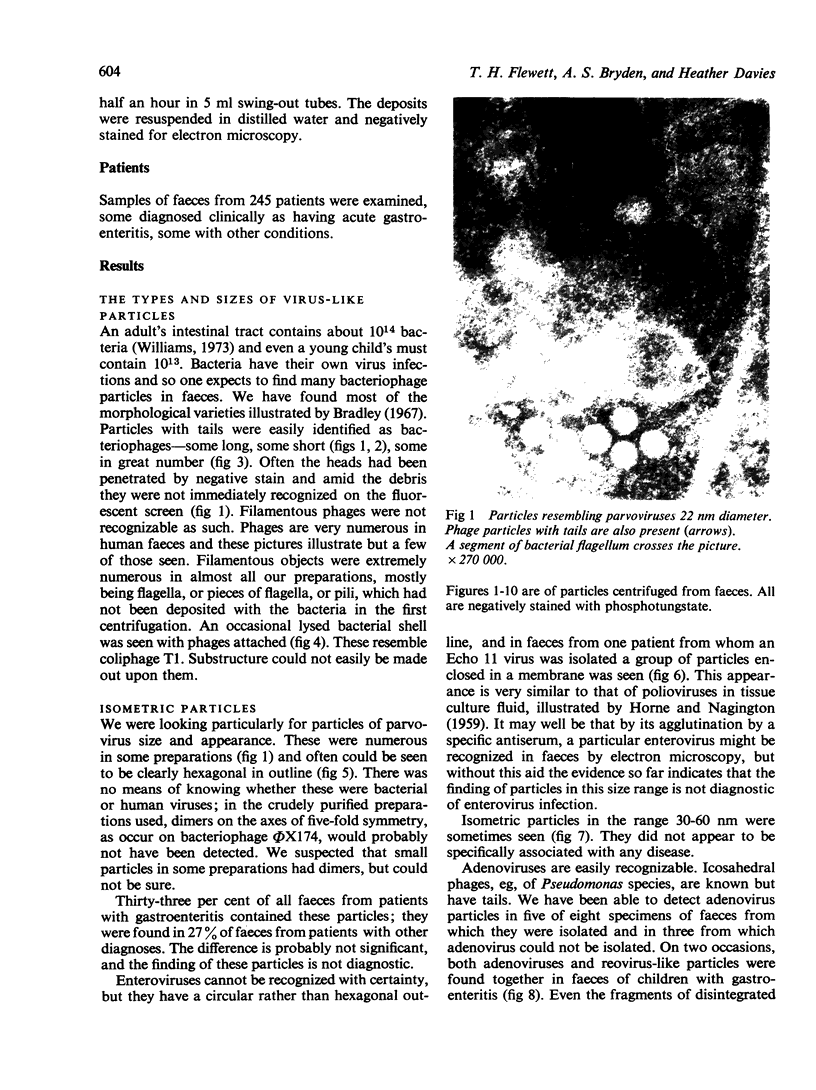
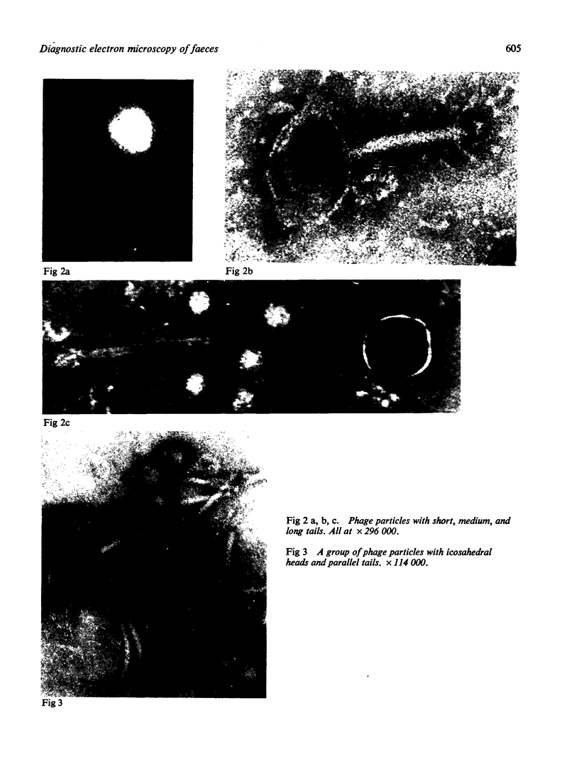
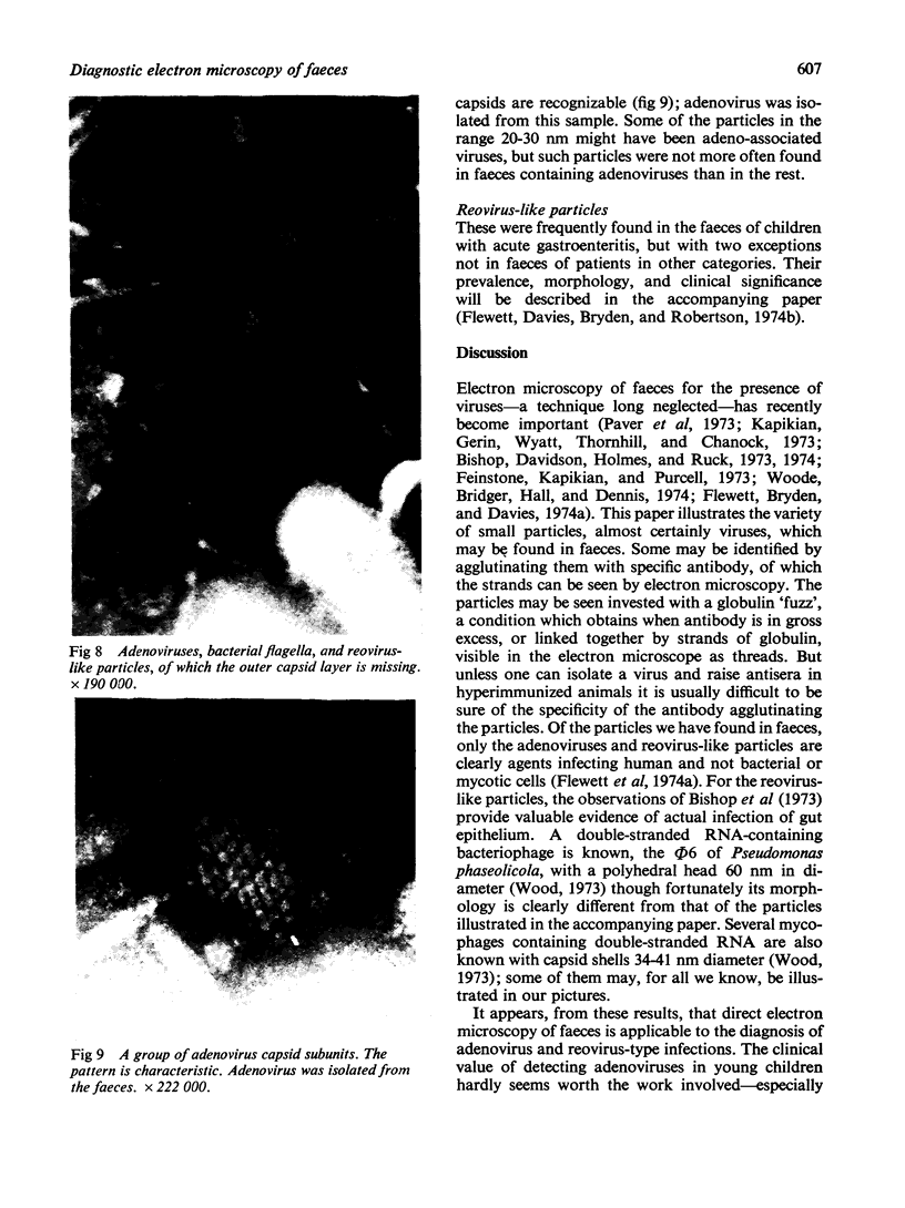
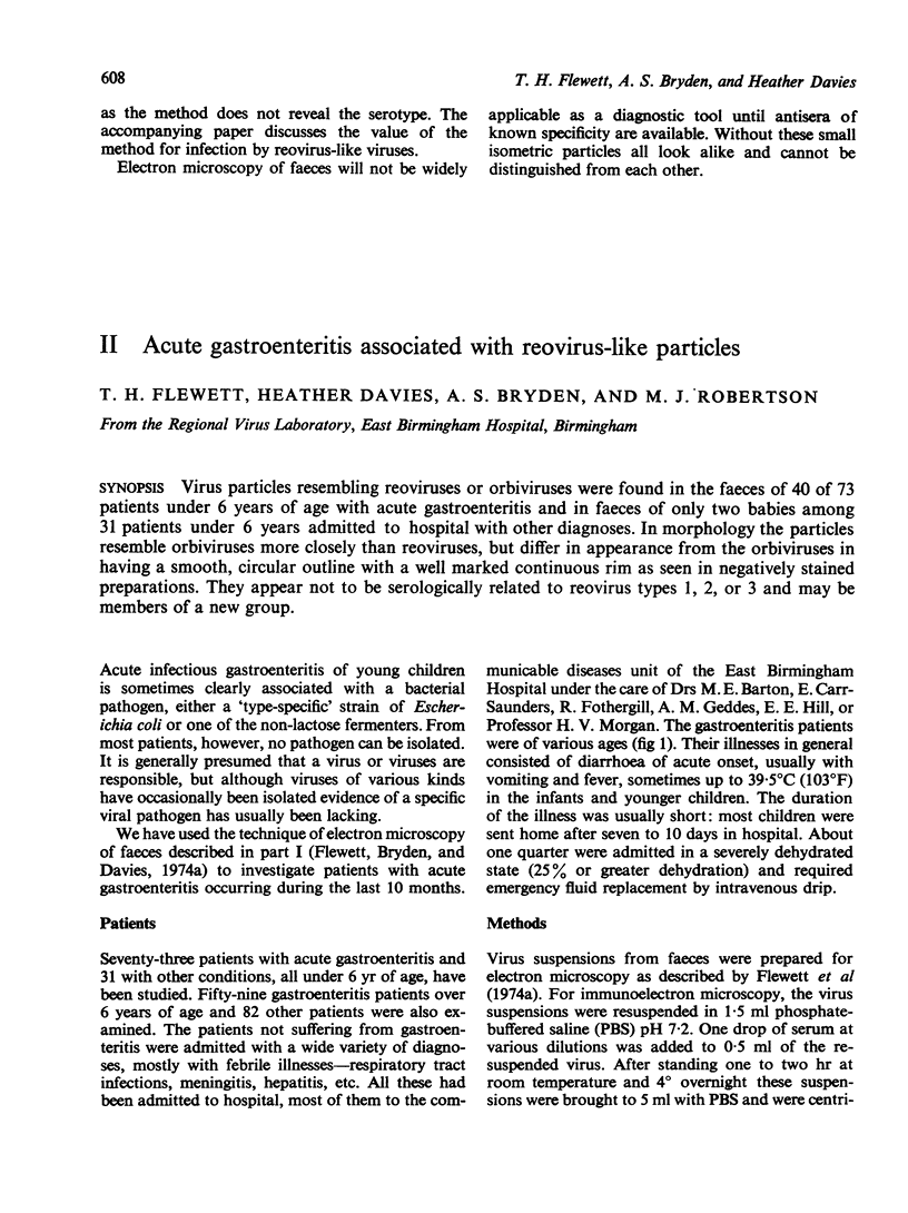
Many of these were probably bacteriophages. Many bacteriophages with tails of various sizes and lengths were found. Adenoviruses were found in five of eight patients from whom they were isolated. Reovirus- (or orbivirus-) like particles were found associated with some gastroenteritis patients.
Full text
PDF