Abstract
Hirschsprung disease (HSCR) is attributed to a failure of neural crest derived cells to migrate, proliferate, differentiate or survive in the bowel wall during embryonic Enteric Nervous System (ENS) development. This process requires a wide and complex variety of molecules and signaling pathways which are activated by transcription factors. In an effort to better understand the etiology of HSCR, we have designed a study to identify new transcription factors participating in different stages of the colonization process. A differential expression study has been performed on a set of transcription factors using Neurosphere-like bodies from both HSCR and control patients. Differential expression levels were found for CDYL, MEIS1, STAT3 and PAX6. A significantly lower expression level for PAX6 in HSCR patients, would suit with the finding of an over-representation of the larger tandem (AC)m(AG)n repeats within the PAX6 promoter in HSCR patients, with the subsequent loss of protein P300 binding. Alternatively, PAX6 is a target for DNMT3B-dependant methylation, a process already proposed as a mechanism with a role in HSCR. Such decrease in PAX6 expression may influence in the proper function of signaling pathways involved in ENS with the confluence of additional genetic factors to the manifestation of HSCR phenotype.
Hirschsprung disease (HSCR, (OMIM 142623), the most common neurocristopathy in humans (1:5000 newborns), is characterized by the absence of enteric ganglia along variable lengths of the distal gastrointestinal tract, resulting in severe intestinal dysfunction1. It appears either with a familial basis or, most often, sporadically exhibiting a complex pattern of inheritance with low sex-dependent penetrance and variable expression. Such aganglionosis is attributed to a failure to fully colonize the gastrointestinal tract by enteric neural precursor crest derived cells (ENCCs) during embryogenesis. The Enteric Nervous System (ENS) development is a process finely directed by cell surface receptors and their ligands, transcription factors that regulate their expression and proteins transmitting different signals2. Differentiation of the ENCCs during migration is one of the key processes required to achieve the dynamic and complex circuit of neurons and glia in the course of the ENS formation3,4,5. This complex process is finely regulated by a large number of transcription and signaling factors and alterations throughout such processes can lead to drastic consequences as evidenced by the aganglionosis observed in HSCR.
The RET proto-oncogene (OMIM 164761) is the main gene associated to HSCR with differential contributions of its coding and non-coding mutations6,7. In addition, this phenotype has been associated with mutations in sets of genes involved in survival, proliferation, differentiation and migration processes, either in isolated cases or syndromic presentations of the disease which suits with the complex nature of the ENS formation7,8.
Several transcription factors are critical for the correct time course of ENS development. In humans SOX10, PHOX2B and NKX2-1 (TTIF1), previously associated to some isolated or syndromic forms of HSCR, act as potential regulators of RET expression and SOX10 also modulate EDNRB expression9,10,11,12. In enteric precursors from mice and human, Pax3 is required for the activation of Ret transcription co-operatively acting with Sox1013,14. In addition, it has been shown that the ablation of Zfhx1b in mice neural crest prevents ENCCs migration beyond the proximal duodenum, and mutations in this gene are a cause of the Mowat-Wilson syndromic form of HSCR15.
Nevertheless, mechanisms underlying enteric precursor cell fate decisions during colonization of the bowel are relatively poorly understood. In an effort to better understand the etiology of HSCR and the signals required for a proper ENS development, we have designed a study to identify new transcription factors participating in different stages of the colonization process. With such purpose, we have performed a large-scale real-time PCR expression study of transcription factors participating in Embryonic Stem Cell networks that might be involved in ENS development, specially focusing on cell proliferation and/or differentiation process. For this study we have used human enteric precursor cells isolated from colon tissue of HSCR patients and control individuals as Neurospheres-like bodies (NLBs).
Results
Characterization of enteric neural precursor cells. Different expression of transcription factors in HSCR patients versus Control individuals
We have evaluated the gene expression profile of 48 transcription factors, all of them previously described as cell proliferation or differentiation modulators, which may be potential participants in the interactome network of enteric precursor cells during ENS development. Analysis and display of quantitative gene expression assay was positive for 14 transcription factors (Supplementary Fig. S1, Table S1 and Table S2 online), four of which showed statistically significant differential expression patterns (>2-fold change) between HSCR-NLBs and Control-NLBs (Table 1). These genes were CDYL, MEIS1, STAT3 (up-regulated) and PAX6 (down-regulated). Among these results, the most significant finding was the reduction by 4.67 fold of (cDNA)PAX6 expression in HSCR-NLBs compared with Control-NLBs (p value = 0.0006). PAX6 is a regulator required for a correct differentiation and proliferation in the signaling and formation of the Central Nervous System (CNS). Therefore, in light of these results we further focused on PAX6 as a candidate gene in the pathogenesis of HSCR.
Table 1. Genes with a significantly different expression in HSCR-NLBs versus control-NLBs.
| Genes | Expression | P value |
|---|---|---|
| CDYL | Up-regulated | 0.02 |
| MEIS1 | Up-regulated | 0.008 |
| STAT3 | Up-regulated | 0.006 |
| PAX6 | Down-regulated | 0.0006 |
PAX6 expression was reduced in HSCR-NLBs. The expression ratio PAX6/PAX6(5a) vary within narrow limits in NLBs
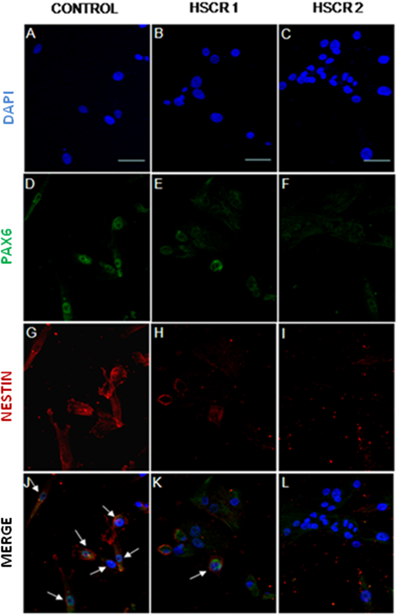
The reduced expression levels of (cDNA) PAX6 in HSCR-NLBs compared to Control-NLBs were also verified by immunocytochemistry. The percentage of cells expressing PAX6 was 36% in HSCR compared to controls, which represents a 64% of reduction in the basal values of expression (Fig. 1). It is relevant to note, that positive PAX6 cells express a marker from neural precursor cells (NESTIN) supporting that these cells are neural precursors. We have also analyzed the conditional expression of the two major isoforms of PAX6 in HSCR-NLBs, given that changes in the relative concentrations of these isoforms of PAX6 [PAX6/PAX6(5a)] during development and neurogenesis, result in changes in the functions of its downstream regulated genes. However, we did not detect different expression levels between cDNA of PAX6/PAX6 (5a) (HSCR-NLBs Ct-PAX6:32.7 and Ct-PAX6(5a):33.16; Control-NLBs Ct-PAX6:23 and Ct-PAX6(5a):22). The (cDNA) PAX6/PAX6(5a) values after normalization were 1,0175 and 1,09 in HSCR and controls respectively.
Figure 1. Expression of PAX6 in HSCR-NBLs compared with control-NLBs.
Confocal images of protein expression of PAX6 in neural precursors derived from controls (A,D,G,J) and HSCR patients (B,C,E,F,H,I,K,L). Immunostaining for PAX6 is shown in green and for NESTIN is shown in red. Cell nuclei were counterstained with 4´,6-diamidino-2-phenylindole (blue). Scale bars = 25 μM.
Identification of variants within PAX6 sequence
Mutational screening of PAX6 sequence in 196 HSCR patients, revealed no pathogenic variants, ruling out therefore the presence of deleterious mutations in this gene as a mechanism leading to HSCR (Supplementary Table S3 online).
Significant different STR allelic and genotypic distribution at the PAX6 P1 promoter region in HSCR versus controls
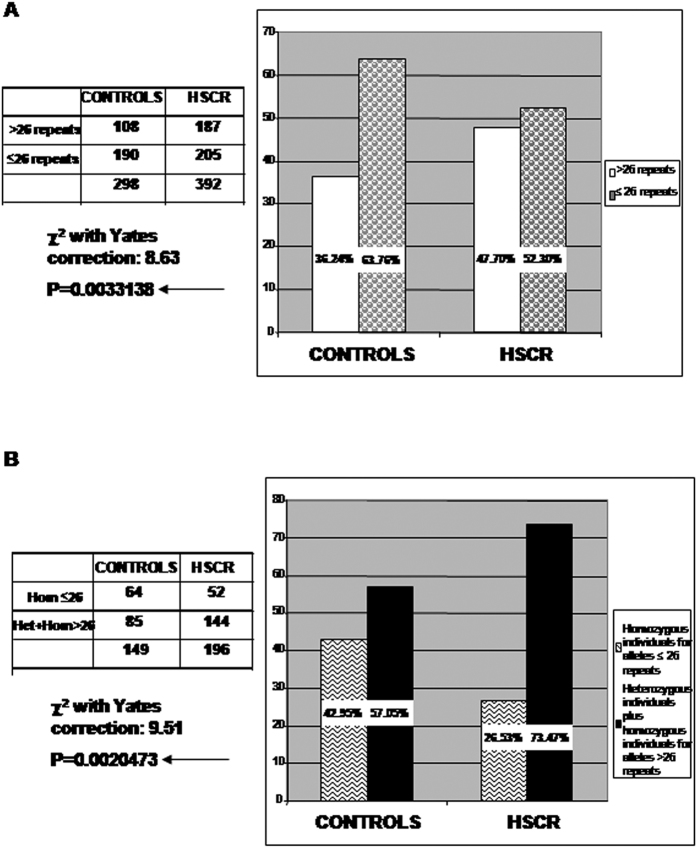
The analysis of the allelic distribution for the (AC)m(AG)n repeat at the PAX6 P1 promoter revealed a total of 13 different alleles for HSCR, ranging from 23 to 35 repeats, and 10 different alleles for controls, ranging from 24 to 33 repeats, the allele frequency can be found as Supplementary Fig. S2 online. In general, allelic distribution showed to be significantly different between HSCR and controls (x2 = 21.00, p = 0.0072). The most common allele for both groups was the one with 26 repeats, although its frequency was 11.4% lower in HSCR in comparison to controls (44.89% vs 56.3%). Moreover, when analyzing the distribution of each specific allele, we found significant under-representation of the 26 repeats allele (x2 = 8.98, p = 0.0027) and over-representation of the 29 repeats allele (x2 = 10.23, p = 0.0014) in the group of patients. These findings led us to set up a cut-off point at 26 repeats, and a detailed inspection showed that alleles with 26 or less repeats accounted for 52.29% of patients and 63.76% of controls, whereas the >26 repeats alleles accounted for 47.71% of patients and 36.24% of controls (x2 = 8.63, p = 0.0033) (Fig. 2A). In addition, when analyzing the genotypic distribution among both groups, we also found a significantly different distribution of homozygous individuals for ≤26 repeats alleles (HSCR: 26.53%, controls: 42.95%), heterozygous individuals (HSCR: 51.53%, controls: 41.61%) and homozygous individuals for >26 repeats alleles (HSCR: 21.94%, controls: 15.44%) (χ2 = 10.42, p = 0.0055). Statistical significance was maintained when analyzing the genotypic distribution after assembling heterozygous individuals and homozygous individuals with >26 repeats alleles (χ2 = 9.51, p = 0.0020) (Fig. 2B). These findings are concordant with an over-representation of HSCR patients carrying at least one or both alleles with >26 repeats (73.47%) in comparison with controls (57.05%).
Figure 2. Allelic and genotypic distribution of (AC)m(AG)n repeats within the PAX6 P1 promoter region.
(A) Distribution of PAX6 alleles with >26 (AC)m(AG)n repeats in HSCR versus controls. (B) Distribution of individuals carrying at least one PAX6 allele with >26 (AC)m(AG)n repeats. Heterozygous and homozygous individuals with alleles >26 repeats were grouped.
To further investigate the possible impact of the (AC)m(AG)n differential representation at PAX6 P1 promoter, we used in silico approaches aiming to analyze any differential binding of regulator molecules. We submitted different alleles containing variable repeats to TFSEARCH and, noteworthy, the analysis revealed a loss of the binding of E1A Binding Protein P300 (P300) for PAX6 alleles containing ≥29 repeats. Furthermore, such loss was observed with different combinations of (AC) and (AG) units leading to alleles of 29 or more repeats.
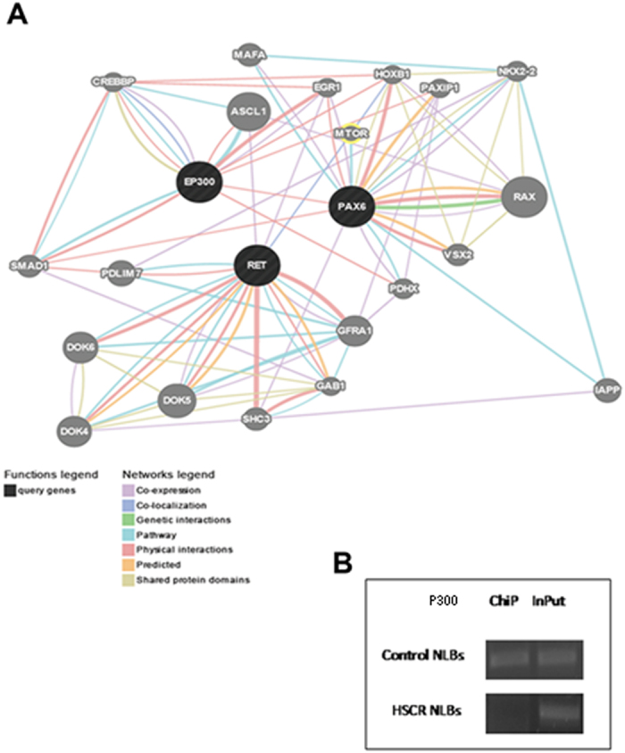
Gene-Mania and Var-Elect tools were then used to analyze interactions between PAX6 and P300, and predictions revealed a direct physical interaction. Moreover, both proteins were found to be indirectly related to RET and other proteins encoded by genes previously associated to HSCR (Fig. 3A).
Figure 3. Interactions of PAX6 with P300 and other HSCR-related proteins.

(A) Report of GeneMANIA search, which shows a physical interaction between PAX6 and P300 and indirect interactions with RET. (B) Amplification bands corresponding to the region (AC)m(AG)n repeat at the PAX6 P1 promoter, isolated from the inmunoprecipitated samples with P300 antibody (ChiP) from Control- NLBs (up) and HSCR-NLBs (down). The amplification band of the coresponding InPut samples were considered as positive controls.
PAX6 is a target of P300
To further investigate the role of P300 in the interplay of HSCR and its relation with PAX6, we checked first for the expression of P300 in HSCR and control NLBs obtaining positive expression (Supplementary Table S4). In order to study the physical interaction between P300 and PAX6 which in silico analysis suggested, ChIP-PCR analysis was performed using anti-P300 antibody in human NLBs from one control and one HSCR patient. This assay confirmed the binding of P300 to the region encompasses the (AC)m(AG)n tandem in control-NLBs. However, the band was dramatically reduced in the HSCR patient (Fig. 3B). DNA isolated from both samples was afterward genotyped; the HSCR patient carried out both alleles with 28 repeats and the control harbored alleles with different number of repeats (26/27).
According to data from the ALGGEN-PROMO bioinformatics tool, the specific binding sites for P300 were identified just flanking the repeating dinucleotide within PAX6 sequence. The ability of P300 to interact with PAX6 might be affected by the distance between these regions. The increasing number of repeats may influence the conformation of that particular sequence modifying the access of the protein to its binding site, thus we found differences between patient and control related to P300 binding affinity depending on the number of repeats.
PAX6 is a target of the Methyltransferase DNMT3B
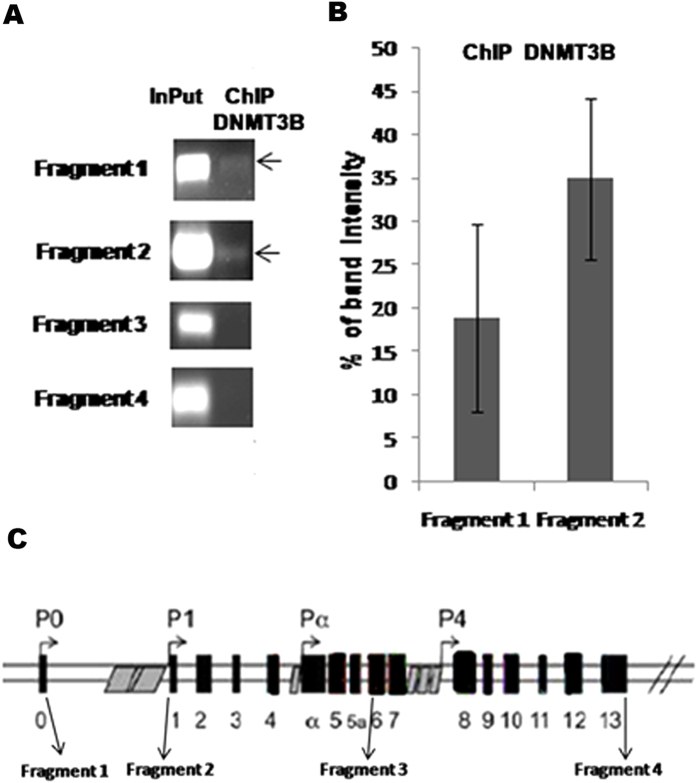
The methyltransferase DNMT3B is essential for the de novo methylation process that occurs during neurogenesis in the embryonic development. As previously reported by Torroglosa et al., HSCR-NLBs displayed lower DNA methylation and less expression of (cDNA) DNMT3B and (cDNA) PAX6 compared with Control-NLBs16. Based on these results, we aimed to elucidate if PAX6 is a direct target of DNMT3B and therefore its expression might be regulated by methylation. For this purpose, ChiP-PCR assay was performed in NLBs from mice. Samples after ChIP assay with anti-Dnmt3b antibody were amplified by PCR. Two out of four Pax6 studied regions were detected in ChiP samples (Fig. 4). These fragments correspond to regions that show a high degree of homology (59,75% and 85,18% respectively) with humans and they are also within the PAX6 human CpG islands called CpG35 and CpG464 annotated by the UCSC Genome Browser database (http://genome.ucsc.edu/) (Supplementary Fig. S3 online).
Figure 4. Identification of Pax6 as a target of Dnmt3b methylation by ChiP-PCR.
(A) Amplification bands corresponding to a selection of different regions of Pax6, isolated from the immunoprecipitated samples with Dnmt3b antibody. Amplification was observed for fragments 1 and 2. (B) Graph showing the relative mean intensity values of the corresponding amplification bands after normalization with the InPut. (C) Scheme of the Pax6 gene showing precise regions for analized fragments.
Discussion
The ENS arises from neural crest-derived cells with multipotency and migratory capabilities leading to the formation of a complex network of enteric neurons and glial cells that colonize the gastrointestinal tract. This complex process is carefully regulated by interacting signals and transcription factors that confer cell characteristics in each moment of the ENS development, and it is accepted that failures in this process are responsible for HSCR pathogenesis. Understanding underlying transcriptional programs required for enteric precursors normal development might help us to identify new genes and pathways involved in HSCR pathogenesis. With this aim, through the expression study performed among HSCR and controls NLBs, four new transcription factors (CDYL, MEIS1, STAT3 and PAX6) that might be involved in human enteric precursor cells interactome networks have been identified. It comes as no surprise that all transcription factors found to be deregulated in ENS precursors from HSCR, are implicated in the transition of proliferation/differentiation state, a critical process for the correct ENS formation. CDYL is a chromodomain-containing transcriptional co-repressor ubiquitously expressed. A study conducted in mice suggests that during neural development, Cdyl might be inhibiting the neuronal differentiation of induced pluripotent stem cells (iPS)17,18. MEIS1 is a conserved transcription factor that specifically acts as a direct molecular regulator of PAX6 expression during vertebrate’s development19,20. STAT3 is part of the JAK-STAT signaling pathway and plays an important role in the coordination of the succession of steps along the cell cycle and the differentiation process during neural crest cell specification21,22. Finally, we also obtained deregulated expression levels for the transcription factor PAX6. This gene is a highly conserved transcription factor belonging to the family of the so-called “paired-box” with functions in the eye, central nervous system and pancreas development. PAX6 is a regulator of the neural precursor proliferation and differentiation which acts by modulating the expression of different downstream effectors. Its main role is the generation of new neurons from stem cells and neural progenitors during the initial stages of the CNS development23,24,25. In the developing brain, PAX6 initially modify the proliferation of progenitor cells and later neural differentiation in a highly context-dependent manner. This changing role might be due to the relative balance of the two major isoforms PAX6 and PAX6(5a) expression levels during development26. Nevertheless, the enteric precursor cells showed no differences between expression levels of both (cDNA)PAX6 isoforms, leading to the conclusion that the ratio PAX6/PAX6(5a) does not seem to be essential for the enteric precursor decision (proliferation/differentiation).
Deregulated expression levels for PAX6 confirmed data previously obtained by Torroglosa et al., 2014, which prompted us to explore additional and different mechanisms underlying the PAX6 lower expression observed in HSCR-NLBs.
Several human diseases are caused by non-coding variations at regulatory regions of the respective genes, whose mechanism consists of the disruption of a proper regulation of the gene expression. In Friedreich ataxia (FRDA), for instance, GAA-repeat expansion in the first intron of FRDA gene results in decreased levels of frataxin due to inhibition of transcriptional elongation. Another example is FRAXA, where the expansion of a CGG-repeat in the 5′UTR of the FMR1 gene, leads to hypermethylation of the CpG Island, transcriptional silencing and loss of the protein product.
PAX6 expression is regulated by alternate usage of two promoters (P0 and P1) which are differentially regulated in a tissue-specific manner27 and its activation is positively related to PAX6 transcripts expression. P1 promoter contains distinct cis elements and several potential binding sites for transcription factors involved in tissue-specific expression in the eye, central nervous system and pancreas28,29,30. One of these elements is a polymorphic (AC)m(AG)n repeat located about 1 kb from the transcription start site which influences the transcriptional promoter efficiency in brain31,32. The analysis of the allelic and genotypic distribution of this repetitive element in HSCR patients versus control individuals, showed an over-representation of >26 repeats alleles in HSCR patients in comparison with control subjects. In silico analyses revealed the existence of two putative binding sites, just flanking the repetitive sequence, recognized by P300. This protein is a histone acetyltransferase essential in the processes of cell proliferation and differentiation regulating transcription via chromatin remodeling. Subsequently, ChIP analyses revealed the existence of that physical interaction between PAX6 and the protein P300 in enteric precursors. However, the ability of P300 to bind its target sequence was dramatically reduced when the number of repeats was increased. Changes in the repeat number might alter the distance between P300 binding sites, thus modifying this protein binding affinity. This specific DNA region within PAX6 promoter sequence must undergo structural changes to either facilitate or hinder the accessibility to P300. The increase of repeats may also have influence on the conformation of the sequence and generate different structures for other proteins recognition. This mechanism might contribute to a lower expression of PAX6 and, probably leads to a decrease in regulation of the signaling pathways related to the ENS development in which this protein is involved. Regarding to the complex genetic basis of HSCR disease, we propose this mechanism as a necessary event contributing to PAX6 down-regulation observed in patients, even though other conditions are required to completely elucidate this finding.
Epigenetic programs are essentials to drive the transcriptional profiles required for a normal neural development. As described by Torroglosa et al., an aberrant methylation status in HSCR-NLBs, was proposed as a mechanism responsible for incorrect gene expression patterns in these patients16. Regulation of gene expression through methylation is one of several ways to modulate many cellular processes during development, particularly in neurogenesis33. Mammalian postnatal neurons express at high levels DNA methyltransferases and DNA methylation along these cells is extremely dynamic34.
Furthermore, it has been shown that PAX6 expression levels may be disturbed by methylation35,36. With the aim of studying if PAX6 is a target of DNMT3B and therefore susceptible of being regulated by methylation, we performed a new ChIP-PCR analysis. Pax6 was shown to be a target for Dnmt3b, as well, as we identified two specific regions of this gene to where Dnmt3b is attached. Such binding sites are within CpG-rich regions in the human PAX6 sequence.
We hypothesize that enteric precursors from HSCR-NLBs seem to be non-responsive to external signals that lead to the correct neuronal differentiation, so that this event cannot occur at the right time, regardless of the balance between PAX6 isoforms as it has been discussed previously.
We have evaluated the gene expression profile of transcription factors which may be potential participants in the gene regulatory network of enteric precursor cells in the context of ENS development and HSCR. Findings of reduced PAX6 expression levels in HSCR-NLBs supports that PAX6 operates and regulates transcriptional network during the ENS development. We report the first association study of the transcription factor PAX6 with HSCR and that its low expression levels may result in an aberrant neurogenesis, which is directly related with manifestation of HSCR phenotype.
Methods
Generation of ENS NLBs
ENS progenitor cells were obtained from human postnatal tissues of ganglionic gut of six sporadic non-related patients diagnosed with isolated HSCR (L-HSCR:S-HSCR = 2:4; male: female = 3:3) and six patients of other gastrointestinal disorders undergoing gut resection surgery at Hospital were used as controls (4 males and 2 females). For both HSCR patients and control individuals, age range was between 3 months and 3 years.
The extraction of ENS progenitor cells from ganglionic bowels in mice was carried out in the same way as in humans, as previously described in Torroglosa et al.16.
Written informed consent for surgery, clinical and molecular genetic studies was obtained from all participants. The study was approved by the Ethics Committee for clinical research of the University Hospital Virgen del Rocío (Seville, Spain) and complies with the tenets of the declaration of Helsinki.
All procedures involving mice were performed in accordance with European Union guidelines (2010/63/EU) and Spanish law (R.D. 53/2013 BOE 34/11370-420, 2013) concerning the care and use of laboratory animals and were approved by the Animal Experimentation Ethics Committee (EAEC/IEC) of University Hospital Virgen del Rocío / Institute of Biomedicine of Seville (IBIS).
Gene Expression Study by Quantitative Real-Time PCR (qRT-PCR)
A differential expression study in cultures of NLBs has been performed in a set of transcription factors implicated in the regulatory networks of embryonic stem cells (Supplementary Table S5 online). Purification and synthesis of cDNA were performed using the protocol provided by μMACS mRNA isolation Kit and μMACS cDNA Synthesis Kit in a thermo MAKSTM Separator (MACS Miltenyi Biotec, Germany). Expression studies were carried out in an Applied Biosystems 7900HT system (Life Technologies, USA) through the TaqMan® Human Transcriptional Regulatory Network in Embryonic Stem Cell Array Plate (Life Technologies, USA) and SYBR Green method (Bio-Rad, USA). Amounts of 1000 ng (10 ng/μL) of cDNA (converted from total RNA) were used per fill reservoir for the amplification reactions through the Taqman Low Density Array. For the amplification studies performed with SYBR, 2000 ng (100 ng/μL) of cDNA (converted from total RNA) were added per well, following manufacturer recommendations.
Analysis was performed using the RQ Manager Software (Life Technologies, USA) based on the comparative Ct (ΔΔCt) method. GAPDH or B-ACTIN was used as endogenous control respectively. Following the software recommendations, the upper limit of the cycle threshold (Ct) was set at 32 for the TaqMan Array Gene Signature Plates and 35 for the SYBR Green assay. Positive expression was exclusively considered when Ct values were lower than values described above.
Statistical analyses for gene expression study
Data are presented as the mean ± SEM (Standard Error Mean) of values obtained from at least three experiments. Comparisons between values obtained in Control-NLBs and HSCR-NLBs were analyzed using the Student´s t test. Differences were considered significant when p value < 0.05.
Immunocytochemistry
For immunocytochemical studies, cells derived from NLBs were seeded onto coverslips fibronectin-poly D lysine coated and fixed with 4% (wt/vol) paraformaldehyde in 0.1 M PBS. Then, they were incubated for one hour in 2.5% (wt/vol) bovine serum albumin (BSA) in PBS and with primary and secondary antibodies. After washing, the coverslips were mounted on slides with Fluoro-Gel (EMS, Hatfield, PA, USA) and fluorescent signals were detected using a Leica Spectra confocal microscope. The primary antibodies used were anti-PAX6 (rabbit polyclonal, 1:20) and anti-NESTIN (goat polyclonal, 1:400) (Santa Cruz Biotechnology, Inc). The secondary antibodies used were anti-rabbit IgG labeled with Cy2 (1:200) and anti-goat IgG labeled with Cy5 (1:200) (Jackson Immuno Research Laboratories, Inc.). The nuclei of cells were counterstained with DAPI. For the quantification, ten images were taken per condition in which at least a number of 700 cells were counted.
Molecular Analysis of PAX6
For molecular studies we included a total of 196 Spanish HSCR patients (124 males/72 females) comprising sporadic and apparently isolated cases. A total of 158 cases were short segment forms (S-HSCR), 31 were long segment forms (L-HSCR) and 7 presented with total colonic aganglionosis (TCA). In addition, we also analyzed a group of 150 normal control individuals comprising unselected, unrelated, race-, age-, and sex-matched individuals. This study was approved by our Ethics Committee for clinical research and conformed to the tenets of the declaration of Helsinki. Fully written informed consent for molecular genetic studies was obtained from all the participants. Genomic DNA was extracted using standard protocols from peripheral blood leukocytes of all individuals included in the study.
The mutational screening of the complete coding sequence of PAX6 (NM-001127612) was performed by direct sequencing using an ABI Prism 3730 Genetic Analyzer and SeqScape v2.5 software (Life Technologies, Carlsbad, CA; conditions available on request).
On the other hand, genotyping of the (AC)m(AG)n STR, located approximately 1 kb upstream of the transcription initiation site and associated with promoter P1 of PAX6, was performed by fluorescent PCR and subsequent analysis in the ABI Prism 3730 Genetic Analyzer with the Genemapper software (Life Technologies, Carlsbad, CA; conditions available on request). Statistical analysis of the allelic and genotypic distribution in HSCR patients versus controls was performed using Pearson or Chi Squared analysis, with statistical significance set at p < 0.05. Data were analyzed employing the Statistical Package for Social Sciences (SPSS) Version 14.0 for Windows.
To perform in silico predictions of transcription factors binding sites we have used TFSEARCH (http://diyhpl.us/~bryan/irc/protocol-online/protocol-cache/TFSEARCH.html). ALGGEN-PROMO (http://alggen.lsi.upc.es/cgi-bin/promo_v3/promo/promoinit.cgi?dirDB = TF_8.3), was used to predict transcription factor binding sites in DNA sequences. Putative interactions between different genes and their participation in signaling pathways were analyzed with both Gene-Mania (http://www.genemania.org) and Var-Elect (http://www.genecards.org/?path = /Search/VarElect).
Chromatin Immunoprecipitation Assay (ChIP) and PCR
Enteric NLBs from mice and human were cross-linked with formaldehyde 1%. Cell breakage from NLBs was performed in lysis buffer (SDS, Lysis buffer-ChIP Assay, Kit Millipore Corporation, USA) and sonicated in a Bioruptor® system (Diagenode Inc., USA). Immunoprecipitation was performed with P300 and Dnmt3b antibody (Abcam, UK) following the instructions of ChIP Assay Kit (Millipore Corporation, USA) in two different assays. Amplification of the region (AC)m(AG)n STR within the PAX6 promoter and four different regions that cover the full gene (Pax6, NM_013627) (Supplementary Table S6 and S7 online) were performed in each input and inmunoprecipitated sample. Finally, the amplification products were separated in 2% agarose gel by electrophoresis.
Additional Information
How to cite this article: Enguix-Riego, M. V. et al. Identification of different mechanisms leading to PAX6 down-regulation as potential events contributing to the onset of Hirschsprung disease. Sci. Rep. 6, 21160; doi: 10.1038/srep21160 (2016).
Supplementary Material
Acknowledgments
This work was supported by the Instituto de Salud Carlos III (ISCIII), Spanish Ministry of Economy and Competitiveness, Spain (PI1301560) and Regional Ministry of Innovation, Science and Enterprise of the Autonomous Government of Andalucia (CTS-7447). MVE-R is supported by fellowship PI11/00533 from ISCIII. We would like to thank all the patients that participated in this study.
Footnotes
Author Contributions M.V.E.-R. and A.T. have designed the study. They participated in all the lab tasks carrying out all the assays and the analysis and interpretation of data. Finally they have drafted the manuscript. R.M.F. and G.A. have drafted the manuscript and revised it critically for important intellectual content. M.J.M.-J. and J.C.A. have provided the clinical history and have recruited all the patients involved in this study.S.B. has coordinated and supervised all the analyses, and provided the final version of the manuscript. All authors have read the manuscript and they have given their final approval of the version to be published.
References
- Chakravarti A. & Lyonnet S. In The Metabolic and Molecular Bases of Inherited Disease, 8th ed (eds Beaudet A. R., Scriver C. R., Sly W., Valle D. ) Ch. 251 (McGraw-Hill, 2001). [Google Scholar]
- Lake J. I. & Heuckeroth R. O. Enteric nervous system development: migration, differentiation, and disease. Am. J. Physiol. Gastrointest. Liver Physiol. 305(1), G1–24 (2013). [DOI] [PMC free article] [PubMed] [Google Scholar]
- Young H. M., Turner K. N. & Bergner A. J. The location and phenotype of proliferating neural-crest-derived cells in the developing mouse gut. Cell Tissue Res. 320, 1–9 (2005). [DOI] [PubMed] [Google Scholar]
- Henion P. D. & Weston J. A. Timing and pattern of cell fate restrictions in the neural crest lineage. Development. 124, 4351–4359 (1997). [DOI] [PubMed] [Google Scholar]
- Newgreen D. & Young H. M. Enteric nervous system: development and developmental disturbances–part 2. Pediatr. Dev. Pathol. 5, 329–349 (2002). [DOI] [PubMed] [Google Scholar]
- Emison E. S. et al. Differential contributions of rare and common, coding and noncoding Ret mutations to multifactorial Hirschsprung disease liability. Am. J. Hum. Genet. 87, 60–74 (2010). [DOI] [PMC free article] [PubMed] [Google Scholar]
- Amiel J. et al. Hirschsprung disease, associated syndromes and genetics: a review. J. Med. Genet. 45, 1–14 (2008). [DOI] [PubMed] [Google Scholar]
- Borrego S., Ruiz-Ferrer M., Fernández R. M. & Antiñolo G. Hirschsprung’s disease as a model of complex genetic etiology. Histol. Histopathol. 28, 1117–36 (2013). [DOI] [PubMed] [Google Scholar]
- Leon T. Y. et al. Transcriptional regulation of RET by Nkx2-1, Phox2b, Sox10, and Pax3. J. Pediatr. Surg. 44, 1904–1912 (2009). [DOI] [PubMed] [Google Scholar]
- Pattyn A., Morin X., Cremer H., Goridis C. & Brunet J. F. The homeobox gene Phox2b is essential for the development of autonomic neural crest derivatives. Nature. 399, 366–370 (1999). [DOI] [PubMed] [Google Scholar]
- Garcia-Barcelo M. M. et al. Evaluation of the thyroid transcription factor-1 gene (TITF1) as a Hirschsprung’s disease locus. Ann. Hum. Genet. 71, 746–754 (2007). [DOI] [PubMed] [Google Scholar]
- Zhu L. et al. Spatiotemporal regulation of endothelin receptor-B by SOX10 in neural crest-derived enteric neuron precursors. Nat. Genet. 36, 732–737 (2004). [DOI] [PubMed] [Google Scholar]
- Lang D. et al. Pax3 is required for enteric ganglia formation and functions with Sox10 to modulate expression of c-ret. J. Clin. Invest. 106, 963–971 (2000). [DOI] [PMC free article] [PubMed] [Google Scholar]
- Lang D. & Epstein J. A. Sox10 and Pax3 physically interact to mediate activation of a conserved c-RET enhancer. Hum. Mol. Genet. 12, 937–945 (2003). [DOI] [PubMed] [Google Scholar]
- Van de Putte T., Francis A., Nelles L., van Grunsven L. A. & Huylebroeck D. Neural crest-specific removal of Zfhx1b in mouse leads to a wide range of neurocristopathies reminiscent of Mowat-Wilson syndrome. Hum. Mol. Genet. 16, 1423–1436 (2007). [DOI] [PubMed] [Google Scholar]
- Torroglosa A. et al. Involvement of DNMT3B in the pathogenesis of Hirschsprung disease and its possible role as a regulator of neurogenesis in the human enteric nervous system. Genet. Med. 16, 703–710 (2014). [DOI] [PubMed] [Google Scholar]
- Lui V. C. et al. Perturbation of hoxb5 signaling in vagal neural crests down-regulates ret leading to intestinal hypoganglionosis in mice. Gastroenterology. 134, 1104–1115 (2008). [DOI] [PubMed] [Google Scholar]
- Wan L. et al. Generation and neuronal differentiation of induced pluripotent stem cells in Cdyl-/- mice. Neuroreport. 24, 114–119 (2013). [DOI] [PubMed] [Google Scholar]
- Zhang X., Friedman A., Heaney S., Purcell P. & Maas R. L. Meis homeoproteins directly regulate Pax6 during vertebrate lens morphogenesis. Genes Dev. 16, 2097–2107 (2002). [DOI] [PMC free article] [PubMed] [Google Scholar]
- Zhang X. et al. Pax6 is regulated by Meis and Pbx homeoproteins during pancreatic development. Dev. Biol. 300, 748–757 (2006). [DOI] [PubMed] [Google Scholar]
- Nichane M., Ren X. & Bellefroid E. J. Self-regulation of Stat3 activity coordinates cell-cycle progression and neural crest specification. EMBO J. 29, 55–67 (2010). [DOI] [PMC free article] [PubMed] [Google Scholar]
- Yoshimatsu T. et al. Non-cell-autonomous action of STAT3 in maintenance of neural precursor cells in the mouse neocortex. Development. 133, 2553–2563 (2006). [DOI] [PubMed] [Google Scholar]
- Jang E. S. & Goldman J. E. Pax6 expression is sufficient to induce a neurogenic fate in glial progenitors of the neonatal subventricular zone. PLoS One. 6(6), e20894 (2011) [DOI] [PMC free article] [PubMed] [Google Scholar]
- Kanakubo S. et al. Abnormal migration and distribution of neural crest cells in Pax6 heterozygous mutant eye, a model for human eye diseases. Genes Cells. 11, 919–933 (2006). [DOI] [PubMed] [Google Scholar]
- Osumi N., Shinohara H., Numayama-Tsuruta K. & Maekawa M. Concise review: Pax6 transcription factor contributes to both embryonic and adult neurogenesis as a multifunctional regulator. Stem Cells. 26, 1663–1672 (2008). [DOI] [PubMed] [Google Scholar]
- Pinson J., Mason J. O., Simpson T. I. & Price D. J. Regulation of the Pax6 : Pax6(5a) mRNA ratio in the developing mammalian brain. BMC Dev. Biol. 5, 13 (2005). [DOI] [PMC free article] [PubMed] [Google Scholar]
- Xu Z. P. & Saunders G. F. Transcriptional regulation of the human PAX6 gene promoter. J Biol Chem. 272, 3430–6 (1997). [DOI] [PubMed] [Google Scholar]
- Anderson T. R., Hedlund E. & Carpenter E. M. Differential Pax6 promoter activity and transcript expression during forebrain development. Mech Dev. 114, 171–5 (2002). [DOI] [PubMed] [Google Scholar]
- Zheng J. B., Zhou Y. H., Maity T., Liao W. S. & Saunders G. F. Activation of the human PAX6 gene through the exon 1 enhancer by transcription factors SEF and Sp1. Nucleic Acids Res. 29, 4070–8 (2001). [DOI] [PMC free article] [PubMed] [Google Scholar]
- Griffin C., Kleinjan D. A., Doe B. & van Heyningen V. New 3′ elements control Pax6 expression in the developing pretectum, neural retina and olfactory region. Mech Dev. 112, 89–100 (2002). [DOI] [PubMed] [Google Scholar]
- Okladnova O., Syagailo Y. V., Mossner R. & Lesch K. P. Regulation of PAX-6 gene transcription: alternate promoter usage in human brain. Brain Res. Mol. Brain Res. 60, 177–192 (1998). [DOI] [PubMed] [Google Scholar]
- Okladnova O. et al. A promoter-associated polymorphic repeat modulates PAX-6 expression in human brain. Biochem. Biophys. Res. Commun. 248, 402–405 (1998). [DOI] [PubMed] [Google Scholar]
- Trowbridge J. J. & Orkin S. H. DNA methylation in adult stem cells: new insights into self-renewal. Epigenetics. 5, 189–938 (2010). [DOI] [PubMed] [Google Scholar]
- Feng J., Chang H., Li E. & Fan G. Dynamic expression of de novo DNA methyltransferases Dnmt3a and Dnmt3b in the central nervous system. J Neurosci Res. 79, 734–46 (2005). [DOI] [PubMed] [Google Scholar]
- Martins-Taylor K., Schroeder D. I., Lasalle J. M., Lalande M. & Xu R. H. Role of DNMT3B in the regulation of early neural and neural crest specifiers. Epigenetics. 7, 71–81 (2012). [DOI] [PMC free article] [PubMed] [Google Scholar]
- Balmer N. V. et al. Epigenetic changes and disturbed neural development in a human embryonic stem cell-based model relating to the fetal valproate syndrome. Hum Mol Genet. 21, 4104–14 (2012). [DOI] [PubMed] [Google Scholar]
Associated Data
This section collects any data citations, data availability statements, or supplementary materials included in this article.


