

Abstract
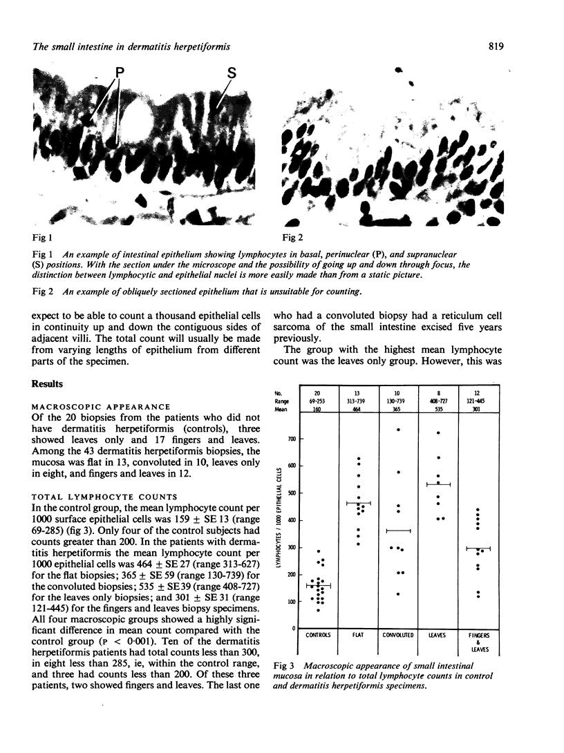
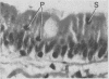
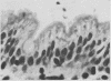
Small intestinal biopsies from 43 patients with dermatitis herpetiformis have been studied. The diagnosis of dermatitis herpetiformis was made on clinical and histological criteria and the presence of IgA deposits in the uninvolved skin. The macroscopic appearance of the intestinal biopsy was flat in 13, convoluted in 10, leaves only in eight, and fingers and leaves in 12. Twenty small intestinal biopsies from patients who did not have dermatitis herpetiformis or gastrointestinal disorder showed leaves only in three and fingers and leaves in 17. The mean total lymphocyte count per 1000 epithelial cells for this control group was 159 ± SE 13; for the dermatitis herpetiformis patients with flat biopsies it was 464 ± SE 27; for the convoluted biopsies 365 ± SE 59; for leaves only 535 ± SE 39; and for the fingers and leaves biopsies 301 ± SE 31. The counts for all four groups are significantly greater than the control group (P < 0.001). Three of the 43 patients with dermatitis herpetiformis had lymphocyte counts below 200 per 1000 epithelial cells, and four of our controls had counts greater than 200 but none above 300.
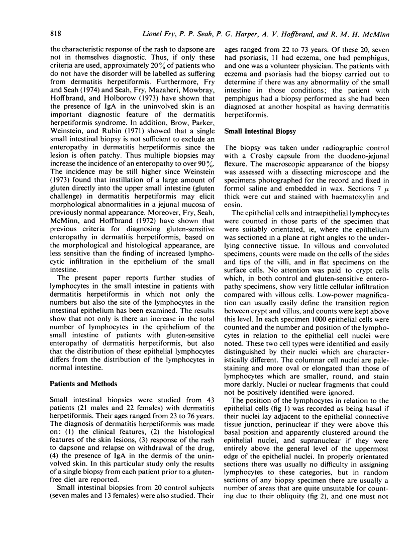
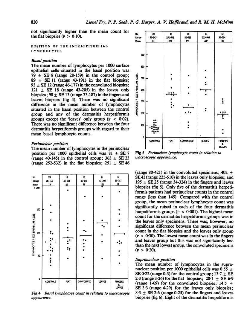
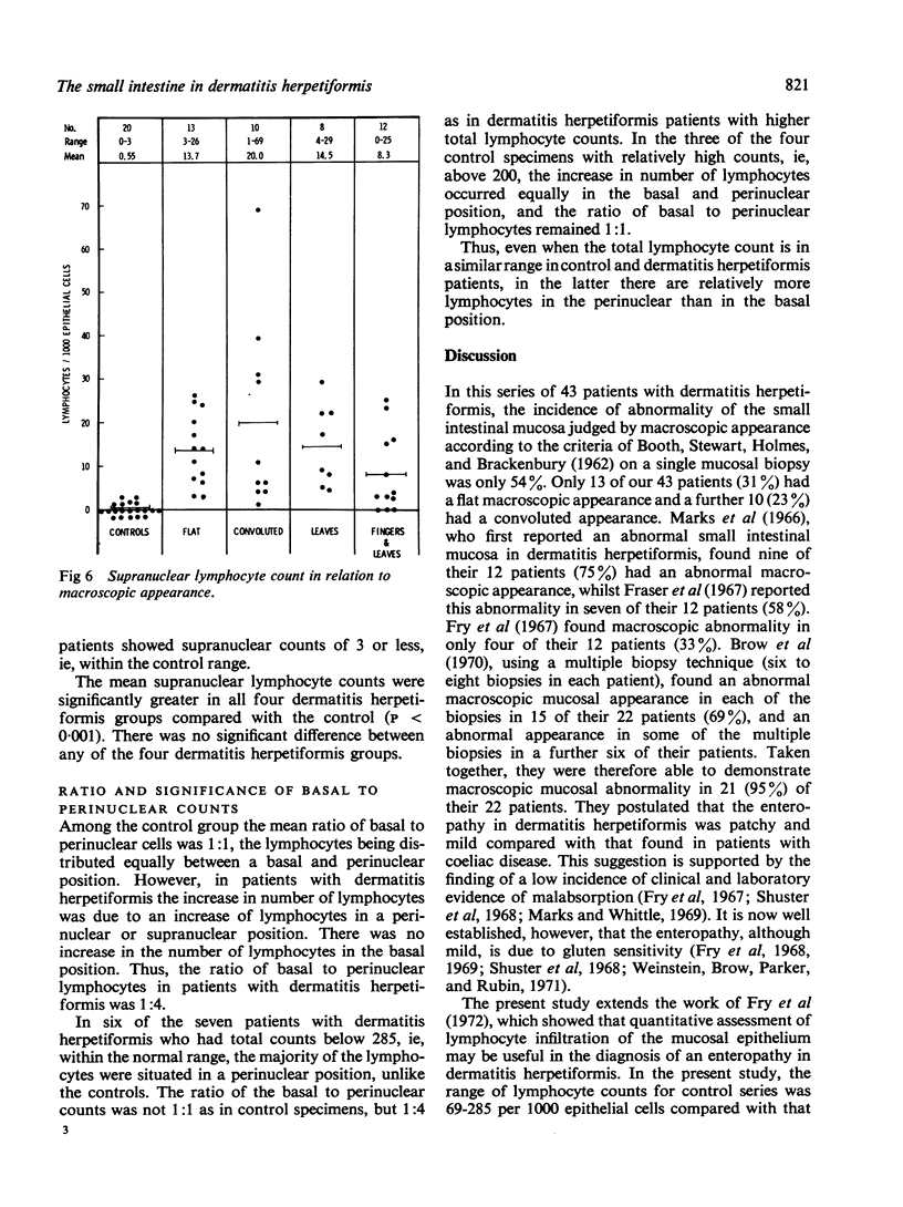
In the control group the mean counts for lymphocytes per 1000 epithelial cells in the basal position of the intestinal epithelium was 79 ± SE 8; in the dermatitis herpetiformis flat biopsies it was 89 ± SE 11; for the convoluted biopsies 93 ± SE 12; for the leaves only biopsies 121 ± SE 18 and for the fingers and leaves 98 ± SE 13. However, the mean lymphocyte count in the perinuclear position was 81 ± SE 7 for the controls, 362 ± SE 23 for the flat dermatitis herpetiformis biopsies; 251 ± SE 46 for the convoluted specimens; 402 ± SE 43 for the leaves only specimens, and 195 ± SE 25 for the fingers and leaves biopsies. The mean lymphocyte count for the supranuclear position was 0·55 ± SE 0·22 for the control group; 13·7 ± SE 2·3 for the flat dermatitis herpetiformis biopsies; 20·0 ± SE 6·9 for the convoluted biopsies; 14·5 ± SE 3·3 for the leaves only biopsies; and 8·3 ± SE 2·6 for the fingers and leaves biopsies. Thus in gluten-sensitive enteropathy the increase in lymphocytes in the intestinal epithelium is in the perinuclear and supranuclear position. The ratio of basal lymphocytes to peri- and supranuclear lymphocytes appears to be 1:1 in normal intestinal epithelium, but approximately 1:4 in the gluten-sensitive enteropathy of dermatitis herpetiformis.
Full text
PDF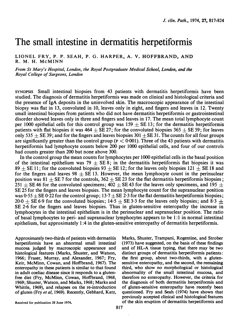
Images in this article
Selected References
These references are in PubMed. This may not be the complete list of references from this article.
- Brow J. R., Parker F., Weinstein W. M., Rubin C. E. The small intestinal mucosa in dermatitis herpetiformis. I. Severity and distribution of the small intestinal lesion and associated malabsorption. Gastroenterology. 1971 Mar;60(3):355–361. [PubMed] [Google Scholar]
- Ferguson A., Murray D. Quantitation of intraepithelial lymphocytes in human jejunum. Gut. 1971 Dec;12(12):988–994. doi: 10.1136/gut.12.12.988. [DOI] [PMC free article] [PubMed] [Google Scholar]
- Fraser N. G., Murray D., Alexander J. O. Structure and function of the small intestine in dermatitis herpetiformis. Br J Dermatol. 1967 Oct;79(10):509–518. doi: 10.1111/j.1365-2133.1967.tb11405.x. [DOI] [PubMed] [Google Scholar]
- Fry L., Keir P., McMinn R. M., Cowan J. D., Hoffbrand A. V. Small-intestinal structure and function and haematological changes in dermatitis herpetiformis. Lancet. 1967 Oct 7;2(7519):729–733. doi: 10.1016/s0140-6736(67)91942-3. [DOI] [PubMed] [Google Scholar]
- Fry L., McMinn R. M., Cowan J. D., Hoffbrand A. V. Effect of gluten-free diet on dermatological, intestinal, and haematological manifestations of dermatitis herpetiformis. Lancet. 1968 Mar 16;1(7542):557–561. doi: 10.1016/s0140-6736(68)92830-4. [DOI] [PubMed] [Google Scholar]
- Fry L., McMinn R. M., Cowan J. D., Hoffbrand A. V. Gluten-free diet and reintroduction of gluten in dermatitis herpetiformis. Arch Dermatol. 1969 Aug;100(2):129–135. [PubMed] [Google Scholar]
- Fry L., Seah P. P. Dermatitis herpetiformis: an evaluation of diagnostic criteria. Br J Dermatol. 1974 Feb;90(2):137–146. doi: 10.1111/j.1365-2133.1974.tb06377.x. [DOI] [PubMed] [Google Scholar]
- Fry L., Seah P. P., McMinn R. M., Hoffbrand A. V. Lymphocytic infiltration of epithelium in diagnosis of gluten-sensitive enteropathy. Br Med J. 1972 Aug 12;3(5823):371–374. doi: 10.1136/bmj.3.5823.371. [DOI] [PMC free article] [PubMed] [Google Scholar]
- GOUGH K. R., READ A. E., NAISH J. M. Intestinal reticulosis as a complication of idiopathic steatorrhoea. Gut. 1962 Sep;3:232–239. doi: 10.1136/gut.3.3.232. [DOI] [PMC free article] [PubMed] [Google Scholar]
- Gebhard R. L., Katz S. I., Marks J., Shuster S., Trapani R. J. HL-A antigen type and small-intestinal disease in dermatitis herpetiformis. Lancet. 1973 Oct 6;2(7832):760–762. doi: 10.1016/s0140-6736(73)91039-8. [DOI] [PubMed] [Google Scholar]
- Harris O. D., Cooke W. T., Thompson H., Waterhouse J. A. Malignancy in adult coeliac disease and idiopathic steatorrhoea. Am J Med. 1967 Jun;42(6):899–912. doi: 10.1016/0002-9343(67)90071-x. [DOI] [PubMed] [Google Scholar]
- Marks J., Shuster S., Watson A. J. Small-bowel changes in dermatitis herpetiformis. Lancet. 1966 Dec 10;2(7476):1280–1282. doi: 10.1016/s0140-6736(66)91692-8. [DOI] [PubMed] [Google Scholar]
- Marks R., Whittle M. W. Results of treatment of dermatitis herpetiformis with a gluten-free diet after one year. Br Med J. 1969 Dec 27;4(5686):772–775. doi: 10.1136/bmj.4.5686.772. [DOI] [PMC free article] [PubMed] [Google Scholar]
- Meader R. D., Landers D. F. Electron and light microscopic observations on relationships between lymphocytes and intestinal epithelium. Am J Anat. 1967 Nov;121(3):763–773. doi: 10.1002/aja.1001210318. [DOI] [PubMed] [Google Scholar]
- Seah P. P., Fry L., Mazaheri M. R., Mowbray J. F., Hoffbrand A. V., Holborow E. J. Alternate-pathway complement fixation by IgA in the skin in dermatitis herpetiformis. Lancet. 1973 Jul;2(7822):175–177. doi: 10.1016/s0140-6736(73)93006-7. [DOI] [PubMed] [Google Scholar]
- Shuster S., Watson A. J., Marks J. Coeliac syndrome in dermatitis herpetiformis. Lancet. 1968 May 25;1(7552):1101–1106. doi: 10.1016/s0140-6736(68)90181-5. [DOI] [PubMed] [Google Scholar]
- Toner P. G., Ferguson A. Intraepithelial cells in the human intestinal mucosa. J Ultrastruct Res. 1971 Feb;34(3):329–344. doi: 10.1016/s0022-5320(71)80076-x. [DOI] [PubMed] [Google Scholar]
- Weinstein W. M., Brow J. R., Parker F., Rubin C. E. The small intestinal mucosa in dermatitis herpetiformis. II. Relationship of the small intestinal lesion to gluten. Gastroenterology. 1971 Mar;60(3):362–369. [PubMed] [Google Scholar]