INTRODUCTION
The congenital diaphragmatic hernia (CDH) is defined as an anatomical defect on diaphragm, which permits the herniation of abdominal viscera into the thorax4. The hernia occurs due to an incomplete occlusion of the pleuroperitoneal channel during the embrionary period. The main cause of the incomplete closure can be a genetic mutation, a teratogen or both.
In terms of anatomic location, the CDH can be classified as Bochdalek type when an incomplete pleuroperitoneal channel occlusion is found posterolaterally; as Morganni type, while the defect is seen retrosternally; and yet as a congenital transhiatal esophagic type hernia. Among them, the Bochdalek type is the most common, found in 78-90% of patients; the Morganni type, in 1,5-6% of cases; and transhiatal, 14-24%11.
In most cases, the clinical impact occurs in the neonatal period, since only 10% of hernias are diagnosed after this period7. In neonates, the clinical presentation is acute, providing a higher morbidity and mortality. In adulthood, symptoms, if any, are more insidious, vague and intermittent, affecting not only the pulmonary dynamics, but also the gastrointestinal function5.
Chest X-ray and CT scan may be used2 , 8 , 12.Nevertheless, CDH findings are incidental when performing radiological examinations for other reasons, with the right-sided Bochdalek hernia accounting for 68% of cases.9 , 13
In elective situations, the minimally invasive surgery, either via laparoscopic or thoracoscopic can be used, but with limited application in cases of right-sided hernia3 , 8. Minor defects, technically easier to fix, can be sutured normally; in the case of larger apertures, or even hemidiaphragmatic agenesis, the use of nonabsorbable polypropylene mesh is the only solution.
CASE REPORT
A forty-five-year-old woman, caucasian, married, admitted into our service, complains of insidious jaundice in the previous five months, associated with itching, choluria and abdominal distension. Eight months before, the patient had a spontaneous abortion during the fourth month of pregnancy. Reported history of cranial malformation at birth, duly corrected surgically.
The patient was underwent to serological laboratory tests for viral hepatitis, resulting all negative. The serum total bilirubin was 4.3 mg/dl due to direct fraction, and creatinine was 1.7 mg/dl. During ultrasound exam, it was shown dilatation of intrahepatic bile ducts and liver enlargement, mainly of the right hepatic lobe. Computed tomography with contrast revealed the partial absence of diaphragmatic dome in the right posterolateral portion, showing an herniation of the right hepatic lobe, the right kidney and right adrenal gland, associated with marked atrophy of the left hepatic lobe and right pulmonary hypoplasia.
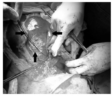
The patient was initially submitted to laparotomy which showed massive hepatomegaly due to the right lobe, whose edge was at the level of the umbilicus scar. Due to the great technical difficulties, thoracophrenolaparotomy was conducted, which showed failure at right diaphragm dome of approximately 10 cm (Figure 1), herniation of the hepatic lobe, kidney, right adrenal, colon, associated to a partial twist of the common bile duct and dilatation of the biliary tract upstream and excessive lateral traction of first and second portions of duodenum, and of the head of the pancreas.
FIGURE 1. - Intraoperatory registry of the diaphragmatic opening (among black arrows).

Posteriorly, the reduction of the hernia contents back into the abdominal cavity was done, with subsequent apposition of polypropylene mesh over the hernia defect and drainage of the chest. Intraoperative cholangiography was performed demonstrating recanalization of the bile duct and satisfying contrast escape into the duodenum.
The postoperative period went on with normalization of bilirubin and renal function. The patient died weeks later due to nosocomial pneumonia and sepsis during hospitalization in intensive care unit.
DISCUSSION
The CDH incidence in general population varies between 2.5-3.8 cases per 10,000 births. There is some difficulty in establishing the prevalence of herniated Bochdalek in adults. A retrospective study of more than 13,000 CT scans of the abdomen showed a prevalence of about 0.17%. Other studies, however, agree with a greater prevalence, at about 6-12%, when computed tomography multislice is used9 , 13.
Studies have found that there is a lower risk of afrodescedent population being CDH carrier, compared to non-hispanic caucasians. There is a relative risk of 50% higher in children of mothers aged over 35, compared to maternal age between 20-24 years4. In this case, the patient is a caucasian descendent; however, maternal age at birth was 23 years.
Retrospective study of 116 cases between 1991 and 2002 in Australia, found a prevalence of 46.6% of clinically significant abnormalities, and 38.8% minimum clinically significant abnormalities, being the most frequent neurological, musculoskeletal, dysmorphic, genitourinary and gastrointestinal1. In this report above, it was mentioned history of cranial malformation, unspecified by the patient.
The left-sided Bochdalek hernia is more prevalent than the right one, because the right-sided dome develops earlier and the liver avoids abdominal viscera protrusion2 , 11.However, hernias during the adulthood through the right dome are more frequent, appearing incidentally in 68% of diagnoses and affects mostly the females13.
The herniary defect varies from 1 cm of diameter until the complete absence of hemidiaphragm8. It was shown during the surgical procedure a diaphragmatic defect of approximately 10 cm in its largest diameter in the right posterolateral portion. In 20% of cases there is an hernial sac, in contrast to the majority of cases where there is a direct communication between the thoracic and abdominal cavities8. In 73% of cases, diaphragmatic hernia contains only visceral fat or omentum7. In the discussed case, it was not found an intraoperatively evidence of hernial sac.
Most CDH are diagnosed during the neonatal period, with only 10% of them discovered after this period7. The symptoms in adults are usually insidious and undefined. They can be not only gastrointestinal symptons, such as nausea, postprandial vomiting, abdominal pain, back pain, post-prandial bloating; but also respiratory complaints, such as dyspnea, chest pain, shoulder referred pain5 , 8. On physical examination, auscultation of typical bowel sounds of peristalsis is a specific signal for diaphragmatic hernia2.Specifically in this related case, the patient denied any symptoms throughout the life, looking for medical care because of a recent jaundice.
In distinction to the reality presented in this case, the most common acute complication, and the most feared, is the hernial incarceration and/or strangulation. The risk of strangulation in the right side is smaller, since the hernial orifice is generally larger than the contralateral side5. Some factors that increase intra-abdominal pressure, such as pregnancy, labor, coughing, sneezing and trauma may increase the risk of hernial content strangulation5 , 13. During the anamnesis of this patient, there are reports of pregnancy, with subsequent abortion three months before the beginning of cholestatic syndrome, a process that may have influenced the increase of the intra-abdominal pressure, twisting of the bile duct and appearance of jaundice.
Different modalities of diagnostic imaging can be used, among which chest x-ray, ultrasound, computadorized tomography, magnetic resonance. The sensitivity of chest x-ray is 70% and is not specific enough to exclude the diagnosis of Bochdalek hernia in case of negative result2 , 8. The gold standard for diagnosing is the double contrast tomography2. During the investigation of jaundice of the case in discussion, it was decided to request ultrasound and abdominal CT with contrast; after diagnosis, there was a complementation with chest tomography.
In urgent cases, the recommended treatment is open surgery with initial abdominal approach, applying for the thoracic via in cases of technical difficulty5 , 8. In elective situations, minimally invasive surgery, either via laparoscopic and/or videothoracoscopic can be used, but with limited application in cases of right-sided hernia3 , 8.
Minor defects, technically easier to fix, can be normally sutured; in situations of larger apertures, or even hemidiaphragmatic agenesis, the use of nonabsorbable mesh is the only way2. If large hernia has been reduced, the intra-abdominal pressure should be intensively monitored postoperatively in order to prevent the appearing of abdominal compartment syndrome8. The postoperative recurrence rate is considered rare4.
Footnotes
Financial source: none
REFERENCES
- 1.Colvin J, Bower C, Dickinson JE, Sokol J. Outcomes of congenital diaphragmatic hernia: a population-based study in Western Australia. Pediatrics. 2005;116:e356–e363. doi: 10.1542/peds.2004-2845. [DOI] [PubMed] [Google Scholar]
- 2.Esmer D, Alvarez Tostado J, Alfaro A, Carmona R, Salas M. Thoracoscopic and laparoscopic repair of complicated Bochdalek hernia in adult. Hernia; [DOI] [PubMed] [Google Scholar]
- 3.Fraser JD, Craft RO, Harold KL, Jaroszewski DE. Minimally invasive repair of a congenital right-sided diaphragmatic hernia in an adult. Surg Laparosc Endosc Percutan Tech. 2009;19:5–7. doi: 10.1097/SLE.0b013e318195c42e. [DOI] [PubMed] [Google Scholar]
- 4.Gaxiola A, Varon J, Valladolid G. Congenital diaphragmatic hernia: an overview of the etiology and current management. Acta Paediatr. 2009;98(4):621–627. doi: 10.1111/j.1651-2227.2008.01212.x. [DOI] [PubMed] [Google Scholar]
- 5.Kanazawa A, Yoshioka Y, Inoi O, Murase J, Kinoshita H. Acute respiratory failure caused by an incarcerated right-sided adult Bochdalek hernia: report of a case. Surg Today. 2002;32:812–815. doi: 10.1007/s005950200156. [DOI] [PubMed] [Google Scholar]
- 6.Laaksonen E, Silvasti S, Hakal T. Right-sided Bochdalek hernia in an adult: a case report. J Med Case Reports. 2009;3:92912–92912. doi: 10.1186/1752-1947-3-9291. [DOI] [PMC free article] [PubMed] [Google Scholar]
- 7.Lee EJ, Lee SY. Fluid shift on chest radiography: Bochdalek hernia. CMAJ. 2010;182:E311–E312. doi: 10.1503/cmaj.091323. [DOI] [PMC free article] [PubMed] [Google Scholar]
- 8.Losanoff JE, Sauter ER. Congenital posterolateral diaphragmatic hernia in an adult. Hernia. 2004;8:83–85. doi: 10.1007/s10029-003-0166-5. [DOI] [PubMed] [Google Scholar]
- 9.Mullins ME, Stein J, Saini SS, Mueller PR. Prevalence of incidental Bochdalek's hernia in a large adult population. AJR Am J Roentgenol. 2001;177:363–366. doi: 10.2214/ajr.177.2.1770363. [DOI] [PubMed] [Google Scholar]
- 10.Owen ME, Rowley GC, Tighe MJ, Wake PN. Delayed diagnosis of infarcted small bowel due to right-sided Bochdalek hernia. Ann R Coll Surg Engl. 2007;89:W1–W2. doi: 10.1308/147870807X160407. [DOI] [PMC free article] [PubMed] [Google Scholar]
- 11.Robinson PD, Fitzgerald DA. Congenital diaphragmatic hernia. Paediatr Respir Rev. 2007;8:323–335. doi: 10.1016/j.prrv.2007.08.004. [DOI] [PubMed] [Google Scholar]
- 12.Rout S, Foo FJ, Hayden JD, Guthrie A, Smith AM. Right-sided Bochdalek hérnia obstructing in an adult: case report and review of the literature. Hernia. 2007;11:359–362. doi: 10.1007/s10029-007-0188-5. [DOI] [PubMed] [Google Scholar]
- 13.Rout S, Foo FJ, Hayden JD, Guthrie A, Smith AM. Right-sided Bochdalek hérnia obstructing in an adult: case report and review of the literature. Hernia. 2007;11:359–362. doi: 10.1007/s10029-007-0188-5. [DOI] [PubMed] [Google Scholar]
- 14.Temizo¨z O, Genc¸hellac¸ H, Yekeler E. Prevalence and MDCT characteristics of asymptomatic Bochdalek hernia in adult population. Diagn Interv Radiol. 2010;16:52–55. doi: 10.4261/1305-3825.DIR.2750-09.1. [DOI] [PubMed] [Google Scholar]
- 15.Yang W, Carmichael SL, Harris JA, Shaw GM. Epidemiologic characteristics of congenital diaphragmatic hernia among 2: 5 million California births, 1989-1997. Birth Defects Res A Clin Mol Teratol. 2006;76:170–174. doi: 10.1002/bdra.20230. [DOI] [PubMed] [Google Scholar]


