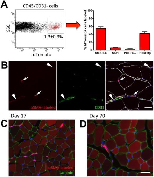
Figure 1. Cell surface marker expression and localization of αSMA labeled cells.
(A) αSMACre/Ai9 mice were injected with tamoxifen to label cells, and two days later, hindlimb muscles were processed for FACS analysis. The labeled population represented around 1.3% of the non-hematopoietic, non-endothelial cell fraction (CD45/CD31−). Expression of cell surface markers of muscle satellite cells (SM/C2.6) and mesenchymal progenitor markers in the tdTomato+ population is indicated. (B) αSMACre/Ai9 muscle two days post labeling was immunostained for CD31 (green) and laminin (white). Some labeled cells are perivascular (arrowheads), while others reside below the basal lamina (indicated by laminin staining) consistent with a muscle satellite cell phenotype (arrows). Lineage tracing initiated in 4-5 week old mice indicates that after (C) 17 days, and (D) 70 days, the majority of muscle fibers are labeled. Images representative of tracing in at least 6 animals are shown, and similar results were seen in male and female mice. Scale bars indicate 20μm (B) or 50μm (C,D).

