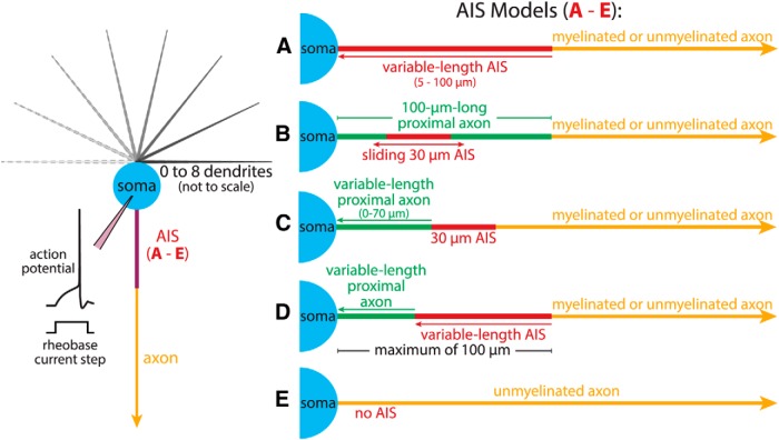
Figure 1.
Diagrams of somatodendritic (left) and axonal (right) compartments in a model neuron. Somata (20 × 20 µm) were attached to a variable number of dendrites (300 µm long, tapering from 2.5 to 0.5 µm in diameter), and to one of five AISs. The AIS in Model A was variable in length (5-100 µm), while AIS Model B had a 30-µm-long AIS that was translocatable (from 0 to 70 µm from the soma) within a 100-µm-long proximal axon segment (1.5 µm diameter) having somatic membrane properties. AIS Model C had a similar 30 µm AIS fixed to the axon proper on one end, and to a variable amount of proximal axon (0-70 µm long, 1.5 µm diameter, with somatic membrane properties) bridging itself and the soma. AIS Model D was a hybrid model combining a variable-length proximal axon with a variable-length AIS (maximum combined length was 100 µm). Finally, AIS Model E lacked an AIS altogether and consisted of an unmyelinated axon attached directly to the soma. AIS Models A through D were attached to myelinated or unmyelinated axons (see Materials and Methods). Inset at left illustrates the simulation setup in which rheobase current injections at the soma (40 ms duration) initiate action potentials.

