Abstract
Endovascular treatment of giant, fusiform and dissecting aneurysms with flow diverter stents is becoming more and more popular. However, very few studies on the follow-up have been published. We describe a patient with a dissecting aneurysm of the right vertebral artery treated with flow diverter stent placement. The patient was followed up with CT angiography (CTA), time-resolved contrast-enhanced MR angiography (CE-MRA) and digital subtraction angiography (DSA). CTA had false negative results in two instances, whereas time-resolved CE-MRA and DSA were the most accurate in depicting the residual flow in the aneurysmal sac. However, in the case of DSA the demonstration of residual flow proved quite difficult and required a very thorough examination with oblique projections. Our 2.5-year experience with this patient led us to believe that time-resolved CE-MRA is a valuable tool in the follow-up of flow diverter-treated stents.
Keywords: Intracranial aneurysm, flow diverter, digital subtraction angiography, CT angiography, contrast-enhanced MR angiography
Introduction
Endovascular treatment of intracranial aneurysms with the use of coils is currently a widely used method and usually preferable to neurosurgical intervention. However, both neurosurgical intervention and coiling are not very effective for the treatment of some categories of aneurysms, i.e. giant (diameter >2.5 cm), fusiform and dissecting aneurysms.1,2
A relatively new method of endovascular treatment for those categories of aneurysms is flow diverter stent placement. These stents have the ability to divert the blood flow, inducing endosaccular stagnation and thrombus formation.1,2
Regarding the follow-up of coiled intracranial aneurysms MR angiography (MRA) has been shown to have an accuracy similar to digital subtraction angiography (DSA).3 To our knowledge, very few studies in the literature have compared different modalities in the follow-up of silk stent-treated intracranial aneurysms.4,5
We describe a case of a dissecting aneurysm of the right vertebral artery treated with silk flow diverter stent and followed up with CT angiography (CTA), time-resolved contrast-enhanced MR angiography (CE-MRA) and DSA.
Case Report
We present a 58-year-old man treated with a flow diverter stent (BALT SILK 4.5x50) for a dissecting aneurysm of the distal part of the right vertebral artery.
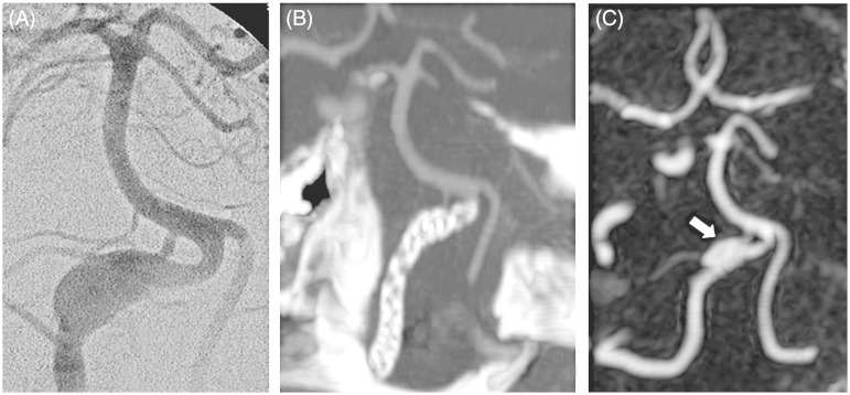
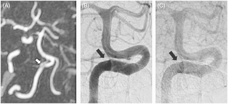
The patient underwent MRA nine days after the procedure. The examination was performed in a 3T scanner (GE HDxt, General Electric Healthcare, Milwaukee WI, USA). The sequences acquired were 3D time of flight (TOF) and dynamic time-resolved CE-MRA (time-resolved imaging of contrast kinetics, TRICKS). Blood flow was detected in a small part of the distal aneurysmal sac. The lumen of the stent was patent. One month later CTA (Philips Brilliance 16, Philips Health Care, Best, The Netherlands) demonstrated findings suggestive of total exclusion of the aneurysmal sac from the blood circulation. The patient subsequently underwent time-resolved CE-MRA demonstrating residual flow in the distal part of the aneurysmal sac. An increase in the thrombotic component was also noted (Figure 1). Another CTA three months later revealed no residual flow. Two consecutive CE-MRAs proved the finding to be false negative once more. Then a DSA was performed to verify the MRA findings. This examination confirmed that there was leakage in the distal part of the stent (Figure 2). An MRA performed 2.5 years after the intervention showed a decrease in the residual flow within the aneurysmal sac.
Figure 1.
A) Antero-posterior DSA shows a dissecting aneurysm of the distal part of the right vertebral artery. B) Coronal maximum intensity projection (MIP) reconstruction of a CTA, 1 month after endovascular treatment, shows an apparent exclusion of the aneurysm from the circulation. The stent is clearly appreciated. C) Coronal MIP reconstruction of time-resolved CE-MRA (TRICKS) 2 months after the CTA demonstrates residual flow in the distal part of the aneurysmal sac (arrow).
Figure 2.
A) Coronal maximum intensity projection (MIP) reconstruction of time-resolved CE-MRA (TRICKS) 2 years after stenting shows a decrease in the residual flow within the aneurysmal sac (white arrow). B) In the subsequent DSA it is hard to discern the residual flow seen in the CE-MRA because the leakage was distributed around the stent and there was overlap. C) In a later phase an area of slower flow can be appreciated around the stent, representing the residual flow within the sac (black arrow).
Discussion
Many studies have compared the effectiveness of DSA, CTA and MRA in the follow-up of coiled intracranial aneurysms. The results are in favour of MRA versus CTA.
Several authors compared MRA to DSA demonstrating that MRA is at least as sensitive as DSA in the visualization of neck remnants.6–8 A few of them have even suggested that CE-MRA may be superior to DSA.9,10
The use of flow diverters is a much newer technique than coil embolization. Despite the fact that the technique is more widely used there are very few studies in the literature on post treatment follow-up.3–5
Our experience in the patient described herein is consistent with the findings of the studies on the follow-up of endovascular coiling, i.e. the modality which best depicted the residual flow in the aneurysmal sac was MRA and in particular time-resolved CE-MRA.
In our case, the CTA was false negative twice. This was probably due to the fact that the CTA was used as a static examination, which cannot evaluate the contrast enhancement as a function of time. Despite that we must be aware that CTA could also enhance its temporal resolution by using a time-resolved technique or by performing a delayed acquisition. These latter techniques might have revealed the residual flow within the aneurysm as we did with time-resolved CE-MRA. In this sense, we must bear in mind that the sensitivity in the detection of the iodinated contrast medium within an already opacified aneurysmal sac is quite low. Moreover multiple CT acquisitions could raise substantial concerns about the increasing radiation dose to the patient.
After the treatment of aneurysms with flow diverter stents, there is slow flow and delayed contrast filling within the aneurysmal sac. Therefore it is of fundamental importance to use a method able to evaluate flow dynamics in the follow-up.
Thanks to its very high temporal and spatial resolution, time-resolved CE-MRA gives a clear depiction of the residual flow in the aneurysmal sac. In addition, the fact that it employs a subtraction technique efficiently reduces T1 hyperintense artifacts that could give false positive results. A complementary morphological MR study is very helpful in the evaluation of the sac, the progression of the thrombosis and the compression or alteration of adjacent structures (Figure 1).
In our patient, CE-MRA was even more accurate than DSA. In order to confirm the small residual flow we had detected in CE-MRA, we had to perform a very thorough DSA and even so the finding was barely perceptible (Figure 2).
This single case cannot prove that CE-MRA is the modality of choice in the follow-up of flow diverter-treated aneurysms. Nevertheless, it illustrates quite clearly that prospective follow-up studies should include time-resolved CE-MRA in their protocol.
Conclusion
Our case report focuses on the use of CTA, time-resolved CE-MRA and DSA in the follow-up of a flow diverter-treated aneurysm. Our aim was to compare the usefulness of each technique in the detection of residual flow and the evaluation of the thrombosis of the aneurysmal sac. The most effective modalities were the dynamic techniques.
We believe that time-resolved CE-MRA can be very valuable in the detection of residual flow, possibly more than DSA and has the characteristics to become the modality of choice in the follow-up of aneurysms treated with flow diverters. Large multimodality studies are required to confirm this finding.
Funding
This research received no specific grant from any funding agency in the public, commercial or not-for-profit sectors.
Conflict of interest
The authors declare no conflict of interest.
References
- 1.Cirillo L, Dall'olio M, Princiotta C, et al. The use of flow-diverting stents in the treatment of giant cerebral aneurysms: preliminary results. Neuroradiol J 2010; 23(2): 220–224. [DOI] [PubMed] [Google Scholar]
- 2.Leonardi M, Cirillo L, Toni F, et al. Treatment of intracranial aneurysms using flow-diverting silk stents (BALT): a single centre experience. Interv Neuroradiol 2011; 17(3): 306–315. [DOI] [PMC free article] [PubMed] [Google Scholar]
- 3.Agid R, Schaaf M, Farb RI. CE-MRA for follow-up of aneurysms post stent-assisted coiling. Interv Neuroradiol. 2012; 18(3): 275–283. [DOI] [PMC free article] [PubMed] [Google Scholar]
- 4.Toni F, Marliani AF, Cirillo L, et al. 3T MRI in the evaluation of brain aneurysms treated with flow-diverting stents: preliminary experience. Neuroradiol J 2009; 22(5): 588–599. [DOI] [PubMed] [Google Scholar]
- 5.Boddu SR, Tong FC, Dehkharghani S. Contrast-enhanced time-resolved MRA for follow-up of intracranial aneurysms treated with the Pipeline embolization device. Am J Neuroradiol. 2014. doi: 10.3174/ajnr.A4008. [Epub ahead of print]. doi: 10.3174/ajnr.A4008. [DOI] [PMC free article] [PubMed]
- 6.Majoie CB, Sprengers ME, van Rooij WJ, et al. MR angiography at 3T versus digital subtraction angiography in the follow-up of intracranial aneurysms treated with detachable coils. Am J Neuroradiol 2005; 26(6): 1349–1356. [PMC free article] [PubMed] [Google Scholar]
- 7.Lubicz B, levivier M, Sadeghi N, et al. Immediate intracranial aneurysm occlusion after embolization with detachable coils: a comparison between MR angiography and intra-arterial digital subtraction angiography. J Neuroradiol 2007; 34(3): 190–197doi: 10.1016/j.neurad.2007.05.002. [DOI] [PubMed] [Google Scholar]
- 8.Cirillo M, Scomazzoni F, Cirillo L, et al. Comparison of 3D TOF-MRA and 3D CE-MRA at 3T for imaging of intracranial aneurysms. Eur J Radiol 2013; 82(12): e853–859doi: 10.1016/j.ejrad.2013.08.052. [DOI] [PubMed] [Google Scholar]
- 9.Yamada N, Hayashi K, Murao K, et al. Time-of-flight MR angiography targeted to coiled intracranial aneurysms is more sensitive to residual flow than is digital subtraction angiography. Am J Neuroradiol 2004; 25(7): 1154–1157. [PMC free article] [PubMed] [Google Scholar]
- 10.Agid R, Willinsky RA, Lee SK, et al. Characterization of aneurysm remnants after endovascular treatment: contrast-enhanced MR angiography versus catheter digital subtraction angiography. Am J Neuroradiol 2008; 29(8): 1570–1574doi: 10.3174/ajnr.a1124. [DOI] [PMC free article] [PubMed] [Google Scholar]


