Abstract
Background
Aneurysmal dilation of the right ventricular outflow tract complicates assessment of right ventricular function in patients with repaired tetralogy of Fallot. Tricuspid annular plane systolic excursion is commonly used to estimate ejection fraction. We hypothesized that tricuspid annular plane systolic excursion measured by cardiac magnetic resonance imaging approximates global and segmental right ventricular function, specifically right ventricular sinus ejection fraction, in pediatric patients with repaired tetralogy of Fallot.
Methods
Tricuspid annular plane systolic excursion was measured retrospectively on cardiac magnetic resonance images in 54 patients with repaired tetralogy of Fallot. Values were compared with right ventricular global, sinus, and infundibular ejection fractions. Tricuspid annular plane systolic excursion was: 1) indexed to body surface area, 2) converted into a fractional value, and 3) converted into published pediatric Z-scores.
Results
Tricuspid annular plane systolic excursion measurements had good agreement between observers. Right ventricular ejection fraction did not correlate with the absolute or indexed tricuspid annular plane systolic excursion and correlated weakly with fractional tricuspid annular plane systolic excursion (r=0.41 and p=0.002). Segmental right ventricular function did not appreciably improve correlation with any of the tricuspid annular plane systolic excursion measures. Pediatric Z-scores were unable to differentiate patients with normal and abnormal right ventricular function.
Conclusions
Tricuspid annular plane systolic excursion measured on cardiac magnetic resonance imaging correlates poorly with global and segmental right ventricular ejection fraction in pediatric patients with repaired tetralogy of Fallot. Tricuspid annular plane systolic excursion is an unreliable approximation of right ventricular function in this patient population.
Keywords: Tricuspid annular plane systolic excursion (TAPSE), Right ventricular function, Tetralogy of Fallot, Pediatric Cardiology
INTRODUCTION
Non-invasive assessment of right ventricular function is difficult due to its complex geometry.1 Right ventricular function is especially difficult to evaluate in patients with repaired tetralogy of Fallot due to aneurysmal right ventricular outflow tract dilatation. These aneurysms are non-contractile and have paradoxical motion, increasing right ventricular volume while simultaneously decreasing the right ventricular ejection fraction. In order to account for this, Kutty et al used cardiac MRI to segment the right ventricle in patients with repaired tetralogy of Fallot and analyzed the right ventricular sinus and infundibulum separately.1, 2 This allowed for assessment of right ventricular sinus ejection fraction, which is presumably a measure of “true” right ventricular myocardial function in repaired tetralogy of Fallot. However, this method of segmentation is time-consuming, and a fast and reproducible method to approximate right ventricular sinus function would be beneficial.
Tricuspid annular plane systolic excursion is a commonly used objective method of estimating right ventricular function. Tricuspid annular plane systolic excursion can be performed on both echocardiographic and cardiac MRI images. Although cardiac MRI has decreased temporal resolution, it provides excellent spatial resolution and cardiac MRI tricuspid annular plane systolic excursion correlates well with tricuspid annular plane systolic excursion measured by echocardiography.3–6 Tricuspid annular plane systolic excursion validation in pediatric and adult patients with repaired tetralogy of Fallot has revealed disappointing results.7–10 We hypothesized that the poor correlation between tricuspid annular plane systolic excursion and right ventricular ejection fraction in repaired tetralogy of Fallot is due to aneurysmal dilatation of the right ventricular outflow tract and that tricuspid annular plane systolic excursion may be a surrogate for right ventricular sinus ejection fraction.
MATERIALS AND METHODS
Patient population
This cross-sectional, retrospective study was approved by the Institutional Review Board. Patients with repaired tetralogy of Fallot who underwent cardiac MRI between 2007 and 2012 were identified from the cardiac MRI database. The first available cardiac MRI was used if patients had undergone multiple MRIs. Exclusion criteria were the following: over 18 years of age at time of MRI, poor image quality, tricuspid regurgitation greater than moderate, and original anatomy of pulmonary atresia or isolated pulmonary stenosis.
Image Acquisition
Images were obtained on either a 1.5 Tesla Siemens Avanto (Siemens Healthcare Sector, Erlangen, Germany) or a 1.5 Tesla Philips Intera (Philips Medical Systems, Best, The Netherlands) using a standard imaging protocol.11 Steady-state free precession sequences were obtained in a short axis stack. Typical imaging parameters were: slice thickness 8mm, no gap, field of view 360mm x 360mm, matrix 208 x 256, spatial resolution <2.2mm x 2.0mm x 8mm, flip angle 80 degrees, temporal resolution < 55ms, minimum echo time and repetition time; the sequences were breath-holds, the cardiac gating was retrospective and parallel imaging with GRAPPA (Siemens) and SENSE (Philips) with an acceleration factor of 2 was used.
Image Analysis
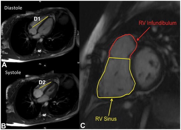
Right ventricular volume and ejection fraction were calculated with manual contouring of the endocardial borders in end-diastole and end-systole using the Leonardo Workstation (Siemens Healthcare Sector, Erlangen, Germany) or Diagnosoft Virtue (Diagnosoft, Durham, North Carolina). Cardiac MRI tricuspid annular plane systolic excursion was measured by 3 observers (JS, EU, DP) in repaired tetralogy of Fallot patients as described previously (Figure 1).5, 12 The distance from the tricuspid annulus to right ventricular apex in systole (D2) was subtracted from the distance from the tricuspid annulus to right ventricular apex in diastole (D1). In order to account for differences in body size in pediatric patients, the tricuspid annular plane systolic excursion was indexed to body surface area. A fractional tricuspid annular plane systolic excursion was also calculated using the following equation:
Figure 1.
(A, B) Tricuspid annular plane systolic excursion was calculated by subtracting the distance from the tricuspid annulus to the apex in systole (D2) from the same distance in diastole (D1). (C) Right ventricular (RV) segmentation in patients with tetralogy of Fallot. The septal and parietal bands were used to divide the right ventricle into the sinus and infundibular portions.
In addition, tricuspid annular plane systolic excursion measurements were assigned a Z-score based on published values for age.13
Right ventricular segmentation was performed in the repaired tetralogy of Fallot patients as described by Kutty et al.2 Using the septal and parietal bands, the right ventricle was divided into the sinus and infundibular portions (Figure 1) then manually contoured. This allowed for calculation of the right ventricular infundibular and sinus volumes and ejection fractions. These measurements were performed by a single observer (JS) with a random sample of 24 contoured by a second observer (DP).
Statistical analysis
Agreement between readers was assessed using intraclass correlation coefficients. The mean tricuspid annular plane systolic excursion measurements from all readers were obtained and used to calculate average, indexed, and fractional tricuspid annular plane systolic excursion. The correlations between the measures of tricuspid annular plane systolic excursion and global and segmental right ventricular function were assessed using a Spearman correlation. Statistical analysis was performed using SPSS 21 (IBM, Armonk, New York).
RESULTS
A total of 87 patients were identified and 54 met inclusion and exclusion criteria. The majority of cardiac MRIs were performed on the Siemens scanner (n=46). Demographics of the patients are listed in Table 1. The most common palliative surgery was a right Blalock-Taussig shunt. The majority of patients had a history of transannular patch repair.
Table 1.
Demographics
| (N=54) | |
|---|---|
| Age (years) | 13.5 ± 3.4 |
| Body surface area (m2) | 1.4 ± 0.36 |
| Gender (male) | 37% |
|
| |
| Indexed RVEDV1 (ml/m2) | 133 ± 36 |
| Indexed RVESV2 (ml/m2) | 64 ± 26 |
| RVEF3 (%) | 52.8% ± 8.9 |
|
| |
| Patients requiring 1 or more palliative procedures | 22.2% (N=12) |
| Right Blalock-Taussig shunt | 13% (N=7) |
| Left Blalock-Taussig shunt | 9.3% (N=5) |
| Balloon valvuloplasty | 3.7% (N=2) |
| Median age at time of palliative surgery (days) | 8 |
|
| |
| Definitive operation | |
| Transannular patch | 70.4% (N=38) |
| Valve-sparing repair | 13% (N=7) |
| RV-PA4 conduit | 3.7% (N=2) |
| Unknown | 13% (N=7) |
| Median age at definitive repair (days) | 213 |
Right ventricular end diastolic volume (RVEDV)
Right ventricular end systolic volume (RVESV)
Right ventricular ejection fraction (RVEF)
Right ventricular to pulmonary artery conduit (RV-PA)
Cardiac MRI tricuspid annular plane systolic excursion measurements had good agreement between all three observers (Table 2). Right ventricular segmentation in repaired tetralogy of Fallot patients had good agreement for the right ventricular sinus but only adequate agreement for the right ventricular infundibulum. Table 3 lists the cardiac MRI tricuspid annular plane systolic excursion values obtained in the cohort.
Table 2.
Interobserver variability of tricuspid annular plane systolic excursion (TAPSE) measurements and right ventricular segmentation using intraclass correlation coefficient
| TAPSE Measurements
| |
|---|---|
| Measurement | ICC (95% CI) |
| Distance from apex to annulus in diastole | 0.965 (0.924–0.982) |
| Distance from apex to annulus in systole | 0.959 (0.908–0.979) |
| Right Ventricular Segmentation, (N=24)
| |
|---|---|
| Measurement | ICC (95% CI) |
| RVEDVsinus1 | 0.945 (0.873–0.976) |
| RVESVsinus2 | 0.963 (0.895–0.985) |
| RVEDVinfundibulum3 | 0.652 (0.184–0.850) |
| RVESVinfundibulum4 | 0.707 (0.336–0.872) |
Right ventricular sinus end diastolic volume (RVEDVsinus)
Right ventricular sinus end systolic volume (RVESVsinus)
Right ventricular infundibular end diastolic volume (RVEDVinfundibulum)
Right ventricular infundibular end systolic volume (RVESVinfundibulum)
Table 3.
Mean tricuspid annular plane systolic excursion (TAPSE) values
| N=54 | |
|---|---|
| Distance from apex to annulus in diastole (cm) | 9.4 ± 0.15 |
| Distance from apex to annulus in systole (cm) | 7.6 ± 0.13 |
| TAPSE (cm) | 1.8 ± 0.04 |
| Indexed TAPSE (cm/m2) | 1.3 ± 0.05 |
| Fractional TAPSE (%) | 18.8 ± 0.4 |
Correlations are shown in Table 4. The brand of MRI scanner did not appreciably affect the correlations. Tricuspid annular plane systolic excursion did not correlate significantly with global right ventricular ejection fraction (r=0.23, p=0.099), though it did correlate weakly with the right ventricular end diastolic volume (r=0.29, p=0.034). The indexed tricuspid annular plane systolic excursion also had no correlation with global right ventricular ejection fraction and only weak correlation with the indexed right ventricular end diastolic volume (r=0.34, p=0.011). Fractional tricuspid annular plane systolic excursion had a weak correlation with global right ventricular ejection fraction (r=0.41, p=0.002).
Table 4.
Correlation between tricuspid annular plane systolic excursion (TAPSE) measurements and measures of right ventricular global and segmental size and function
| Global
| |||
|---|---|---|---|
| Measurement | r | r2 | p-value |
| TAPSE | |||
| RVEF1 (%) | 0.23 | 0.05 | p=0.099 |
| RVEDV2 (ml) | 0.29 | 0.08 | p=0.034 |
| RVESV3 (ml) | 0.15 | 0.02 | p=0.280 |
|
| |||
| Indexed TAPSE | |||
| RVEF (%) | 0.01 | 0.00 | p=0.945 |
| Indexed RVEDV (ml/m2) | 0.34 | 0.12 | p=0.011 |
| Indexed RVESV (ml/m2) | 0.15 | 0.02 | p=0.275 |
|
| |||
| Fractional TAPSE | |||
| RVEF (%) | 0.41 | 0.17 | p=0.002 |
| Indexed RVEDV (ml/m2) | −0.13 | 0.02 | p=0.347 |
| Indexed RVESV (ml/m2) | −0.32 | 0.10 | p=0.018 |
| Segmental
| |||
|---|---|---|---|
| Measurement | r | r2 | p-value |
| TAPSE | |||
| RV sinus EF4 (%) | 0.24 | 0.06 | p=0.076 |
| Indexed RVEDVsinus5 (ml/m2) | 0.04 | 0.00 | p=0.758 |
| Indexed RVESVsinus6 (ml/m2) | −0.13 | 0.02 | p=0.365 |
| RV infundibular EF7 (%) | 0.15 | 0.02 | p=0.282 |
| Indexed RVEDVinfundibulum8 (ml/m2) | 0.14 | 0.02 | p=0.330 |
| Indexed RVESVinfundibulum9 (ml/m2) | 0.08 | 0.01 | p=0.581 |
|
| |||
| Indexed TAPSE | |||
| RV sinus EF (%) | −0.01 | 0.00 | p=0.950 |
| Indexed RVEDVsinus (ml/m2) | 0.36 | 0.13 | p=0.007 |
| Indexed RVESVsinus (ml/m2) | 0.22 | 0.05 | p=0.103 |
| RV infundibular EF (%) | −0.24 | 0.06 | p=0.086 |
| Indexed RVEDVinfundibulum (ml/m2) | 0.19 | 0.04 | p=0.173 |
| Indexed RVESVinfundibulum (ml/m2) | 0.27 | 0.07 | p=0.046 |
|
| |||
| Fractional TAPSE | |||
| RV sinus EF (%) | 0.30 | 0.09 | p=0.028 |
| Indexed RVEDVsinus (ml/m2) | −0.20 | 0.04 | p=0.152 |
| Indexed RVESVsinus (ml/m2) | −0.36 | 0.13 | p=0.008 |
| RV infundibular EF (%) | 0.25 | 0.06 | p=0.072 |
| Indexed RVEDVinfundibulum (ml/m2) | −0.07 | 0.00 | p=0.637 |
| Indexed RVESVinfundibulum (ml/m2) | −0.12 | 0.01 | p=0.395 |
evaluated using Spearman correlation
Right ventricular ejection fraction (RVEF)
Right ventricular end diastolic volume (RVEDV)
Right ventricular end systolic volume (RVESV)
Right ventricular sinus ejection fraction (RV sinus EF)
Right ventricular sinus end diastolic volume (RVEDVsinus)
Right ventricular sinus end systolic volume (RVESVsinus)
Right ventricular infundibular ejection fraction (RV infundibular EF)
Right ventricular infundibular end diastolic volume (RVEDVinfundibulum)
Right ventricular infundibular end systolic volume (RVESVinfundibulum)
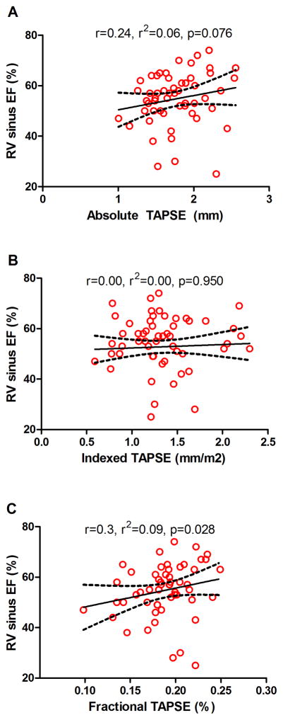
Tricuspid annular plane systolic excursion and indexed tricuspid annular plane systolic excursion did not correlate with right ventricular sinus ejection fraction (r=0.24, p=0.076 and r=−0.01, p=0.950, respectively). Fractional tricuspid annular plane systolic excursion, however, correlated weakly with right ventricular sinus ejection fraction (r= 0.3, p=0.028) (Figure 2). Indexed tricuspid annular plane systolic excursion correlated weakly with indexed right ventricular sinus end diastolic volume (r=0.36, p=0.007) and fractional tricuspid annular plane systolic excursion correlated weakly with the indexed right ventricular sinus end systolic volume (r=−0.36, p=0.008). Otherwise, the correlations between measures of cardiac MRI tricuspid annular plane systolic excursion and segmental right ventricular chamber sizes were not significant.
Figure 2.
Scatterplots demonstrate (A) no correlation between cardiac MRI tricuspid annular plane systolic excursion (TAPSE) and right ventricular (RV) sinus ejection fraction (EF), (B) no correlation between indexed cardiac MRI TAPSE and RV sinus EF, and (C) poor correlation between fractional cardiac MRI TAPSE and RV sinus EF.
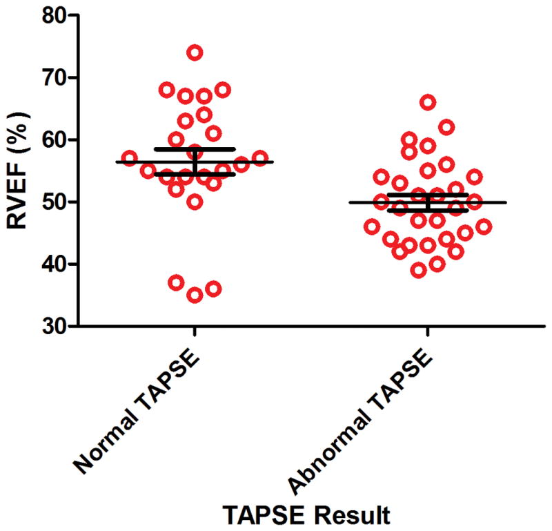
Tricuspid annular plane systolic excursion Z-scores were calculated based on published values for age. There was weak correlation between tricuspid annular plane systolic excursion Z-scores and both global and right ventricular sinus ejection fraction (r=0.3, p=0.027 and r=0.31, p=0.021, respectively). Patients were dichotomized to either normal tricuspid annular plane systolic excursion (Z-score of > −2) or abnormal tricuspid annular plane systolic excursion (Z-score of ≤−2). This dichotomization was unable to adequately differentiate between patients with normal and abnormal right ventricular function by cardiac MRI (Figure 3).
Figure 3.

Scatterplot comparing right ventricular ejection fraction (RVEF) in patients with normal and abnormal tricuspid annular plane systolic excursion (TAPSE) based on pediatric Z-scores demonstrates significant overlap between groups.
DISCUSSION
We demonstrate that the measurement of tricuspid annular plane systolic excursion by cardiac MRI is reproducible but correlates poorly with global right ventricular ejection fraction and right ventricular sinus ejection fraction in pediatric patients with repaired tetralogy of Fallot. These negative results hold true for indexed tricuspid annular plane systolic excursion and fractional tricuspid annular plane systolic excursion, two different methods of accounting for varying patient size in pediatric populations. This is clinically significant because multiple investigators use tricuspid annular plane systolic excursion to estimate right ventricular function with both echocardiography and cardiac MRI. These data suggest that cardiac MRI tricuspid annular plane systolic excursion is a poor surrogate of global or segmental right ventricular ejection fraction in patients with repaired tetralogy of Fallot.
The poor correlation between tricuspid annular plane systolic excursion and global right ventricular ejection fraction has been reported in other studies.8–10 This is likely secondary to complex right ventricular geometry and right ventricular outflow tract aneurysmal dilatation. The poor correlation with segmental function, however, is more difficult to explain. It may be related to the complex mechanics of right ventricular contraction. Although many studies have demonstrated the importance of longitudinal contraction in assessing right ventricular function,14 a recent analysis demonstrated that transverse contraction correlated more strongly with right ventricular ejection fraction than tricuspid annular plane systolic excursion in adults with pulmonary hypertension.15 In addition, tricuspid annular plane systolic excursion represents longitudinal wall motion along one plane in the right ventricle and cannot account for segmental wall motion abnormalities, which are frequently present in patients with repaired tetralogy of Fallot.16
The poor correlation also may be related to varying size in pediatric patients. We corrected for this by indexing the cardiac MRI tricuspid annular plane systolic excursion, using a fractional cardiac MRI tricuspid annular plane systolic excursion, and by using previously published Z-scores, but it is possible that these are suboptimal correction methods. Tricuspid annular plane systolic excursion may also reflect left ventricular function, thus decreasing its specificity for, and correlation with, right ventricular ejection fraction.17, 18 A better method of evaluating global longitudinal contraction, such as strain or strain rate, may provide a better correlation.
Although tricuspid annular plane systolic excursion measured by cardiac MRI may provide data regarding longitudinal motion, our study suggests these tricuspid annular plane systolic excursion values are not estimates of global or segmental right ventricular ejection fraction in pediatric patients with repaired tetralogy of Fallot. This should be taken into consideration when interpreting cardiac MRI tricuspid annular plane systolic excursion values. In addition, given the good correlation reported between cardiac MRI tricuspid annular plane systolic excursion and echocardiographic tricuspid annular plane systolic excursion, this study questions the utility of tricuspid annular plane systolic excursion in repaired tetralogy of Fallot using either modality.
Limitations
Although cardiac MRI has decreased temporal resolution, it provides excellent spatial resolution and cardiac MRI tricuspid annular plane systolic excursion correlates well with tricuspid annular plane systolic excursion measured on echocardiography.6 In addition, cardiac MRI tricuspid annular plane systolic excursion has the advantage of being performed at nearly the same time as right ventricular ejection fraction, minimizing any differences in right ventricular function caused by changes in preload or afterload. Therefore, cardiac MRI tricuspid annular plane systolic excursion has potential to demonstrate better correlation with right ventricular ejection fraction than echocardiographic tricuspid annular plane systolic excursion. While it is possible that the relationship between tricuspid annular plane systolic excursion and right ventricular ejection fraction is non-linear, a non-linear relationship should also be detected using a Spearman correlation. Tricuspid annular plane systolic excursion in this study was measured from the annulus to the apex, which is the standard measurement performed in previous cardiac MRI studies.
Conclusions
Cardiac MRI tricuspid annular plane systolic excursion has poor correlation with global and segmental right ventricular ejection fraction in pediatric patients with repaired tetralogy of Fallot.
Acknowledgments
FINANCIAL SUPPORT
The project was supported by the National Center for Research Resources, Grant UL1 RR024975-01, and is now at the National Center for Advancing Translational Sciences, Grant 2 UL1 TR000445-06.
Footnotes
CONFLICTS OF INTEREST
None
ETHICAL STANDARDS
The authors assert that all procedures contributing to this work comply with the ethical standards of the Belmont report and with the Helsinki Declaration of 1975, as revised in 2008, and has been approved by the Vanderbilt Institutional Review Board.
The content is solely the responsibility of the authors and does not necessarily represent the official views of the NIH.
Contributor Information
Emem Usoro, Email: eusoro11@email.mmc.edu.
Li Wang, Email: li.wang@vanderbilt.edu.
David A. Parra, Email: david.parra@vanderbilt.edu.
References
- 1.Geva T, Powell AJ, Crawford EC, Chung T, Colan SD. Evaluation of regional differences in right ventricular systolic function by acoustic quantification echocardiography and cine magnetic resonance imaging. Circulation. 1998;98:339–345. doi: 10.1161/01.cir.98.4.339. [DOI] [PubMed] [Google Scholar]
- 2.Kutty S, Zhou J, Gauvreau K, Trincado C, Powell AJ, Geva T. Regional dysfunction of the right ventricular outflow tract reduces the accuracy of Doppler tissue imaging assessment of global right ventricular systolic function in patients with repaired tetralogy of Fallot. J Am Soc Echocardiogr. 2011;24:637–643. doi: 10.1016/j.echo.2011.01.020. [DOI] [PubMed] [Google Scholar]
- 3.Kaul S, Tei C, Hopkins JM, Shah PM. Assessment of right ventricular function using two-dimensional echocardiography. Am Heart J. 1984;107:526–531. doi: 10.1016/0002-8703(84)90095-4. [DOI] [PubMed] [Google Scholar]
- 4.Speiser U, Hirschberger M, Pilz G, et al. Tricuspid annular plane systolic excursion assessed using MRI for semi-quantification of right ventricular ejection fraction. Br J Radiol. 2012;85:e716–721. doi: 10.1259/bjr/50238360. [DOI] [PMC free article] [PubMed] [Google Scholar]
- 5.Nijveldt R, Germans T, McCann GP, Beek AM, van Rossum AC. Semi-quantitative assessment of right ventricular function in comparison to a 3D volumetric approach: a cardiovascular magnetic resonance study. Eur Radiol. 2008;18:2399–2405. doi: 10.1007/s00330-008-1017-7. [DOI] [PubMed] [Google Scholar]
- 6.Koestenberger M, Ravekes W, Nagel B, et al. Longitudinal systolic ventricular interaction in pediatric and young adult patients with TOF: a cardiac magnetic resonance and M-mode echocardiographic study. Int J Cardiovasc Imaging. 2013;29:1707–1715. doi: 10.1007/s10554-013-0261-3. [DOI] [PubMed] [Google Scholar]
- 7.Koestenberger M, Nagel B, Ravekes W, et al. Systolic right ventricular function in pediatric and adolescent patients with tetralogy of Fallot: echocardiography versus magnetic resonance imaging. J Am Soc Echocardiogr. 2011;24:45–52. doi: 10.1016/j.echo.2010.10.001. [DOI] [PubMed] [Google Scholar]
- 8.Mercer-Rosa L, Parnell A, Forfia PR, Yang W, Goldmuntz E, Kawut SM. Tricuspid Annular Plane Systolic Excursion in the Assessment of Right Ventricular Function in Children and Adolescents after Repair of Tetralogy of Fallot. J Am Soc Echocardiogr. 2013 doi: 10.1016/j.echo.2013.06.022. [DOI] [PMC free article] [PubMed] [Google Scholar]
- 9.Morcos P, Vick GW, 3rd, Sahn DJ, Jerosch-Herold M, Shurman A, Sheehan FH. Correlation of right ventricular ejection fraction and tricuspid annular plane systolic excursion in tetralogy of Fallot by magnetic resonance imaging. Int J Cardiovasc Imaging. 2009;25:263–270. doi: 10.1007/s10554-008-9387-0. [DOI] [PubMed] [Google Scholar]
- 10.Bonnemains L, Stos B, Vaugrenard T, Marie PY, Odille F, Boudjemline Y. Echocardiographic right ventricle longitudinal contraction indices cannot predict ejection fraction in post-operative Fallot children. Eur Heart J Cardiovasc Imaging. 2012;13:235–242. doi: 10.1093/ejechocard/jer263. [DOI] [PubMed] [Google Scholar]
- 11.Schulz-Menger J, Bluemke DA, Bremerich J, et al. Standardized image interpretation and post processing in cardiovascular magnetic resonance: Society for Cardiovascular Magnetic Resonance (SCMR) Board of Trustees Task Force on Standardized Post Processing. J Cardiovasc Magn Reson. 2013;15:35. doi: 10.1186/1532-429X-15-35. [DOI] [PMC free article] [PubMed] [Google Scholar]
- 12.Caudron J, Fares J, Vivier PH, Lefebvre V, Petitjean C, Dacher JN. Diagnostic accuracy and variability of three semi-quantitative methods for assessing right ventricular systolic function from cardiac MRI in patients with acquired heart disease. Eur Radiol. 2011;21:2111–2120. doi: 10.1007/s00330-011-2152-0. [DOI] [PMC free article] [PubMed] [Google Scholar]
- 13.Koestenberger M, Ravekes W, Everett AD, et al. Right ventricular function in infants, children and adolescents: reference values of the tricuspid annular plane systolic excursion (TAPSE) in 640 healthy patients and calculation of z score values. J Am Soc Echocardiogr. 2009;22:715–719. doi: 10.1016/j.echo.2009.03.026. [DOI] [PubMed] [Google Scholar]
- 14.Brown SB, Raina A, Katz D, Szerlip M, Wiegers SE, Forfia PR. Longitudinal shortening accounts for the majority of right ventricular contraction and improves after pulmonary vasodilator therapy in normal subjects and patients with pulmonary arterial hypertension. Chest. 2011;140:27–33. doi: 10.1378/chest.10-1136. [DOI] [PubMed] [Google Scholar]
- 15.Kind T, Mauritz GJ, Marcus JT, van de Veerdonk M, Westerhof N, Vonk-Noordegraaf A. Right ventricular ejection fraction is better reflected by transverse rather than longitudinal wall motion in pulmonary hypertension. J Cardiovasc Magn Reson. 2010;12:35. doi: 10.1186/1532-429X-12-35. [DOI] [PMC free article] [PubMed] [Google Scholar]
- 16.Vogel M, Sponring J, Cullen S, Deanfield JE, Redington AN. Regional wall motion and abnormalities of electrical depolarization and repolarization in patients after surgical repair of tetralogy of Fallot. Circulation. 2001;103:1669–1673. doi: 10.1161/01.cir.103.12.1669. [DOI] [PubMed] [Google Scholar]
- 17.Lopez-Candales A, Rajagopalan N, Saxena N, Gulyasy B, Edelman K, Bazaz R. Right ventricular systolic function is not the sole determinant of tricuspid annular motion. Am J Cardiol. 2006;98:973–977. doi: 10.1016/j.amjcard.2006.04.041. [DOI] [PubMed] [Google Scholar]
- 18.Lamia B, Teboul JL, Monnet X, Richard C, Chemla D. Relationship between the tricuspid annular plane systolic excursion and right and left ventricular function in critically ill patients. Intensive Care Med. 2007;33:2143–2149. doi: 10.1007/s00134-007-0881-y. [DOI] [PubMed] [Google Scholar]


