Abstract
There is a clinical need for non-invasive biomarkers of tumor hypoxia for prognostic and predictive studies, radiotherapy planning and therapy monitoring. Oxygen enhanced MRI (OE-MRI) is an emerging imaging technique for quantifying the spatial distribution and extent of tumor oxygen delivery in vivo. In OE-MRI, the longitudinal relaxation rate of protons (ΔR1) changes in proportion to the concentration of molecular oxygen dissolved in plasma or interstitial tissue fluid. Therefore, well-oxygenated tissues show positive ΔR1. We hypothesized that the fraction of tumor tissue refractory to oxygen challenge (lack of positive ΔR1, termed “Oxy-R fraction”) would be a robust biomarker of hypoxia in models with varying vascular and hypoxic features. Here we demonstrate that OE-MRI signals are accurate, precise and sensitive to changes in tumor pO2 in highly vascular 786-0 renal cancer xenografts. Furthermore, we show that Oxy-R fraction can quantify the hypoxic fraction in multiple models with differing hypoxic and vascular phenotypes, when used in combination with measurements of tumor perfusion. Finally, Oxy-R fraction can detect dynamic changes in hypoxia induced by the vasomodulator agent hydralazine. In contrast, more conventional biomarkers of hypoxia (derived from blood oxygenation-level dependent MRI and dynamic contrast-enhanced MRI) did not relate to tumor hypoxia consistently. Our results show that the Oxy-R fraction accurately quantifies tumor hypoxia non-invasively and is immediately translatable to the clinic.
Keywords: Biomarker, Hypoxia, Imaging, MRI, Validation
INTRODUCTION
Hypoxia is a common feature of most solid malignancies, resulting from an imbalance between oxygen delivery and consumption (1). Tumor hypoxia is associated with the activation of angiogenesis and with metastatic potential (2). Consequently, tumor hypoxia is an important negative prognostic factor (3-5). Tumor hypoxia also mediates resistance to radiotherapy and to some chemotherapy agents and is an independent predictor of treatment failure. Strategies to counteract tumor hypoxia using either radiosensitizers or hypoxia-activated cytotoxic agents are currently being evaluated (6).
A non-invasive imaging biomarker that identifies the presence of hypoxia and measures its extent and spatial distribution within a tumor would help facilitate personalized medicine. However, no such tool is currently available. Proton (1H) MRI is used routinely in clinical medicine, making it an attractive non-invasive modality for measuring oxygen delivery and hypoxia in tumors. Current 1H MRI methods of imaging hypoxia have focused on either dynamic contrast enhanced MRI (DCE-MRI) or R2*-based intrinsic susceptibility imaging, also referred to as blood oxygenation level dependent (BOLD) imaging (7).
In DCE-MRI, administration of a gadolinium-based contrast agent allows estimation of blood vessel flow and permeability, providing an indirect measurement of oxygen delivery and necrosis (8). In BOLD imaging, paramagnetic deoxyhemoglobin molecules in erythrocytes create magnetic susceptibility perturbations around blood vessels, which increase the local transverse MRI relaxation rate (R2*; units ms−1). The value of tumor R2* decreases when blood oxygen saturation increases following inhalation of hyperoxic gas (9). Unfortunately, both DCE-MRI and BOLD imaging have significant limitations which have hindered implementation as clinical biomarkers of hypoxia (10). Neither measure hypoxia directly. Further, BOLD measurements are affected by presence of hemorrhage, by change in vessel geometry and by artifact in air/soft tissue interfaces, such as in the lungs and bowel (7).
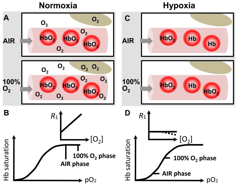
Oxygen enhanced MRI (OE-MRI) is a distinct 1H MRI method for quantifying tumor oxygen delivery. Here, the MRI longitudinal relaxation rate (R1; units s−1) is sensitive to changes in the level of molecular oxygen (O2) dissolved in blood plasma or interstitial tissue fluid (11, 12). When hyperoxic gas is inhaled, excess oxygen is carried in the blood plasma in tissues with adequate perfusion. Since well oxygenated tissue has near complete saturation of hemoglobin molecules (13), the excess delivered oxygen remains dissolved in blood plasma and interstitial tissue fluid, where it increases the R1 value (Figure 1a). The change in R1 (ΔR1) observed is theoretically proportional to magnitude of change in dissolved O2 concentration for a given voxel (11) (Figure 1b). Several previous studies have reported positive average values of ΔR1 following oxygen inhalation in preclinical models of cancer (14-22) and in human tumors (20, 23-25). Importantly, while both oxygen relaxivity, baseline R1 and signal-to-noise ratios show some variation with field strength, the technique is feasible on both preclinical and clinical MRI platforms (22, 26).
Figure 1. OE-MRI distinguishes between normoxic and hypoxic tissue.
Each box represents an imaging voxel which contains erythrocytes (red spheres) in blood vessels (pink cylinders); tumor cells (gray ellipse); and surrounding interstitial space (white box). A. In normoxic tissue, hemoglobin molecules in erythrocytes are oxygen saturated and form oxyhemoglobin (HbO2) molecules. Dioxygen molecules (O2) are dissolved in the plasma. Inhalation of hyperoxic gas markedly increases the amount of dissolved plasma O2 but HbO2 concentration is essentially unaltered. B. Increased pO2 in interstitial fluid and plasma increases tissue longitudinal relaxation rate (R1), which is detected by MRI. C. In hypoxic tissue, hemoglobin molecules are not fully oxygen saturated and exist as deoxyhemoglobin (Hb) molecules. Inhalation of hyperoxic gas increases the HbO2 to Hb ratio but has negligible effect on plasma O2. D. Since there is negligible change in pO2 the R1 remains little changed (straight black line). Since Hb has a slightly higher longitudinal relaxivity than HbO2, tissue R1 may even decrease (dotted black line).
Measuring positive ΔR1 in OE-MRI quantifies and maps oxygen delivery by identifying tissue with fully saturated hemoglobin, but does not directly identify tissue hypoxia per se. Tumor sub-regions refractory to oxygen challenge are considered to have low hemoglobin oxygen saturation and so excess delivered O2 molecules bind preferentially to hemoglobin molecules but do not significantly alter plasma pO2 (10) (Figure 1c). Several preclinical OE-MRI studies have reported that some tumor sub-regions are refractory to hyperoxic gas challenge since they had no positive ΔR1 change (14, 19-21). If these regions are perfused yet lacking in oxygen enhancement then OE-MRI should identify regions of tumor hypoxia (Figure 1d). In this study, we tested the hypothesis that measuring the fraction of each tumor refractory to oxygen challenge – determined by having absent positive ΔR1 (hereafter termed “Oxy-R fraction”) – would enable the identification, quantification and mapping of tumor hypoxia with MRI in vivo.
MATERIALS AND METHODS
Phantom validation of R1 measurement
In vitro validation experiments were performed on the same 7T system as used for the in vivo studies. The phantom consisted of four 5mm NMR tubes (Sigma- Aldrich, UK) with solutions of gadopentetate dimeglumine (Magnevist; Schering, Berlin, Germany) serially diluted in water to yield final concentrations of 0.01, 0.05, 0.07 and 0.09mM gadolinium and an expected range of T1 between 1400-2500ms based on the relaxivity of Magnevist at 7T at 37°C (27). NMR tubes were placed inside a plastic container filled with dental paste to reduce susceptibility effects and to ensure efficient heat transfer for constant temperature. Three repeated measurements of R1 were measured for each tube on three separate days.
Tumor implantation
All experiments were performed in compliance with licenses issued under the UK Animals (Scientific Procedures) Act 1986 and following local ethical review. Studies were compliant with the United Kingdom National Cancer Research Institute guidelines for animal welfare in cancer research (28) and with the ARRIVE guidelines (29).
Three cell lines exhibiting differing vascular and hypoxic phenotypes in vivo were used. Parental 786-0 renal carcinoma cells (ATCC, LCG Standards, Teddington, UK; purchased 2011; 786-0-par), and cells established from a sunitinib refractory subcutaneous 786-0 xenograft (786-0-R) (30), were cultured in RPMI supplemented with 10% (v/v) fetal calf serum (Gibco, Life Technologies, Paisley, UK). Tumors were propagated by injecting 3×10⁶ cells in 100μl of sterile PBS into the flanks of 8 week old female C.B17-scid mice under isoflurane anesthesia. SW-620 colorectal carcinoma cells (ATCC; lot #8924081, purchased 2005) were cultured in DMEM supplemented with 10% fetal calf serum. Tumors were propagated by injecting 5 ×10⁶ cells in 100μl of sterile PBS into the flanks of 8 week old female athymic NCr-Foxn1nu mice. Immediately prior to in vivo implantation, all cells tested negative for mycoplasma infection and the number of short tandem repeats (STRs) present at 7-10 loci were assessed by PCR to provide STR profiles, from which cell line authenticity was confirmed.
Tumor size was monitored using callipers and the formula for ellipsoid volume, (π/6) × L × W × D, where L, W and D are the largest orthogonal dimensions of the ellipsoid. Tumors were typically used for experimentation at a volume of approximately 400mm3.
Measurement of tissue pO2
The fiber optic oxygen sensing device OxyLite™ (Oxford Optronix, Oxford, UK) (31) was used to measure tumor partial pressure of oxygen (pO2; units mm Hg). A heat mat maintained core body temperature and gas delivery was at 2l/min via a nosepiece. Two dual pO2 and temperature probes were positioned within the tumor tissue. Animals initially breathed medical air, followed by 100% oxygen to mimic the gas changes induced during the MRI experiments. Data acquisition and averaging were performed using Chart version 5 (AD Instruments, Castle Hill, Australia). Tissue pO2 data were collected the day after corresponding MRI data.
MRI data acquisition
Anesthesia was induced with a 10 ml/kg intraperitoneal injection of fentanyl citrate (0.315 mg/ml) plus fluanisone (10mg/ml) (Hypnorm; Janssen Pharmaceutical Ltd, High Wycombe, UK), midazolam (5mg/ml) (Hypnovel; Roche, Welwyn Garden City, UK) and sterile water (at 1:1:2 ratio) (16). Mice were positioned in a 3cm birdcage coil on a custom built platform to isolate the tumor, which was surrounded by dental paste (3M; Bracknell, UK) to minimize motion and susceptibility artifacts. Gas delivery (medical air or 100% oxygen) was continuous at 2l/min through a nose piece. Warm air maintained animal core temperature at 37°C. Lateral tail vein cannulation was performed with a heparinised 27G butterfly catheter (Venisystems, Hospira, Royal Leamington Spa, UK) to enable the remote intravenous administration of gadolinium contrast agent in DCE-MRI studies or when vasomodulator was administered.
All MRI data were acquired on a 7T horizontal bore microimaging system (Bruker Instruments, Ettlingen, Germany). Localization was performed using a multi-slice turboRARE T2-weighted sequence and was followed by shimming over the tumor (32). All sequences were acquired using 30mm × 30mm FOV (128 × 128 matrix; in plane resolution 0.234mm) for a single 1 mm thick slice. Experimental protocols are detailed in Supplementary Figure S1. Sequences used were:
OE-MRI
Inversion recovery (IR) True-FISP images were used to calculate R1 (TR 2.4ms, scan TR 10s, 48 inversion times spaced 38.8ms apart with initial inversion time of 106.2ms; TE 1.2ms; α 60°). High signal-to-noise (SNR) images were used to obtain highly accurate R1; these required 8 signal averages and took 10min 40s to perform. Dynamic images were used to quantify the temporal onset of R1 changes induced by switching between air and 100% oxygen. The dynamic images required 2 signal averages and took 2min 40s to perform.
BOLD
Multiple gradient echo (MGE) images were used to calculate R2* (TR 200ms; 8 echo times, TE 6.2 to 28.2ms with 3.1ms echo spacing; 8 signal averages); duration 3min 25s.
DCE-MRI
Data were collected using a modified True-FISP sequence (TR 2.4ms, scan TR 10s, 8 inversion times spaced 155ms apart, initial inversion time 108ms, TE 1.2ms; α 60°; one signal average, temporal resolution 20s). After five baseline measurements, 0.1mmol/kg bolus of the gadolinium-based contrast agent Magnevist (2ml/kg 50mM solution) was injected intravenously at 2ml/min using a power injector; duration 10mins.
MRI analysis
Regions of interest (ROIs) were drawn around the tumor on the T2-weighted images by an experienced operator (YJ). ROI were transferred to all OE-MRI, BOLD and DCE-MRI data. Voxel-wise and median values of R1 and R2* were calculated for each map using a Bayesian maximum a posteriori approach, with in-house software.
For OE-MRI the voxel wise ΔR1 was calculated, where ΔR1 = R1 (O2) – R1 (air). The initial R1 (air) was derived from the average of the first two high SNR R1 maps. The R1 (O2) was derived from the high SNR R1 map acquired during oxygen breathing. Oxygen enhancement was measured as 2 x tumor baseline R1 x cohort CoV. Voxels were then classified as enhancing (termed “Oxy-E) or refractory (termed “Oxy-R”) to oxygen challenge. Voxel-wise BOLD ΔR2* was calculated, where ΔR2* = R2* (O2) – R2* (air).
Where DCE-MRI was performed, the initial area under the contrast agent concentration curve from 0 to 60 seconds (IAUC60) was calculated (16) and voxels were classified as enhancing when IAUC60 >0. Where both OE-MRI and DCE-MRI were acquired, data were combined to distinguish three tumor sub-regions from one another: (1) perfused Oxy-E voxels; (2) perfused Oxy-R voxels; and (3) non-perfused voxels.
Hydralazine challenge
Hydralazine acts directly on vascular smooth muscle in vessels of normal tissues, causing vasodilation and reduced mean arterial blood pressure. Tumor blood vessels lacking smooth muscle do not dilate in response to hydralazine. Hence, blood is redistributed away from the tumor, reducing blood flow and increasing hypoxia within 30 minutes (33-36). Hydralazine challenge has, therefore, been used as a tool to manipulate acute hypoxia.
An initial air–to–oxygen gas challenge was performed. Then gas delivery was switched back to air breathing and intravenous injection of either 5mg/kg hydralazine hydrochloride (Sigma-Aldrich Co., Dorset, UK) or saline was performed. Finally, a second air–to–oxygen gas challenge was performed. Spatial differences between the ΔR1 defined Oxy-E and Oxy-R voxels on the two air–to–oxygen challenges were assessed by mismatch mapping. Formal randomisation was not employed, rather during each day’s scanning mice were prospectively assigned with intent to balance treatment groups according to tumour size.
Immunofluorescence analysis
Intraperitoneal injection of 60mg/kg pimonidazole (Hypoxyprobe, Burlington, MA) was performed 55 minutes before 100% O2 inhalation began to allow for maximal bioreduction of the agent in hypoxic tumor regions. 15mg/kg Hoechst 33342 (Sigma-Aldrich Co., Dorset, UK) was administered intravenously one minute prior to rapid tumor excision. Tumors were excised whole and bisected along the imaging plane so that the cut surface approximated to the MRI region of interest. Half the tumor was snap frozen and half was formalin fixed and paraffin embedded.
Frozen tissue sections (5μm) were obtained from snap frozen tumor material and scanned using fluorescent microscopy on a Panoramic 250 Flash system (3DHistech, Budapest, Hungary) to determine the number of Hoechst-stained (perfused) vessels (excitation 350nm/emission 480nm). Pimonidazole binding was determined in the same sections using Hypoxyprobe-1 (Hypoxyprobe, Burlington, MA), a mouse-monoclonal, followed by rabbit anti-mouse-fluorescein isothiocyanate (FITC) conjugated secondary antibody (excitation 488nm/emission 525nm). Paraffin embedded sections (5μm) were obtained from tumor tissue dehydrated after fixation in 10% phosphate-buffered formalin. Sections were stained with hematoxylin and eosin (H&E). Data was analyzed using ImageJ software (NIH, Bethesda, MD). The perfused vessel area, hypoxic fraction and percentage necrosis were calculated, as described elsewhere (37).
Statistical analysis
IBM SPSS Statistics v.22 (Armonk, NY) was used for all statistical analysis. In all cases, p values of <0.05 were considered significant. Comparison of median values of R1 during medical air only breathing and during oxygen challenge experiments were evaluated using a one way analysis of variance (ANOVA) with a post hoc Bonferroni correction for multiple comparisons.
Data comparing median values of R1, R2* and IAUC60 and fractions of Oxy-E or Oxy-R between different tumor cohorts (786-0-R, 786-0-par or SW620) were evaluated with independent t-tests corrected for multiple comparisons. Data for different cohorts were assumed to be distributed normally and have unequal variance. Correlations between MRI and pathology were assessed by the non-parametric Spearman’s rho. Formal sample size calculations were not performed.
RESULTS
OE-MRI signals are accurate, precise, stable and sensitive to pO2 change
We sought to demonstrate OE-MRI signal precision, stability and sensitivity to altered oxygen tension, because these factors have not been well documented. Since OE-MRI signal changes are based on the longitudinal relaxation rate, R1, we compared the expected R1 against the measured R1 in an in vitro phantom. These data showed that our R1 measurement technique is accurate (Supplementary Figure S2).
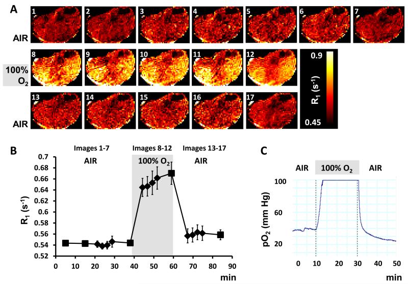
To test measurement precision and stability in vivo, we then performed an experiment in four mice implanted with subcutaneous 786-0-R tumors (786-0-R is a fast-growing subline of the 786-0 renal cancer cell line). Mice initially breathed medical air for 40 minutes. This was followed by 100% oxygen challenge for 20 minutes. Finally the mice breathed medical air again for 25 minutes. Multiple R1 maps were acquired (Supplementary Figure S1a). The within-scan co-efficient of variation (CoV) of baseline voxel-wise R1 was 0.41% in vivo, indicating high measurement precision. No appreciable difference was observed in the spatial mapping of tumor R1 during air breathing (see time points 1-7 in Figure 2a). No significant change was seen in tumor R1 in the four mice (Figure 2b), indicating signal stability while breathing air.
Figure 2. OE-MRI signals are precise, stable and sensitive to pO2 change.
A, R1 maps in one 786-0-R xenograft tumor while the mouse breathed air (top row), 100% oxygen (middle row) and back to air (bottom row). B, R1 measurement was stable in four mice with 786-0-R xenografts during the initial 45 minutes of air breathing. Symbols: ■ 10m40s IR-TrueFISP acquisition, ◆ 2m40s acquisition. Rapid and significant changes in median R1 were induced by 100% oxygen challenge in the same animals and rapid return to baseline values was observed once air breathing resumed. Mean of individual tumor medians are shown ± 1 s.e.m. C, OE-MRI signal changes mirror the time course of change in tumor pO2 as shown in a sample Oxylite™ trace from one xenograft (maximum to the detection range was 100mmHg).
Challenge with 100% oxygen was then performed in the same mice, to increase tumor pO2. All four tumors showed rapid, heterogeneous and significant increase in median R1 following oxygen challenge. These changes were clearly visible in the first map after 2min 40s of oxygen breathing and persisted for the duration of the oxygen challenge (p<0.008 for all time points 8-12 compared with air breathing) showing consistent spatial arrangement (Figure 2a-b). Across all four tumors, 89.3% of voxels were oxygen enhancing (Oxy-E) and 10.7% of voxels were oxygen refractory (Oxy-R). Sample traces of ΔR1 change are shown for Oxy-E and Oxy-R voxels (Supplementary Figure S3). OxyLite™ measurement in the same tumors showed pO2 increase (beyond the limit of detection of the OxyLite™ equipment, namely at 100mmHg) in 7 out of 8 measured regions over the same time frame (sample trace in Figure 2c). The OxyLite™ data provides independent evidence that OE-MRI detects real time increases in the oxygen concentration in tumor interstitial tissue fluid. BOLD MRI, performed in the same mice, revealed a significant reduction in R2* during oxygen challenge (mean reduction 39.8 ms−1; SE 11.7 ms−1; p=0.043), consistent with previous studies (38).
Tumors showed rapid reversal of R1 changes when oxygen challenge ended and air breathing was resumed (see time points 13-17 in Figure 2a). There was no significant difference between baseline and end-of-study mean R1 values for the cohort. OxyLite™ measurement showed pO2 return to pre-challenge levels over a period of 2 to 6 minutes mirroring ΔR1 change. Collectively these data confirm that R1 measurements used for this study are accurate, precise, stable in the absence of perturbation and sensitive to change in tumour pO2.
Oxy-R fraction detects differential levels of hypoxia in an isogenic system
We investigated whether Oxy-R fraction (the fraction of each tumor refractory to oxygen challenge) could detect different levels of hypoxia in cancer models. We measured the Oxy-R fraction in two isogenic cell lines: the slow growing parental 786-0 cell line (786-0-par), which took 206.3 days ± 66.2 SD to reach a tumor volume of ~400 mm3 (n = 8 mice), and the much faster growing 786-0 subline (786-0-R), which only took only 32.1 days ± 6.9 SD to reach the same tumor volume (n = 9 mice). From these mice we acquired OE-MRI, ΔR2* and DCE-MRI data along with histopathological analysis of pimonidazole adduct formation (for hypoxia) and Hoechst 33342 uptake (for perfusion) (Supplementary Figure S1b).
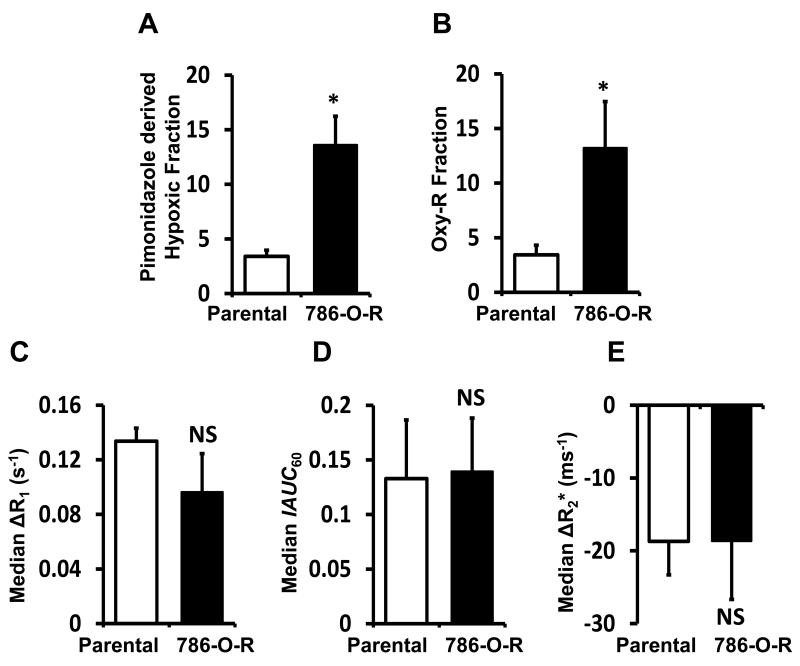
Staining for pimonidazole adduct formation showed a significantly higher hypoxic fraction in 786-0-R tumors compared to the 786-0-par tumors (p=0.008) (Figure 3a). This finding was mirrored by the Oxy-R fraction being significantly higher in the 786-0-R xenografts compared with the 786-0-par xenografts (p=0.047) (Figure 3b). In distinction, there was no significant difference in the median values of ΔR1, IAUC60 or ΔR2* between the 786-0-R and 786-0-par xenografts (Figure 3c-e). No significance difference was seen in the perfused vessel area measured by Hoechst 33342 staining (data not shown). These data confirm that Oxy-R fraction is sensitive to differential levels of hypoxia but that median values of ΔR1, IAUC60 and ΔR2* were insensitive to this difference.
Figure 3. Oxy-R fraction identifies differential levels of hypoxia.
A, 786-0-R xenografts had significantly greater pimonidazole adduct derived hypoxic fraction than 786-0-par xenografts, mirrored in B, Oxy-R fraction tumor. No difference was seen in C, median ΔR1, D, median IAUC60 or E, median ΔR2* between the two cell lines. All error bars are 1 s.e.m., *p<0.05.
Perfused Oxy-R quantifies hypoxic fraction
We sought to test if Oxy-R fraction could estimate tumor hypoxic fraction in two tumor models originating from different cell lines. In particular, we wanted to test if the presence of avascular or necrotic tumor tissue – which does not receive any delivery of oxygen gas in the blood plasma – would confound the relationship between Oxy-R and hypoxia. Therefore we analyzed data from the perfused portion of each tumor.
To do this, we first used the 786-0-R data already collected. We examined the within cohort correlation between Oxy-R fraction and the pimonidazole adduct formation based measurement of hypoxic fraction. This model is known to be highly vascularized. Further, there was a wide dynamic range of hypoxia in this model. We then performed an equivalent experiment using SW620 tumors. This model was chosen because these tumors are relatively poorly vascularized (39).
Anticipated differences in the perfusion status of the two models were confirmed; 786-0-R xenografts had significantly greater perfused tumor area compared with SW620 xenografts, measured by DCE-MRI (p=0.011) (Table 1) and by Hoechst 33342 (p=0.004). Pathology analysis showed that perfused vessel area measured by Hoechst 33342 (Supplementary Figure S4a-b) correlated with perfused Oxy-E fraction (those voxels showing positive enhancement with both oxygen and gadolinium) and that non-perfused fraction correlated with necrosis on H&E in both models (Supplementary Figure S4c-d).
Table 1.
Proportions of Oxy-E and Oxy-R tumor (with standard deviation; SD), subdivided by voxel perfusion status
| % Oxy-E voxels | % Oxy-R voxels | |||
|---|---|---|---|---|
| Perfused | Not perfused | Perfused | Not perfused | |
| 786-O-R | 78.2 (SD18.3) | 7.2 (SD 5.9) | 13.2 (SD 12.9) | 1.2 (SD 1.6) |
| Parental 786-O | 84.7 (SD 14.0) | 11.0 (SD 12.2) | 3.4 (SD 2.5) | 0.9 (SD 0.8) |
| SW620 | 51.5 (SD 13.8) | 25.2 (SD 14.9) | 15.7 (SD 6.2) | 7.6 (SD 4.3) |
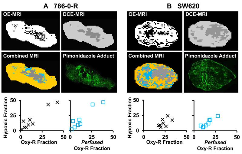
Oxy-R fraction correlated strongly with pathological hypoxic fraction in 786-0-R tumours (rho 0.810, p=0.028) (Figure 4a), irrespective of whether the entire tumor was analyzed or if analysis was restricted to gadolinium-enhancing tumor only (perfused Oxy-R fraction). In SW620 tumours the Oxy-R fraction did not correlate with hypoxic fraction (measured by pimonidazole adduct formation). However, as anticipated the perfused Oxy-R fraction and hypoxic fraction (measured by pimonidazole adduct formation) correlated strongly (rho 0.929, p=0.002) (Figure 4b). In both models, visual inspection revealed that perfused Oxy-R voxels were located at an interface between the peripheral perfused Oxy-E voxels and the centrally located non-perfused voxels. Collectively, these data provide evidence that Oxy-R fraction can quantify hypoxic fraction accurately, but requires perfusion data in poorly vascularized tumors.
Figure 4. Perfused Oxy-R fraction measures hypoxic fraction in multiple tumor models.
Representative OE-MRI and DCE-MRI maps from A, 786-0-R and B, SW620 tumors are binarized to show enhancing and non-enhancing voxels. For combined MRI data the voxels are color coded as perfused Oxy-E (yellow), perfused Oxy-R (blue) and non-perfused (gray). Immunofluoresence was performed to map tumor hypoxia by pimonidazole adduct formation. In 786-0-R tumors Oxy-R fraction correlated with hypoxic fraction (and this relationship was maintained when only perfused voxels were assessed). In SW620 tumors, Oxy-R fraction did not correlate significantly with hypoxic fraction, which it overestimated. However when analysis is restricted to perfused Oxy-R voxels (blue squares) a significant correlation was observed.
Oxy-R fraction is sensitive to dynamic change in hypoxia
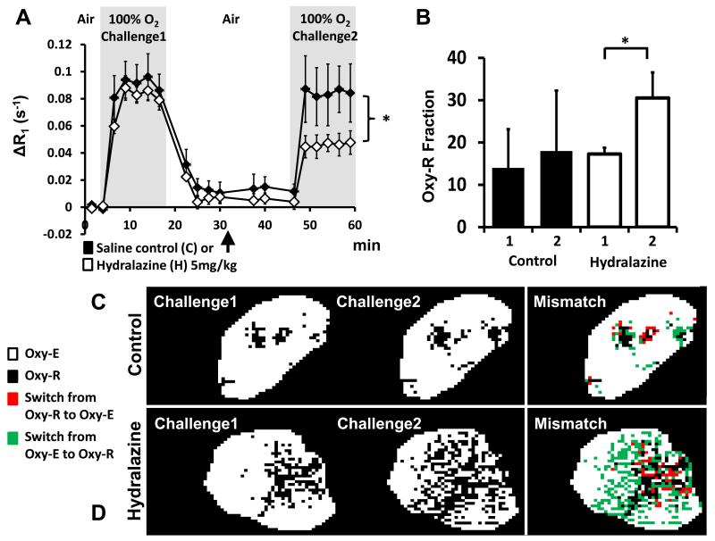
In order to provide evidence that Oxy-R fraction can detect reduced oxygen delivery to tumors, we administered the vasomodulator hydralazine to a cohort of mice bearing 786-0-R xenografts (n=4 mice) and compared with control mice receiving saline (n=6 mice). We performed an air–to–oxygen challenge before and after the administration of hydralazine or control (Supplementary Figure S1c). In the initial air–to–oxygen challenge, overall ΔR1 changes were positive, as expected in all ten mice (Figure 5a).
Figure 5. Oxy-R detects reduced oxygen delivery induced by hydralazine.
A, Time course of the ΔR1 in 786-0-R xenografts challenged with 100% oxygen, before and after intravenous injection of either saline control or 5mg/kg hydralazine hydrochloride. Mean of initial two air breathing R1 values taken as baseline for subsequent ΔR1. All error bars are 1 s.e.m. B, Hydralazine-treated tumors showed increase in the Oxy-R fraction with the second oxygen challenge, relative to control. Oxy-E (white) and Oxy-R (black) voxels are mapped for each oxygen challenge in example control and hydralazine treated tumors. C, Mismatch mapping reveals that approximately equal numbers of voxels switch from Oxy-R to Oxy-E (red) and from Oxy-E to Oxy-R (green) in the control tumor. Most voxels remain either Oxy-E (white) or Oxy-R (black) throughout. D, In distinction, Oxy-R fraction increases on the second oxygen challenge with hydralazine due to substantially more voxels switching from Oxy-R to Oxy-E (red) than from Oxy-E to Oxy-R (green).
Importantly, the second air–to–oxygen challenge was initiated 15 minutes after administration of hydralazine or saline. The magnitude of ΔR1 was reduced significantly in mice receiving hydralazine, relative to control (Figure 5a) (p<0.01). This difference in hydralazine-treated tumors was driven by the Oxy-R fraction increasing significantly from 17.3% to 30.5% (p=0.045) (Figure 5b).
To further understand these changes, we used ‘mismatch mapping’ to identify tumor sub-regions that changed oxygen enhancement status. In control tumors, the majority of voxels remained consistently either Oxy-E or Oxy-R. The number of voxels changing from Oxy-E to Oxy-R or vice versa was equal for both directions of change and was small (Figure 5c). In hydralazine treated tumors, substantially more voxels turned from Oxy-E to Oxy-R than vice versa. Some of these newly Oxy-R tumor sub-regions were immediately adjacent to established Oxy-R regions, but other new areas of Oxy-R were detected at spatially distant sub-regions (Figure 5d). These data confirm that OE-MRI can detect dynamic change in tumor hypoxia. Further, tumor regions with appearing or resolving hypoxia can be mapped using this technique.
DISCUSSION
Hypoxia is an important prognostic factor for solid tumors and mediates resistance to radiotherapy and to some chemotherapy agents. Non-invasive clinical imaging has potential to improve patient care by producing biomarkers that alter clinical decision making (40, 41). In the case of hypoxia, a clinically translational imaging biomarker has potential to be prognostic and/or predictive (identifying presence of hypoxia and quantifying hypoxic fraction/volume prior to therapy), to assist treatment planning (mapping targets for radiation boost or adaptive therapy) (42), or to monitor therapy induced changes in tumor hypoxia, for example following radiosensitisation or hypoxia activated cytotoxic therapy (10, 43).
To date, clinical strategies to image tumor hypoxia have been dominated by positron emission tomography (PET) methods (7). Several investigational PET tracers have been evaluated, each with slightly different underlying relationship to tissue oxygen tension (44, 45). However, at present no imaging method identifies and maps the extent of tumor hypoxia that is validated, cost-effective, widely available and used for patient benefit (7).
OE-MRI is an emerging technique that detects the presence of excess dissolved O2 molecules in plasma and tissue fluid. Most OE-MRI studies in cancer have been small cohort based studies, and without corresponding pathology or intervention, tending to report changes in median signal intensity or median ΔR1. In well oxygenated tumor tissue, oxygen-induced ΔR1 signals are positive, similar to signal changes observed in healthy tissues (46, 47). Advances in OE-MRI have focused largely around providing dual ΔR1 and ΔR2* acquisitions (25) or in enhancing the magnitude of oxygen-induced ΔR1, for example by developing methods sensitive to tumor lipid signals (17).
In this study, we adopted a different approach. We focused on the fact that tumors are biologically heterogeneous, composed of sub-regions with distinct pathophysiology (48, 49). Previous OE-MRI studies have suggested that some tumor sub-regions are refractory to the positive ΔR1 induced by oxygen-challenge (14, 20). We built on this observation by partitioning the OE-MRI signal into regions with enhancing (Oxy-E) or refractory (Oxy-R) signatures. We conducted a series of experiments to test the hypothesis that Oxy-R fraction tumor can identify, quantify and map tumor hypoxia.
We first used the renal carcinoma model 786-0-R (rapid growing, vascular tumors with anticipated large oxygen-induced ΔR1) to develop a protocol that was not only stable while breathing medical air, but also sensitive to oxygen-induced signals. This is the first study to provide detailed evidence of measurement stability and sensitivity for serial R1 mapping in OE-MRI. The temporal evolution of oxygen-enhanced positive ΔR1 occurred within 3 minutes and was spatially stable. The OE-MRI data mirrored OxyLite™ measurement of pO2 changes, concurring with data from a previous study where rapid increases in ΔR1 were observed following gas challenge (15) and with data comparing OE-MRI signal changes with 19F oximetry (19).
Next, we demonstrated that Oxy-R fraction could distinguish differences in hypoxic fraction within tumors derived from two isogenic cell lines: parental 786-0 cells and 786-0-R cells. Here, the relatively rapid growing 786-0-R tumors had significantly more hypoxia than the slow growing 786-0-par tumors. Crucially, Oxy-R fraction distinguished between cell lines, mirroring differences observed in values of hypoxic fraction quantified through pimonidazole staining.
We then tested if Oxy-R fraction could estimate hypoxic fraction. We chose the 786-0-R and SW620 models because of their differing perfusion characteristics. Significant correlation was seen between Oxy-R fraction and hypoxic fraction (defined on pimonidazole adduct formation) in the relatively well perfused 786-0-R xenografts. This association was not replicated in the poorly perfused SW620 xenografts because many Oxy-R voxels had negligible perfusion, so received insufficient oxygen gas to alter voxel R1. Having accounted for these voxels using DCE-MRI, we measured Oxy-R fraction in perfused tissue and observed a significant association in the SW620 tumors. We then increased the fraction of tumor hypoxia in a further cohort of 786-0-R tumors using the vasomodulator hydralazine. Oxy-R fraction increased two-fold, providing direct evidence that OE-MRI can track acute changes in tumor oxygenation. Some signal fluctuation was observed in both directions in hydralazine and control tumors. This may reflect changes in acute transient hypoxia (50), but further work is required to understand these signal changes.
In these experiments, median ΔR1 (the most common biomarker derived from OE-MRI data) was unable to distinguish the differing levels of hypoxia between the 786-0 sub-lines and did not correlate with the degree of hypoxic fraction within the SW620 tumors. We also compared the Oxy-R analysis approach with BOLD MRI. Oxygen-induced ΔR2* did mirror changes in tissue pO2, similar to previous reports (38), but ΔR2* did not differ between the 786-0 sub-lines with differing levels of hypoxia, nor did ΔR2* relate to degree of hypoxic fraction in 786-0-R or SW620 tumors, suggesting that R2* measurements were confounded by other factors, such as vessel flow. These findings are consistent with previous reports of inconsistent relationships between ΔR2* and pimonidazole-based assay of hypoxic fraction (38).
Our findings are important for at least four reasons. Firstly, these data demonstrate that positive ΔR1 changes on OE-MRI imaging are sufficiently precise, stable and strong enough to be measured reliably. This enables those portions of tumor refractory to positive signal change (i.e. Oxy-R sub-regions) to be identified with confidence. Secondly, these data suggest that in OE-MRI, Oxy-R fraction relates more closely to tumor hypoxia than median ΔR1. This supports further evaluation of Oxy-R fraction or Oxy-R tumor volume as a prognostic and/or predictive biomarker. Thirdly, Oxy-R voxels were located in spatially distinct sub-regions suggesting that, if replicated in humans, this technique may facilitate the planning of localized boost and adaptive radiotherapy delivery strategies. Fourthly, clinical tumors vary in their extent of perfusion and this may further change with therapy. Our data suggest that subsequent studies of OE-MRI in patients with cancer should evaluate Oxy-R fraction in perfused tumor regions (using a combined analysis with DCE-MRI or an equivalent technique), rather than using OE-MRI in isolation.
In conclusion, these data provide substantial evidence to validate the measurement of Oxy-R fraction to identify, quantify and map tumor hypoxia. Unlike other nuclear magnetic resonance techniques such as electron paramagnetic resonance oximetry, 19F oximetry and Overhauser enhanced MRI – which are either invasive or require use of an investigational diagnostic agent, hindering clinical translation (7) – Oxy-R fraction can be readily quantified on clinical MRI scanners (24), making the technique suitable for rapid clinical translation.
Supplementary Material
Acknowledgements
We thank Allan Thornhill and his staff (ICR) for animal maintenance and Dr Damian McHugh (University of Manchester) for generation of maps used in the figures.
Grant support:
Cancer Research UK (CRUK) Clinician Scientist award (grant C19221/A15267) to J. P. B. O’Connor;
CRUK and EPSRC Cancer Imaging Centre in Cambridge and Manchester funding to The University of Manchester (grant C8742/A18097) to J. P. B. O’Connor, K. J. Williams, A. Jackson, G. J. M. Parker;
CRUK Cancer Imaging Centre funding to The Institute of Cancer Research (grants C1060/A10334 and C1060/A16464) to S. P. Robinson;
The Wellcome Trust funding (grant 091763Z/10/Z) to S. P. Robinson;
Paul O’Gorman Postdoctoral Fellowship funded by Children with Cancer UK (grant 2014/176) to Y. Jamin.
Breakthrough Breast Cancer Senior Fellowship to A. R. Reynolds. Breakthrough Breast Cancer, which has recently merged with Breast Cancer Campaign to form Breast Cancer Now.
Footnotes
Potential Conflict of Interest: GJMP is a director and shareholder in Bioxydyn Limited. No other authors declared a potential conflict of interest.
REFERENCES
- 1.Harris AL. Hypoxia--a key regulatory factor in tumour growth. Nat Rev Cancer. 2002;2:38–47. doi: 10.1038/nrc704. [DOI] [PubMed] [Google Scholar]
- 2.Pugh CW, Ratcliffe PJ. Regulation of angiogenesis by hypoxia: role of the HIF system. Nat Med. 2003;9:677–684. doi: 10.1038/nm0603-677. [DOI] [PubMed] [Google Scholar]
- 3.Hockel M, Schlenger K, Aral B, Mitze M, Schaffer U, Vaupel P. Association between tumor hypoxia and malignant progression in advanced cancer of the uterine cervix. Cancer Res. 1996;56:4509–4515. [PubMed] [Google Scholar]
- 4.Brizel DM, Sibley GS, Prosnitz LR, Scher RL, Dewhirst MW. Tumor hypoxia adversely affects the prognosis of carcinoma of the head and neck. Int J Radiat Oncol Biol Phys. 1997;38:285–289. doi: 10.1016/s0360-3016(97)00101-6. [DOI] [PubMed] [Google Scholar]
- 5.Vergis R, Corbishley CM, Norman AR, Bartlett J, Jhavar S, Borre M, et al. Intrinsic markers of tumour hypoxia and angiogenesis in localised prostate cancer and outcome of radical treatment: a retrospective analysis of two randomised radiotherapy trials and one surgical cohort study. Lancet Oncol. 2008;9:342–351. doi: 10.1016/S1470-2045(08)70076-7. [DOI] [PubMed] [Google Scholar]
- 6.Wilson WR, Hay MP. Targeting hypoxia in cancer therapy. Nat Rev Cancer. 2011;11:393–410. doi: 10.1038/nrc3064. [DOI] [PubMed] [Google Scholar]
- 7.Tatum JL, Kelloff GJ, Gillies RJ, Arbeit JM, Brown JM, Chao KS, et al. Hypoxia: importance in tumor biology, noninvasive measurement by imaging, and value of its measurement in the management of cancer therapy. Int J Radiat Biol. 2006;82:699–757. doi: 10.1080/09553000601002324. [DOI] [PubMed] [Google Scholar]
- 8.O'Connor JP, Jackson A, Parker GJ, Roberts C, Jayson GC. Dynamic contrast-enhanced MRI in clinical trials of antivascular therapies. Nat Rev Clin Oncol. 2012;9:167–177. doi: 10.1038/nrclinonc.2012.2. [DOI] [PubMed] [Google Scholar]
- 9.Howe FA, Robinson SP, Rodrigues LM, Griffiths JR. Flow and oxygenation dependent (FLOOD) contrast MR imaging to monitor the response of rat tumors to carbogen breathing. Magn Reson Imaging. 1999;17:1307–1318. doi: 10.1016/s0730-725x(99)00089-2. [DOI] [PubMed] [Google Scholar]
- 10.Hammond EM, Asselin MC, Forster D, O'Connor JP, Senra JM, Williams KJ. The meaning, measurement and modification of hypoxia in the laboratory and the clinic. Clin Oncol (R Coll Radiol) 2014;26:277–288. doi: 10.1016/j.clon.2014.02.002. [DOI] [PubMed] [Google Scholar]
- 11.Young IR, Clarke GJ, Bailes DR, Pennock JM, Doyle FH, Bydder GM. Enhancement of relaxation rate with paramagnetic contrast agents in NMR imaging. J Comput Tomogr. 1981;5:543–547. doi: 10.1016/0149-936x(81)90089-8. [DOI] [PubMed] [Google Scholar]
- 12.Berkowitz BA. Role of dissolved plasma oxygen in hyperoxia-induced contrast. Magn Reson Imaging. 1997;15:123–126. doi: 10.1016/s0730-725x(96)00230-5. [DOI] [PubMed] [Google Scholar]
- 13.Gray LH, Steadman JM. Determination of the Oxyhaemoglobin Dissociation Curves for Mouse and Rat Blood. J Physiol. 1964;175:161–171. doi: 10.1113/jphysiol.1964.sp007509. [DOI] [PMC free article] [PubMed] [Google Scholar]
- 14.Matsumoto K, Bernardo M, Subramanian S, Choyke P, Mitchell JB, Krishna MC, et al. MR assessment of changes of tumor in response to hyperbaric oxygen treatment. Magn Reson Med. 2006;56:240–246. doi: 10.1002/mrm.20961. [DOI] [PubMed] [Google Scholar]
- 15.Winter JD, Akens MK, Cheng HL. Quantitative MRI assessment of VX2 tumour oxygenation changes in response to hyperoxia and hypercapnia. Phys Med Biol. 2011;56:1225–1242. doi: 10.1088/0031-9155/56/5/001. [DOI] [PubMed] [Google Scholar]
- 16.Burrell JS, Walker-Samuel S, Baker LC, Boult JK, Jamin Y, Halliday J, et al. Exploring DeltaR(2) * and DeltaR(1) as imaging biomarkers of tumor oxygenation. J Magn Reson Imaging. 2013;38:429–434. doi: 10.1002/jmri.23987. [DOI] [PubMed] [Google Scholar]
- 17.Jordan BF, Magat J, Colliez F, Ozel E, Fruytier AC, Marchand V, et al. Mapping of oxygen by imaging lipids relaxation enhancement: A potential sensitive endogenous MRI contrast to map variations in tissue oxygenation. Magn Reson Med. 2013;70:732–744. doi: 10.1002/mrm.24511. [DOI] [PubMed] [Google Scholar]
- 18.Colliez F, Neveu MA, Magat J, Cao Pham TT, Gallez B, Jordan BF. Qualification of a noninvasive magnetic resonance imaging biomarker to assess tumor oxygenation. Clin Cancer Res. 2014;20:5403–5411. doi: 10.1158/1078-0432.CCR-13-3434. [DOI] [PubMed] [Google Scholar]
- 19.Hallac RR, Zhou H, Pidikiti R, Song K, Stojadinovic S, Zhao D, et al. Correlations of noninvasive BOLD and TOLD MRI with pO2 and relevance to tumor radiation response. Magn Reson Med. 2014;71:1863–1873. doi: 10.1002/mrm.24846. [DOI] [PMC free article] [PubMed] [Google Scholar]
- 20.Linnik IV, Scott ML, Holliday KF, Woodhouse N, Waterton JC, O'Connor JP, et al. Noninvasive tumor hypoxia measurement using magnetic resonance imaging in murine U87 glioma xenografts and in patients with glioblastoma. Magn Reson Med. 2014;71:1854–1862. doi: 10.1002/mrm.24826. [DOI] [PubMed] [Google Scholar]
- 21.Zhao D, Pacheco-Torres J, Hallac RR, White D, Peschke P, Cerdan S, et al. Dynamic oxygen challenge evaluated by NMR T1 and T2 * - insights into tumor oxygenation. NMR Biomed. 2015;28:937–947. doi: 10.1002/nbm.3325. [DOI] [PMC free article] [PubMed] [Google Scholar]
- 22.Beeman SC, Shui YB, Perez-Torres CJ, Engelbach JA, Ackerman JJ, Garbow JR. O2 - sensitive MRI distinguishes brain tumor versus radiation necrosis in murine models. Magn Reson Med. 2015 doi: 10.1002/mrm.25821. Published OnlineFirst July 14, 2015. [DOI] [PMC free article] [PubMed] [Google Scholar]
- 23.Arnold JF, Kotas M, Fidler F, Pracht ED, Flentje M, Jakob PM. Quantitative regional oxygen transfer imaging of the human lung. J Magn Reson Imaging. 2007;26:637–645. doi: 10.1002/jmri.21033. [DOI] [PubMed] [Google Scholar]
- 24.O'Connor JP, Naish JH, Parker GJ, Waterton JC, Watson Y, Jayson GC, et al. Preliminary study of oxygen-enhanced longitudinal relaxation in MRI: a potential novel biomarker of oxygenation changes in solid tumors. Int J Radiat Oncol Biol Phys. 2009;75:1209–1215. doi: 10.1016/j.ijrobp.2008.12.040. [DOI] [PubMed] [Google Scholar]
- 25.Remmele S, Sprinkart AM, Muller A, Traber F, von Lehe M, Gieseke J, et al. Dynamic and simultaneous MR measurement of R1 and R2* changes during respiratory challenges for the assessment of blood and tissue oxygenation. Magn Reson Med. 2013;70:136–146. doi: 10.1002/mrm.24458. [DOI] [PubMed] [Google Scholar]
- 26.Blockley NP, Jiang L, Gardener AG, Ludman CN, Francis ST, Gowland PA. Field strength dependence of R1 and R2* relaxivities of human whole blood to ProHance, Vasovist, and deoxyhemoglobin. Magn Reson Med. 2008;60:1313–1320. doi: 10.1002/mrm.21792. [DOI] [PubMed] [Google Scholar]
- 27.Kalavagunta C, Michaeli S, Metzger GJ. In vitro Gd-DTPA relaxometry studies in oxygenated venous human blood and aqueous solution at 3 and 7 T. Contrast Media Mol Imaging. 2014;9:169–176. doi: 10.1002/cmmi.1568. [DOI] [PMC free article] [PubMed] [Google Scholar]
- 28.Workman P, Aboagye EO, Balkwill F, Balmain A, Bruder G, Chaplin DJ, et al. Guidelines for the welfare and use of animals in cancer research. Br J Cancer. 2010;102:1555–1577. doi: 10.1038/sj.bjc.6605642. [DOI] [PMC free article] [PubMed] [Google Scholar]
- 29.Kilkenny C, Browne WJ, Cuthill IC, Emerson M, Altman DG. Improving bioscience research reporting: the ARRIVE guidelines for reporting animal research. PLoS Biol. 2010;8:e1000412. doi: 10.1371/journal.pbio.1000412. [DOI] [PMC free article] [PubMed] [Google Scholar]
- 30.Bridgeman VL, Wan E, Foo S, Nathan MR, Welti JC, Frentzas S, et al. Preclinical evidence that trametinib enhances the response to anti-angiogenic tyrosine kinase inhibitors in renal cell carcinoma. Mol Cancer Ther. 2015 doi: 10.1158/1535-7163.MCT-15-0170. Published OnlineFirst October 20, 2015. [DOI] [PubMed] [Google Scholar]
- 31.Griffiths JR, Robinson SP. The OxyLite: a fibre-optic oxygen sensor. Br J Radiol. 1999;72:627–630. doi: 10.1259/bjr.72.859.10624317. [DOI] [PubMed] [Google Scholar]
- 32.Gruetter R. Automatic, localized in vivo adjustment of all first- and second-order shim coils. Magn Reson Med. 1993;29:804–811. doi: 10.1002/mrm.1910290613. [DOI] [PubMed] [Google Scholar]
- 33.Stratford IJ, Adams GE, Godden J, Nolan J, Howells N, Timpson N. Potentiation of the anti-tumour effect of melphalan by the vasoactive agent, hydralazine. Br J Cancer. 1988;58:122–127. doi: 10.1038/bjc.1988.177. [DOI] [PMC free article] [PubMed] [Google Scholar]
- 34.Horsman MR, Nordsmark M, Hoyer M, Overgaard J. Direct evidence that hydralazine can induce hypoxia in both transplanted and spontaneous murine tumours. Br J Cancer. 1995;72:1474–1478. doi: 10.1038/bjc.1995.532. [DOI] [PMC free article] [PubMed] [Google Scholar]
- 35.Seddon BM, Maxwell RJ, Honess DJ, Grimshaw R, Raynaud F, Tozer GM, et al. Validation of the fluorinated 2-nitroimidazole SR-4554 as a noninvasive hypoxia marker detected by magnetic resonance spectroscopy. Clin Cancer Res. 2002;8:2323–2335. [PubMed] [Google Scholar]
- 36.Zhao D, Constantinescu A, Jiang L, Hahn EW, Mason RP. Prognostic radiology: quantitative assessment of tumor oxygen dynamics by MRI. Am J Clin Oncol. 2001;24:462–466. doi: 10.1097/00000421-200110000-00010. [DOI] [PubMed] [Google Scholar]
- 37.Williams KJ, Telfer BA, Shannon AM, Babur M, Stratford IJ, Wedge SR. Combining radiotherapy with AZD2171, a potent inhibitor of vascular endothelial growth factor signaling: pathophysiologic effects and therapeutic benefit. Mol Cancer Ther. 2007;6:599–606. doi: 10.1158/1535-7163.MCT-06-0508. [DOI] [PubMed] [Google Scholar]
- 38.Baker LC, Boult JK, Jamin Y, Gilmour LD, Walker-Samuel S, Burrell JS, et al. Evaluation and immunohistochemical qualification of carbogen-induced DeltaR(2) as a noninvasive imaging biomarker of improved tumor oxygenation. Int J Radiat Oncol Biol Phys. 2013;87:160–167. doi: 10.1016/j.ijrobp.2013.04.051. [DOI] [PubMed] [Google Scholar]
- 39.Li J, Jamin Y, Boult JK, Cummings C, Waterton JC, Ulloa J, et al. Tumour biomechanical response to the vascular disrupting agent ZD6126 in vivo assessed by magnetic resonance elastography. Br J Cancer. 2014;110:1727–1732. doi: 10.1038/bjc.2014.76. [DOI] [PMC free article] [PubMed] [Google Scholar]
- 40.Parkinson DR, Johnson BE, Sledge GW. Making personalized cancer medicine a reality: challenges and opportunities in the development of biomarkers and companion diagnostics. Clin Cancer Res. 2012;18:619–624. doi: 10.1158/1078-0432.CCR-11-2017. [DOI] [PubMed] [Google Scholar]
- 41.Waterton JC, Pylkkanen L. Qualification of Imaging Biomarkers for Oncology Drug Development. Eur J Cancer. 2012;48:409–415. doi: 10.1016/j.ejca.2011.11.037. [DOI] [PubMed] [Google Scholar]
- 42.Gatenby RA, Silva AS, Gillies RJ, Frieden BR. Adaptive therapy. Cancer Res. 2009;69:4894–4903. doi: 10.1158/0008-5472.CAN-08-3658. [DOI] [PMC free article] [PubMed] [Google Scholar]
- 43.Ahn GO, Brown M. Targeting tumors with hypoxia-activated cytotoxins. Front Biosci. 2007;12:3483–3501. doi: 10.2741/2329. [DOI] [PubMed] [Google Scholar]
- 44.Carlin S, Zhang H, Reese M, Ramos NN, Chen Q, Ricketts SA. A comparison of the imaging characteristics and microregional distribution of 4 hypoxia PET tracers. J Nucl Med. 2014;55:515–521. doi: 10.2967/jnumed.113.126615. [DOI] [PMC free article] [PubMed] [Google Scholar]
- 45.Peeters SG, Zegers CM, Lieuwes NG, van Elmpt W, Eriksson J, van Dongen GA, et al. A comparative study of the hypoxia PET tracers [(1)(8)F]HX4, [(1)(8)F]FAZA, and [(1)(8)F]FMISO in a preclinical tumor model. Int J Radiat Oncol Biol Phys. 2015;91:351–359. doi: 10.1016/j.ijrobp.2014.09.045. [DOI] [PubMed] [Google Scholar]
- 46.O'Connor JPB, Jackson A, Buonaccorsi GA, Buckley DL, Roberts C, Watson Y, et al. Organ-specific effects of oxygen and carbogen gas inhalation on tissue longitudinal relaxation times. Magn Reson Med. 2007;58:490–496. doi: 10.1002/mrm.21357. [DOI] [PubMed] [Google Scholar]
- 47.Winter JD, Estrada M, Cheng HL. Normal tissue quantitative T1 and T2* MRI relaxation time responses to hypercapnic and hyperoxic gases. Acad Radiol. 2011;18:1159–1167. doi: 10.1016/j.acra.2011.04.016. [DOI] [PubMed] [Google Scholar]
- 48.Junttila MR, de Sauvage FJ. Influence of tumour micro-environment heterogeneity on therapeutic response. Nature. 2013;501:346–354. doi: 10.1038/nature12626. [DOI] [PubMed] [Google Scholar]
- 49.O'Connor JP, Rose CJ, Waterton JC, Carano RA, Parker GJ, Jackson A. Imaging intratumor heterogeneity: role in therapy response, resistance, and clinical outcome. Clin Cancer Res. 2015;21:249–257. doi: 10.1158/1078-0432.CCR-14-0990. [DOI] [PMC free article] [PubMed] [Google Scholar]
- 50.Baudelet C, Cron GO, Ansiaux R, Crokart N, Dewever J, Feron O, et al. The role of vessel maturation and vessel functionality in spontaneous fluctuations of T(2)*-weighted GRE signal within tumors. NMR Biomed. 2006;19:69–76. doi: 10.1002/nbm.1002. [DOI] [PubMed] [Google Scholar]
Associated Data
This section collects any data citations, data availability statements, or supplementary materials included in this article.


