Abstract
Purpose
To determine the relationship between peripapillary retinal nerve fiber layer (PRNFL) swelling and eventual PRNFL atrophy, and between PRNFL swelling/atrophy and neural function, in a nonhuman primate model of nonarteritic anterior ischemic optic neuropathy (pNAION).
Methods
pNAION was induced in five normal, adult male rhesus monkeys by laser activation of intravenously injected rose bengal at the optic nerve head. Spectral-domain optical coherence tomography measurements of the PRNFL were performed at baseline; 1 day; 1, 2, and 4 weeks; and several later times over a period of an additional 2 to 3 months. Simultaneous pattern-reversal electroretinograms (PERGs) and visual evoked potentials (VEPs) were recorded and color fundus photographs taken at the same time points.
Results
In all cases, initial PRNFL swelling was associated with atrophy, and the greater the initial swelling, the greater the degree of eventual atrophy (r = 0.65, P = 0.0002). The change in PRNFL thickness closely correlated with VEP amplitude loss (r = 0.90), although this relationship was only a strong trend (P = 0.083). Furthermore, VEP amplitude loss closely correlated with PERG N95 amplitude loss (r = 0.80, P = 0.00002)
Conclusions
In our model of human NAION, the degree of initial PRNFL swelling correlated with the severity of atrophy. Areas that did not swell developed little to no atrophy. The amount of PRNFL loss was reflected in VEP and PERG N95 amplitude reductions.
Keywords: anterior ischemic optic neuropathy, optical coherence tomography, nerve fiber layer thickening, animal model, visual evoked potential
Nonarteritic anterior ischemic optic neuropathy (NAION) is the most common cause of new optic nerve–related vision loss in individuals older than 50 years.1 There currently is no accepted treatment for this disorder. Our laboratory has developed two animal models, one in rodents (rNAION)2 and one in nonhuman primates (pNAION),3 to investigate the pathophysiology of this disorder and to develop treatments for NAION based on these mechanisms of damage.
Improvements in optical coherence tomography (OCT) have made it easier to quantify swelling in the fundus that occurs from various pathologic processes. In NAION, the thickness of the peripapillary retinal nerve fiber layer (PRNFL), the optic nerve head, and the macular retinal ganglion cell–inner plexiform layer (mRGCIPL) complex are among the variables that can be compared with measures of visual function such as visual acuity and visual field sensitivity to assess the extent and progression of damage. In particular, the mRGCIPL has been shown to correlate with visual field loss in acute NAION but not with final visual field loss.4 Optical coherence tomography measures of swelling, in addition to visual acuity and field loss, have become endpoints in small-sample clinical trials exploring the efficacy of new medical treatments.5,6
Among the advantages that our nonhuman primate NAION model has in exploring the relationship between OCT thickness and structural and functional outcome in NAION are the following: (1) the time of pNAION onset is known; (2) the natural history from disease onset can be rigorously followed; (3) many sources of variablity in noninvasive measurements (i.e., OCT, visual evoked potential [VEP], pattern electroretinogram [PERG]) can be controlled, resulting in consistently high data quality; (4) there is no concomitant systemic pathology; and (5) there is no spontaneous improvement. This last consideration is important when the goal is to determine the absolute relationship among variables, as 40% of patients with NAION experience some degree of spontaneous recovery.7 In this study, we report the relationship between pNAION-induced PRNFL swelling and eventual PRNFL atrophy and loss of optic nerve and ganglion cell function.
Methods
Animals
All animal protocols were approved by the UMB Institutional Animal Care and Use Committee, and all animals were handled in accordance with the ARVO Statement for the Use of Animals in Ophthalmic and Vision Research. The induction of pNAION has been described elsewhere,3 but briefly, a normal adult male rhesus monkey (Macacca mulatta) was anesthetized with ketamine (10 mg/kg) and xylazine (2 mg/kg) and his pupils dilated with a combination of 1% tropicamide and 2.5% phenylephrine. The animal then was placed in front of a slit-lamp biomicroscope fitted with an ophthalmic neodymium-yttrium-aluminum-garnet (Nd-YAG) frequency-doubled diode laser (532 nm; Iridex, Mountain View, CA, USA). We replaced the 209-μm-diameter laser fiberoptic cable with a 500-μm-diameter fiberoptic cable that had SMA adapter ends, permitting the standard 532-nm slit-lamp adapter to generate a 1.06-mm spot size when using the 200-μm spot size setting. The optic disc in the eye to be treated was visualized by using a Glasser monkey contact lens (Ocular Instruments, Inc., Bellevue, WA, USA). pNAION was induced by injecting Rose bengal intravenously in a dose of 2.5 mg/kg of lean body weight, followed 30 seconds later by dye activation at the optic disc by using the 1.06-mm spot size, at 200 mW, for times ranging from 8 to 9 seconds. All animals had pNAION induced sequentially in the two eyes, with the second eye induced 1 to 2 months after the first.
Before performing functional measurements, all animals were anesthetized with a mixture of ketamine (10 mg/kg) and xylazine (2 mg/kg). They then were intubated, and assessments were performed while the animals were supported with a continuous infusion of propofol. Data from five animals were used for the study. These animals were part of studies examining natural history and treatment; functional and structural data have been reported elsewhere.3,8
Optical Coherence Tomography
Optical coherence tomography was used to assess the thickness of the PRNFL. We used a Heidelberg Spectralis spectral-domain HRA + OCT (SD-OCT) instrument (Heidelberg Engineering, Heidelberg, Germany) with 5.4b-US software and equipped with an automated real-time eye-tracking system (ART). Before imaging, each animal's pupils were dilated with topical 2.5% phenylephrine and 1.0% tropicamide. For assessment of the PRNFL, the circular scan mode was used, which uses a circle measuring 3.5 mm in diameter. Measurements of PRNFL thickness were analyzed by using an OCT-generated algorithm that measures the thickness in six contiguous peripapillary sectors. One hundred images were averaged. A minimum of three scans was obtained for each eye at the same location; the set with the best quality was used for manual correction of the instrument's automated segmentation.
Optical coherence tomography measurements were obtained before treatment, and again at 1 day, 1 week, 2 weeks, 4 weeks, and several later times over a period of an additional 2 to 3 months. Data from only one eye of each monkey were used. The eye chosen for analysis was that which had the longest follow-up.
Electrophysiological Studies
Simultaneous PERGs and VEPs were recorded by using a modified fundus camera described previously.9 Briefly, a Topcon fundus camera (Topcon Corporation, Tokyo, Japan) was modified by inserting a 2-cm OLED screen from a head-mounted display into the split-viewer pathway. The input to the screen was an alternating (1.9 Hz) black-and-white checkerboard pattern having a luminance of 109 cd/m2 at a nominal contrast of 100%, generated by a LKC UTAS visual diagnostic system (LKC Technologies, Inc., Gaithersburg, MD, USA). The location of the screen was adjusted so that it was conjugate to the plane of the animal's pupil. In this way, when the monkey's retina was in focus, the image of the checkerboard was in focus on the monkey's retina.
The VEP was recorded by using Grass gold cup skin electrodes and EC2 electrode paste (Grass Instruments, Warwick, RI, USA). The active electrode (Oz) was placed above the inion in the midline over the shaved skull; the reference electrode was placed in the midline frontal position (Fz), and the ground electrode was placed on an arm with Grass gold surface electrodes. The PERG was recorded simultaneously with the VEP, with a Burian-Allen bipolar contact lens electrode.
Statistics
Before analysis of the data, each data set was evaluated by the D'Agostino-Pearson omnibus K2 test to determine if the distribution of the data was Gaussian. If it was, a Pearson correlation coefficient was calculated and the statistical significance of that value determined in a 2-tailed test. If the data were close to Gaussian and the subtraction of one or two outliers made the distribution Gaussian, then these data were excluded and a Pearson correlation coefficient was calculated. Two data sets (Figs. 5A, 6) contained too few samples to determine the shape of their distributions. In those cases, Spearman correlation coefficients were computed. A linear curve was fit to each data set by using regression analysis.
Figure 5.
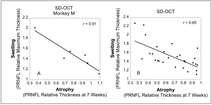
Peripapillary retinal nerve fiber layer maximum thickness divided by baseline thickness, versus minimum thickness (atrophy) at 7 weeks post induction divided by baseline thickness. Data are plotted for all six peripapillary regions for monkey M (A) and for the five study monkeys (B). The linear equation fit to both sets of data indicates that for each initial 1% increase in thickness there is an eventual 0.82% to 1.07% loss in thickness.
Figure 6.
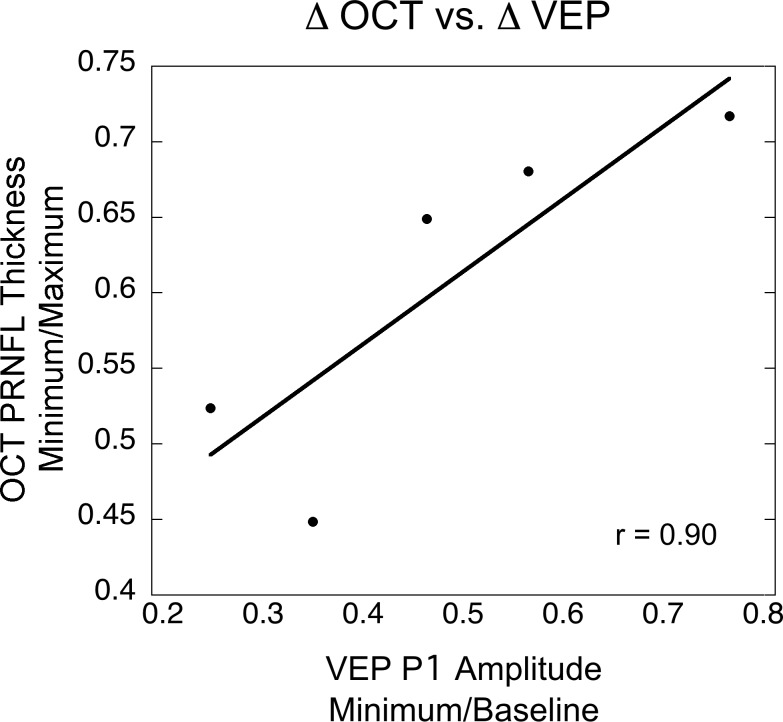
Relative VEP amplitude loss versus change in PRNFL thickness for the five subjects. Global values are plotted.
Results
All subjects had either diffuse or sectoral optic disc and PRNFL swelling at 1 day post induction of pNAION (Fig. 1). Maximum swelling was observed between 1 day and 1 week post induction.
Figure 1.
Fundus photograph of one eye with pNAION from each of five monkeys. Photographs were taken 1 day post induction.
Swollen Nerve Fibers Become Atrophic; Nonswollen Nerve Fibers Remain Normal
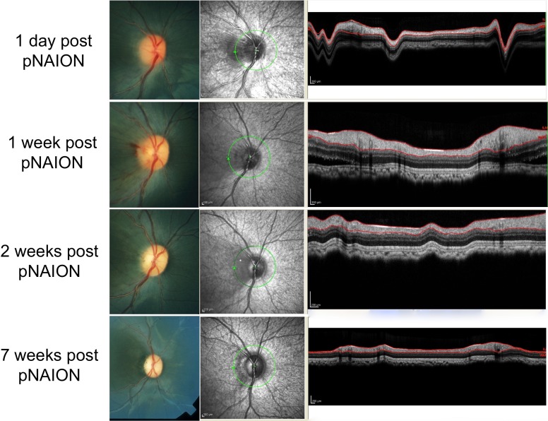
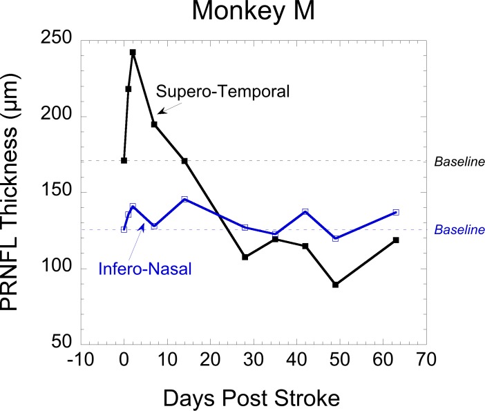
In all eyes, PRNFL regions that were swollen later became atrophic, whereas areas that had little or no swelling remained normal or nearly so. Figures 2 and 3 illustrate the progression from mild to moderate swelling and eventually to atrophy for monkey M.
Figure 2.
Color photographs and OCT in monkey M following induction of pNAION. Top to bottom: 1 day, 1 week, 2 weeks, and 7 weeks post induction. Note progressive loss of the PRNFL associated with resolution of optic disc swelling and development of optic disc pallor.
Figure 3.
Peripapillary retinal nerve fiber layer thickness as a function of time after induction in two peripapillary regions of monkey M. The horizontal dotted lines indicate the baseline thickness measurement for the specific sector. Values above each line indicate increased thickness and values below, decreased thickness (i.e., from atrophy). The graph shows that initial swelling in the superotemporal PRNFL was associated with later atrophy in the same region, whereas the PRNFL in the inferonasal region had very little swelling and subsequently had no significant thinning.
The Extent of PRNFL Swelling Correlates With the Severity of Eventual Atrophy
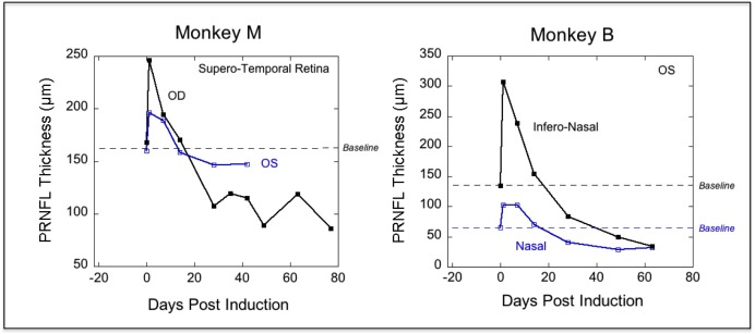
We found that in all eyes with pNAION, the severity of PRNFL swelling correlated with the severity of atrophy. Figure 4 illustrates this relationship in two animals by plotting PRNFL thickness data from two different regions of the PRNFL having different amounts of initial swelling. Regions of the PRNFL that were more swollen after induction of pNAION developed more atrophy. This pattern of atrophy is a consistent feature of pNAION, as demonstrated in Figure 5. In all cases, maximum thickness in our model was observed between 1 day and 1 week post induction, with a return to baseline level at approximately 3 weeks and subsequent thinning leveling off at 5 to 9+ weeks. The greater the degree of initial PRNFL thickening, the longer the thinning continued.
Figure 4.
Peripapillary retinal nerve fiber layer thickness measurements as a function of time after induction of pNAION in two monkeys. Select peripapillary regions from each monkey are shown to illustrate the correlation between the severity of swelling and the severity of subsequent atrophy. For monkey M, the superotemporal sectors of the right (black) and left (blue) eyes are shown, whereas the inferonasal (black) and nasal (blue) sectors of the same eye in monkey B are illustrated. Horizontal dashed lines indicate the baseline value of each sector. Values above the line indicate increases in thickness, whereas values below the line indicate decreases in thickness.
Data for all six regions (superior temporal, temporal, inferior temporal, inferior nasal, nasal, and superior nasal) are shown for monkey M in Figure 5A and for the entire group in Figure 5B. These figures indicate the relationship between PRNFL thickness and atrophy by plotting the maximum thickness for each segment versus the minimum thickness, relative to baseline values. This data normalization is necessary to account for normal variation in baseline thickness both within and among subjects, as can be appreciated from Figures 3 and 4. The correlation between PRNFL swelling and atrophy approached statistical significance for monkey M (r = 0.91, P = 0.058) but was significant for the entire cohort (r = 0.65, P = 0.0002). Areas that swelled became atrophic and areas that did not swell showed little to no atrophy (see Fig. 5B).
We previously have reported that VEP amplitudes are reduced as a consequence of pNAION.3,8 Here we showed a strong trend between VEP amplitude reduction and the extent of PRNFL swelling and eventual atrophy. Figure 6 is a plot of the ratio of the minimum VEP amplitude to the baseline VEP amplitude (i.e., the maximum VEP amplitude) versus the ratio of the minimum PRNFL thickness to the maximum thickness. As with OCT measurements, relative VEP amplitudes were calculated because of the recognized10 large VEP amplitude variability among normal subjects. Global (∼average) values are used for PRNFL thickness here, so there is one datum for each animal. There is a close, but not quite significant, correlation (r = 0.90, P = 0.083) between PRNFL swelling/atrophy and VEP amplitude loss.
Optic Nerve Function Loss Correlates With Ganglion Cell Function Loss
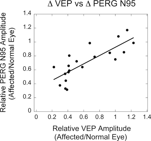
We previously have shown significant and close correlations between PERG and VEP amplitudes in pNAION animals treated with the experimental drug PGJ2.8 We showed the same relationship in this study (Fig. 7). Comparison of the reduction in VEP amplitudes with the reduction in PERG N95 amplitudes provides insight into mechanism of loss. Extrapolation of this linear relationship to the ordinate, discussed below, indicates that 32% of retinal ganglion cells (RGCs), on average, are still responsive at a time when the VEP is totally unrecordable.
Figure 7.
Optic nerve function loss, defined as the VEP amplitude in the affected versus the normal eye, was significantly correlated (r = 0.80, P = 0.00002) with RGC function loss, defined as PERG amplitude ratios similarly calculated.
Discussion
We previously have demonstrated that our pNAION model resembles human NAION in many ways.3 We believe that the results from our current study also translate to the human condition, revealing the relationship between PRNFL thickness and eventual atrophy that would be obvious if measurements could be made in patients before and at NAION onset, and repeated frequently.
The thinning of the PRNFL closely approximated the reduction in VEP amplitude in our animals but did not reach statistical significance (r = 0.90, P = 0.083); however, because the covariance is high, we attribute the lack of statistical significance at the P = 0.05 level to the nonparametric test that was necessary for evaluating a sample containing only five data pairs. This relationship is defined as a strong trend, with the interpretation of the results subject to a little less certainty than with a 95% confidence limit.
Measurement of the OCT mRGCIPL complex has been advocated as a more sensitive means to predict visual outcome in a variety of optic nerve disorders.11,12 In our model, we evaluated macular ganglion cell function by PERG and found that it correlated closely with the pattern VEP. This finding is not surprising as most of the pattern VEP arises from central macular projections. Furthermore, linear extrapolation of the regression line comparing VEP and PERG values suggests that VEP loss precedes RGC loss in our model, provided the relationship remains linear with small values of both.
Our pNAION model differs from the human condition in that in all cases, loss of optic nerve and ganglion cell function occurred from the moment of pNAION induction, whereas in humans, significant PRNFL and optic disc swelling may precede the development of symptoms, at least in some cases,13 even though patients do not present until they become symptomatic. Visual symptoms also may wax and wane over time, suggesting a smouldering or incipient process that may be reflected in changes in PRNFL thickness. Finally, most patients with NAION have coexisting morbidities that may influence their response to treatment. Nevertheless, should our findings showing a correlation between extent of PRNFL swelling and both PRNFL atrophy and electrophysiological evidence of functional loss be found to be present in human NAION, this would provide further evidence that the pNAION model may be a time- and cost-effective way of determining the best candidates for human clinical trials.
Acknowledgments
Supported by National Eye Institute (NEI) Grant R01 EY019529 and a grant from the Hirschhorn Foundation.
Portions of this work previously presented at the annual meeting of the Association for Research in Vision and Ophthalmology, Seattle, Washington, United States, May 2013.
Disclosure: M.A. Johnson, None; N.R. Miller, None; T. Nolan, None; S.L. Bernstein, None
References
- 1. Arnold AC. Ischemic optic neuropathy. : NR Miller,, Newman NJ,, Biousse V,, Kerrison JB, Walsh and Hoyt's Clinical Neuro-Ophthalmology. 6th ed. Vol 1. Baltimore, MD: Lippincott-Williams & Wilkins; 2005: 349–384. [Google Scholar]
- 2. Bernstein SL,, Guo Y,, Kelman SE,, Flower RW,, Johnson MA. Functional and cellular responses in a novel rodent model of anterior ischemic optic neuropathy. Invest Ophthalmol Vis Sci. 2003; 44: 4153–4162. [DOI] [PubMed] [Google Scholar]
- 3. Chen CS,, Johnson MA,, Flower RA,, Slater BJ,, Miller NR,, Bernstein SL. A primate model of nonarteritic anterior ischemic optic neuropathy. Invest Ophthalmol Vis Sci. 2008; 49: 2985–2992. [DOI] [PMC free article] [PubMed] [Google Scholar]
- 4. Park SW,, Ji YS,, Heo H. Early macular ganglion cell-inner plexiform layer analysis in non-arteritic anterior ischemic optic neuropathy [published online ahead of print December 3 2015]. Graefes Arch Clin Exp Ophthalmol. doi:http://dx.doi.org/10.1007/s00417-015-3191-z. [DOI] [PubMed]
- 5. Lyttle DP,, Johnson LN,, Margolin EA,, Madsen RW. Levodopa as a possible treatment of visual loss in nonarteritic anterior ischemic optic neuropath [published online ahead of print October 20 2015]. Graefes Arch Clin Exp Ophthalmol. doi:http://dx.doi.org/10.1007/s00417-015-3230-9. [DOI] [PubMed]
- 6. Sanjari N,, Pakravan M,, Nourinia R,, et al. Intravitreal injection of a Rho-kinase inhibitor (fasudil) for recent onset non-arteritic anterior ischemic optic neuropathy [published online ahead of print October 7, 2015]. J Clin Pharmacol. doi:http://dx.doi.org/10.1002/jcph.655. [DOI] [PubMed]
- 7. Ischemic Optic Neuropathy Decompression Trial Research Group. Ischemic Optic Neuropathy Decompression Trial: twenty-four–month update. Arch Ophthalmol. 2000; 118: 793–797. [PubMed] [Google Scholar]
- 8. Miller NR,, Johnson MA,, Nolan T,, Guo Y,, Bernstein AM,, Bernstein SL. Sustained neuroprotection from a single intravitreal injection of PGJ2 in a nonhuman primate model of nonarteritic anterior ischemic optic neuropathy. Invest Ophthalmol Vis Sci. 2014; 55: 7047–7056. [DOI] [PMC free article] [PubMed] [Google Scholar]
- 9. Johnson MA,, Slater BJ,, Miller NR,, Bernstein SL,, Flower RW. A simple integrated system for electrophysiologic recording in animals. Doc Ophthalmol. 2009; 119: 9–12. [DOI] [PMC free article] [PubMed] [Google Scholar]
- 10. Odom JV,, Bach M,, Brigell M,, Holder GE,, McCulloch DL,, Tormene AP, Vaegan. ISCEV standard for clinical visual evoked potentials (2009 update). Doc Ophthalmol. 2010; 120: 111–119. [DOI] [PubMed] [Google Scholar]
- 11. Larrea BA,, Iztueta MG,, Indart LM,, Alday NM. Early axonal damage detection by ganglion cell complex analysis with optical coherence tomography in nonarteritic anterior ischaemic optic neuropathy. Graefes Arch Clin Exp Ophthalmol. 2014; 252: 1839–1846. [DOI] [PubMed] [Google Scholar]
- 12. Rebolleda G,, Diez-Alvarez L,, Casado A,, et al. OCT: new perspectives in neuro-ophthalmology. Saudi J Ophthalmol. 2015; 29: 9–25. [DOI] [PMC free article] [PubMed] [Google Scholar]
- 13. Hayreh SS,, Zimmerman MB. Incipient nonarteritic anterior ischemic optic neuropathy. Ophthalmology. 2007; 114: 1763–1772. [DOI] [PubMed] [Google Scholar]


