Abstract
An immuno-affinity chromatography technique for purification of infective bluetongue virus (BTV) has been descried using anti-core antibodies. BTV anti-core antibodies (prepared in guinea pig) were mixed with cell culture-grown BTV-1 and then the mixture was added to the cyanogens bromide-activated protein-A Sepharose column. Protein A binds to the antibody which in turn binds to the antigen (i.e. BTV). After thorough washing, antigen–antibody and antibody-protein A couplings were dissociated with 4M MgCl2, pH6.5. Antibody molecules were removed by dialysis and virus particles were concentrated by spin column ultrafiltration. Dialyzed and concentrated material was tested positive for BTV antigen by a sandwich ELISA and the infectivity of the chromatography-purified virus was demonstrated in cell culture. This method was applied for selective capture of BTV from a mixture of other viruses. As group-specific antibodies (against BTV core) were used to capture the virus, it is expected that virus of all BTV serotypes could be purified by this method. This method will be helpful for selective capture and enrichment of BTV from concurrently infected blood or tissue samples for efficient isolation in cell culture. Further, this method can be used for small scale purification of BTV avoiding ultracentrifugation.
Keywords: Anti-core antibody, Bluetongue virus, Immuno-affinity chromatography
Bluetongue virus (BTV), the prototype species of the genus Orbivirus within the family Reoviridae, is the causative agent of bluetongue, a major disease of sheep although the virus can infect all ruminants. BTV genome has ten segments of double-stranded (ds) RNA which encodes seven structural proteins (VP1toVP7) and four non-structural proteins (NS1, NS2, NS3/NS3A and NS4). The outer capsid of the virion is composed of VP2 and VP5 and the internal core is formed by two layers, constituted by VP3 (sub-core) and VP7 (intermediate layer). VP7, whose sequence is relatively well-conserved between isolates, is the most abundant structural protein and it is also the major immunogenic and serogroup-reactive protein [18]. VP2 (encoded by segment-2), the most variable of the BTV proteins, is the key determinant of neutralizing antibody specificity and solely responsible for serotype determination [8]. Presently, BTV has 26 distinct serotypes distributed all over the world.
Purification of virus is essential for understanding the molecular details and functions of its proteins and nucleic acid. A number of ultracentrifugation protocols have been developed for purification of BTV and African horsesickness virus (AHSV), an important member of the Orbivirus genus. Initially, cesium chloride (CsCl) gradient was used for purification of cell culture-grown BTV, but the virus was frequently found to be contaminated with host proteins and non-structural viral proteins. CsCl was also found to convert the virion into core particle by removing the outer capsid proteins due to which the specific infectivity of the BTV particles was greatly reduced [9, 16, 17]. Later, improved methods were developed for purification of whole virion particle, sub-viral particle and core particle of BTV and AHSV by sequential ultracentrifugation through sucrose gradient and CsCl gradient [2, 10]. All these ultracentrifugation-based methods provide flexible means for purification of virus with high degree of purity. However, there are many critical variables to consider when developing and adapting an ultracentrifugation method for optimal performance. It cannot be emphasized enough how important it is to monitor each step in the purification process to ensure one is effectively purifying the virus. The above methods are also cumbersome and time-taking as they require large-scale virus culture, preliminary clarification, treatment with various detergents, and cycles of ultracentrifugation through gradients. The purified virus thus obtained often loses the infectivity due to long exposure to different detergents and chemicals. Therefore, a rapid, simple and less rigorous purification method is needed which will recover bioactive or infective BTV from a small amount of tissue, blood or infected cultured cells and the small quantity of infective virus thus obtained may be used for direct isolation on cell culture or embryonated chicken’s egg. In the present study, we describe a method for purification and concentration of BTV by immuno-affinity chromatography (IAC) using anti-core antibody immobilized to protein-A Sepharose beads and also demonstrate the infectivity of the virus purified by this method.
Anti-core antibody was used to capture BTV. BTV-23 was infected to BHK-21 cells grown in roller culture vessels and harvested between 36 and 48 h post infection when 80 % cells showed cytopathic effects (CPE). From the infected cell lysate and supernatant, BTV core was purified by sucrose density gradient ultracentrifugation [10] and hyperimmune serum (HIS) was produced in guinea pigs against the purified core particles following a standard method. The IgG fraction of the HIS was separated by ammonium sulfate precipitation and dialysis.
Purification of cell culture-grown BTV was done by immuno-affinity chromatography (IAC). Briefly, cyanogen bromide-activated Protein A-Sepharose CL-4B resin (Sigma, St. Louis, MO, USA) was swollen in a buffer (20 mM NaH2PO4, 150 mM NaCl, pH 8.0) for 30 min and washed twice with the same buffer. Purified IgG (against BTV core) was added to the resin and incubated overnight at 4 °C for conjugation. The mixture was transferred to a column and washed twice with the above buffer. To the resin-antibody complex, unpurified cell culture-grown BTV-1 was added and incubated overnight at 4 °C for binding of virus with the antibody. After washing, virus-antibody and resin-antibody couplings were dissociated by using the elution buffer. From the elute, antibody and buffer salts were removed and simultaneously virus preparation was concentrated (to about 200 μl volume) by dialysis against PBS using 300 kDa molecular weight cutoff (MWCO) spin column (Sartorius, Goettingen, Germany). About 50 μl concentrated virus preparation was used for testing the presence of BTV by a sandwich ELISA (s-ELISA) as per the method described by Chand et al. [3]. Dissociation of virus-antibody bonding was optimized by using different elution buffers viz., A (4M MgCl2 with 75 mM HEPES, pH6.5); B (3M KSCN) and C (100 mM Glycine HCl, pH 3.0) and eluted fractions were tested by s-ELISA for the presence of BTV. Elution buffer showing highest efficiency with minimal effect on virus infectivity was selected for IAC purification.
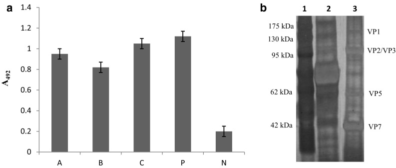
Best result was obtained when 20 mg resin (cyanogen bromide-activated protein A-Sepharose CL-4B), 400 μg IgG and 1 ml of BTV-1 (106 TCID50/ml) were used and incubation was done at 4 °C for overnight. In this condition, coupling of resin-IgG and virus-IgG was found to be optimum because virus was not detectable by s-ELISA in the flow through following the addition of virus to the resin-antibody complex in the column. After addition of virus, unbound materials were removed by washing till A280 of the effluent measured less than 0.05. This required use of about 10 column volume of buffer. In s-ELISA, A492 value of eluted fractions using different elution buffers was more than 0.80 indicating presence of BTV antigen in all the fractions (Fig. 1a). Other quantitative data of IAC have been shown in Table 1.
Fig. 1.

a A492 value for the eluted fraction by different elution buffer in s-ELISA. A (4M MgCl2 with 75 mM HEPES, pH6.5); B (3M KSCN); C (100 mM Glycine HCl, pH 3.0); P positive control; N negative control. b SDS-PAGE analysis of proteins of BTV-1 purified by immuno-affinity chromatography. Viral proteins were resolved on 10 % polyacrylamide gel and stained with silver nitrate. Lane-1 Protein molecular weight marker (#PG500-0500PI, Puregene); Lane-2 BTV-1 infected BHK-21 cell culture lysate showing mainly the cellular proteins; Lane-3 Major structural proteins (VP1, VP2/VP3, VP5, VP7) of BTV-1 purified by immuno-affinity chromatography method
Table 1.
Quantitative data on immuno-affinity chromatographic purification of BTV
| Parameters | Culture supernatant | Elute fraction |
|---|---|---|
| Volume (μl) | 1000 | 200 |
| Protein concentration (mg/ml) | 3.6 | 0.8 |
| Absorbance (260/280) | 1.08 | 1.45 |
| Infectivity of virus (TCID50/ml) | 106 | 102.5–103 |
| s-ELISA (A492) | 1.05 | 0.95 |
After assessing the presence of BTV by s-ELISA, eluted fractions were filtered through 0.22 μm membrane filter and the filtrates were added to BHK-21 cells grown in 24-well plate in Glasgow modified Eagle’s medium (GMEM) supplemented with 10 % fetal bovine serum. After 48 h post inoculation, BTV related CPE was observed only in cells infected with the fraction eluted by using the buffer A (4M MgCl2 with 75 mM HEPES, pH 6.5). Infected cell culture lysate was tested positive for BTV antigen by s-ELISA and viral nucleic acid was detected by a BTV diagnostic RT-PCR [4] (data not shown). No CPE was observed, even after 96 h post infection, in the cells infected with the fractions eluted using buffer B and buffer C. Therefore, buffer A was chosen as an elution buffer, as it efficiently dissociated virus from the antigen–antibody complex and at the same time maintained the infectivity of the virus. Other buffers efficiently eluted the virus but infectivity of the virus was lost.
Specificity of IAC technique for BTV purification was assessed by selectively capturing BTV-1 from a mixture of different viruses that commonly infect the small ruminants, for example, goat pox virus (GTPV, 105 TCID50/ml), sheep pox virus (SPPV, 105 TCID50/ml), peste des petits ruminants virus (PPRV, 106 TCID50/ml) and foot and mouth disease virus (FMDV, 106 TCID50/ml). To the resin-antibody complex, about one ml of each of the BTV-1, PPRV, FMDV, SPPV and GTPV was added, mixed gently and incubated overnight at 4 °C. This was loaded on column for IAC as described above. Eluted fractions were tested by PCR or RT-PCR for presence of nucleic acid of GTPV [7], SPPV [7], PPRV [5] and FMDV [6] which yielded negative results. Only the BTV nucleic acid could be detected in the elutes by RT-PCR. Virus isolation from the elutes also resulted recovery of BTV only, which was confirmed by RT-PCR and s-ELISA in three successive passages in cell culture. BTV-1 purified by IAC method was resolved in 10 % SDS-PAGE and stained with silver nitrate which revealed distinct band of viral proteins VP1, VP2, VP3, VP5 and VP7 (Fig. 1b).
In the present study, we used high concentration of magnesium (4M MgCl2 with 75 mM HEPES, pH6.5), a chaotropic agent (3M KSCN) and low pH (100 mM Glycine HCl, pH 3.0) for dissociation of virus (BTV)-antibody bond. High concentration of magnesium (5M MgCl2) has earlier been used to dissociate virus-antibody bond for purification of equine infectious anemia virus by affinity chromatography [14]. Elution buffer containing 4M MgCl2 with 75 mM HEPES was also used for purification of type-specific FMDV antibody from antigen coated to affinity matrix without affecting the viral antigen [1]. After elution with this buffer, elute was found positive for BTV antigen in s-ELISA in the present study (Fig. 1a) and virus present in elute produced CPE upon inoculation to BHK-21 cells. However, after elution with 3M KSCN and 100 mM Glycine HCl, pH 3.0, elutes were found to be positive for BTV but the infectivity of the virus was completely lost as no CPE was observed in BHK-21 cell upon inoculation. This may be due to inactivation of BTV at low pH [15] and denaturation of viral proteins by KSCN, a chaotropic agent [12]. Therefore, 4M MgCl2 with 75 mM HEPES, pH6.5 has been used as an elution buffer for purification of infective BTV by IAC. In the eluted fraction, virus and antibody remain dissociated in the presence of 4M MgCl2; so it is essential to remove antibody before inoculation of virus onto cell. Traces of antibody may neutralize the virus or block the viral ligands for attachment with cell surface receptors. Antibody molecules (molecular weight approximately 150 kDa) could be removed by dialysis/ultrafiltration using 300 kDa MWCO spin column in the presence of 4M MgCl2, followed by washing with PBS. BTV (diameter 80–90 nm) is big enough to be retained in the spin column. IAC described in this study specifically captured BTV leaving the other viruses. BTV core is mainly composed of group-specific VP7 and as the capture antibody was against the core particles, it is expected that strains of all BTV serotypes could be captured by this technique. Indeed, BTV-1 has been captured by using BTV-23 anti-core antibodies. However, viruses of other heterologous BTV serotypes need to be purified using the same anti-core antibodies to compare the efficiency of this technique.
Mixed infections of small ruminants with PPRV, BTV, orf virus and capripox virus are occasionally seen not only in India [11, 13]. In our laboratory, FMDV have been isolated in several occasions from BTV antigen-positive sheep blood samples while attempting BTV isolation directly in BHK-21 cells (unpublished observations). From such a blood sample, it will be very difficult to isolate BTV directly in BHK-21 cells, because FMDV is more cytocidal than BTV. FMDV completes its replication within 16–20 h post inoculation after which hardly any cell will be left for BTV replication which generally takes 2–3 days. In such situations, selective capture and purification of infective BTV, as described in the present study, will be helpful for direct isolation in cell culture or embryonated chicken’s egg.
In conclusion, a simple and rapid method has been described for purification of infective BTV by an immuno-affinity chromatography technique using BTV anti-core antibodies. Strains of all BTV serotypes could selectively be captured and purified from a small amount of co-infected blood or tissue material for direct isolation on cell culture.
Acknowledgments
This work has partly been supported by an ICAR funded project—All India Network Program on Bluetongue. Authors are grateful to the Director and Head Virology Division of IVRI for providing facilities to carry out this work.
References
- 1.Bayry J, Prabhudas K, Bist P, Reddy GR, Suryanarayana VVS. Immuno affinity purification of foot and mouth disease virus type specific antibodies using recombinant protein adsorbed to polystyrene wells. J Virol Methods. 1999;81:21–30. doi: 10.1016/S0166-0934(99)00059-2. [DOI] [PubMed] [Google Scholar]
- 2.Burroughs JN, O’Hara RS, Smale CJ, Hamblin C, Walton A, Armstrong R, Mertens PPC. Purification and properties of virus particles, infectious subviral particles, cores and VP7 crystals of African horsesickness virus serotype 9. J Gen Virol. 1994;75:1849–1857. doi: 10.1099/0022-1317-75-8-1849. [DOI] [PubMed] [Google Scholar]
- 3.Chand K, Biswas SK, De A, Sing B, Mondal B. A polyclonal antibody-based sandwich ELISA for the detection of bluetongue virus in cell culture and blood of sheep infected experimentally. J Virol Methods. 2009;2009(160):189–192. doi: 10.1016/j.jviromet.2009.04.032. [DOI] [PubMed] [Google Scholar]
- 4.Dangler CA, De Mattos CA, De Mattos CC, Osburn BI. Identifying bluetongue virus ribonucleic acid sequences by polymerase chain reaction. J Virol Methods. 1990;28:281–292. doi: 10.1016/0166-0934(90)90121-U. [DOI] [PubMed] [Google Scholar]
- 5.George A, Dhar P, Sreenivasa BP, Singh RP, Bandyopadhyay SK. The M and N genes-based simplex and multiplex PCRs are better than the F or H gene-based simplex PCR for Peste-des-petits-ruminants virus. Acta Virol. 2006;50:217–222. [PubMed] [Google Scholar]
- 6.Giridharan P, Hemadri D, Tosh C, Sanyal A, Bandyopadhyay SK. Development and evaluation of a multiplex PCR for differentiation of foot-and-mouth disease virus strains native to India. J Virol Methods. 2005;126:1–11. doi: 10.1016/j.jviromet.2005.01.015. [DOI] [PubMed] [Google Scholar]
- 7.Heine HG, Stevens MP, Foord AJ, Boyle DB. A capripoxvirus detection PCR and antibody ELISA based on the major antigen P32, the homolog of the vaccinia virus H3L gene. J Immunol Methods. 1999;227:187–196. doi: 10.1016/S0022-1759(99)00072-1. [DOI] [PubMed] [Google Scholar]
- 8.Kahlon J, Sugiyama K, Roy P. Molecular basis of bluetongue virus neutralization. J Virol. 1983;48:627–632. doi: 10.1128/jvi.48.3.627-632.1983. [DOI] [PMC free article] [PubMed] [Google Scholar]
- 9.Martin SA, Zweerink HJ. Isolation and characterization of two types of bluetongue virus particles. Virology. 1972;50:495–506. doi: 10.1016/0042-6822(72)90400-X. [DOI] [PubMed] [Google Scholar]
- 10.Mertens PPC, Burrough JN, Anderson J. Purification and properties of viral particles, infectious subviral particles and core of Bluetongue virus serotype 1 and 4. Virology. 1987;157:375–386. doi: 10.1016/0042-6822(87)90280-7. [DOI] [PubMed] [Google Scholar]
- 11.Mondal B, Sen A, Chand K, Biswas SK, De A, Rajak KK, Chakravarti S. Evidence of mixed infection of peste des petits ruminants virus and bluetongue virus in a flock of goats as confirmed by detection of antigen, antibody and nucleic acid of both the viruses. Trop Anim Health Prod. 2009;41:1661–1667. doi: 10.1007/s11250-009-9362-3. [DOI] [PubMed] [Google Scholar]
- 12.Prusiner SB, Groth F, McKinley MP, Cochran SP, Bowman KA, Kasper KC. Thiocyanate and hydroxyl ions inactivate the scrapie agent. Proc Natl Acad Sci USA. 1981;78:4606–4610. doi: 10.1073/pnas.78.7.4606. [DOI] [PMC free article] [PubMed] [Google Scholar]
- 13.Saravanan P, Balamurugan V, Sen A, Sarkar J, Sahay B, Rajak KK, Hosamani M, Yadav MP, Singh RK. Mixed infection of peste des petits ruminants and orf on a goat farm in Shahjahanpur, India. Vet Rec. 2007;2007(160):410–412. doi: 10.1136/vr.160.12.410. [DOI] [PubMed] [Google Scholar]
- 14.Sugiura T, Nakajima H. Purification of equine infectious anemia virus antigen by affinity chromatography. J Clin Microbiol. 1977;5:635–639. doi: 10.1128/jcm.5.6.635-639.1977. [DOI] [PMC free article] [PubMed] [Google Scholar]
- 15.Svehag SE, Leendertsen L, Gorham JR. Sensitivity of bluetongue virus to lipid solvents, trypsin and pH changes and its serological relationship to arboviruses. J Hyg (Lond) 1966;64:339–346. doi: 10.1017/S0022172400040614. [DOI] [PMC free article] [PubMed] [Google Scholar]
- 16.Verwoerd DM. Purification and characterization of bluetongue virus. Virology. 1969;38:203–212. doi: 10.1016/0042-6822(69)90361-4. [DOI] [PubMed] [Google Scholar]
- 17.Verwoerd DW, Els HJ, De Villiers EM, Huismans H. Structure of the bluetongue virus capsid. J Virol. 1972;10:783–794. doi: 10.1128/jvi.10.4.783-794.1972. [DOI] [PMC free article] [PubMed] [Google Scholar]
- 18.Wilson WC, Ma HC, Venter EH, van Djik AA, Seal BS, Mecham JO. Phylogenetic relationships of bluetongue viruses based on gene S7. Virus Res. 2000;67:141–151. doi: 10.1016/S0168-1702(00)00138-6. [DOI] [PubMed] [Google Scholar]


