A 66-year-old woman who had undergone heart transplantation 19 years ago for dilated cardiomyopathy presented to the ambulatory clinic with weight gain and worsening dyspnea on exertion. She had experienced an excellent post-transplant course that was free of significant rejection or graft dysfunction. The patient had undergone annual angiography with no evidence of significant epicardial coronary artery disease, and annual right ventricle biopsy revealed only a single abnormal demonstration of mild cellular vacuolization in 2008. Her immunosuppressive regimen had initially included cyclosporine, prednisone, and azathioprine, which was ultimately transitioned to mycophenolate mofetil.
Physical examination revealed a regular pulse of 92 beats per minute, a blood pressure of 130/86 mm Hg, and a weight of 209 pounds (increased 7 pounds from a year prior). Cardiac auscultation was remarkable for an S4 gallop with no murmurs. The lungs were clear to auscultation, and her extremities were warm and without pitting edema.
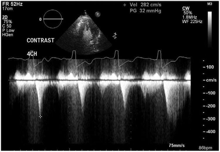
Electrocardiography revealed normal sinus rhythm, an rSR′ pattern suggestive of right ventricle conduction delay, voltage criteria for left ventricular (LV) hypertrophy with marked repolarization abnormalities, and ST-T wave abnormalities in the anterolateral leads. She had been followed with serial echocardiography over her posttransplant course, which revealed normal findings until 2008 when it had been noted that her LV wall thickness had increased. Over the course of the following 5 years, her wall thickness continued to increase in a predominantly apical and midventricular pattern, and her most recent echocardiogram demonstrated a hyperdynamic left ventricle with a midcavitary peak gradient of 32 mm Hg (Figure 1).
Figure 1.

Transthoracic echocardiogram. Four-chamber view with continuous-wave Doppler demonstrating a left ventricle cavitary peak gradient of 32 mm Hg.
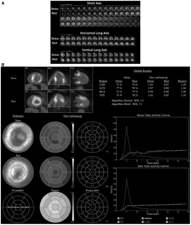
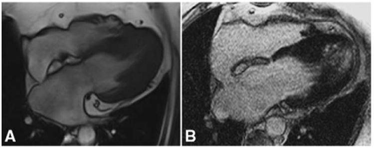
She was referred for a regadenoson N-13 ammonia rest-stress myocardial perfusion positron emission tomography scan, which demonstrated marked transient LV dilatation during stress imaging, and severe diffuse microvascular dysfunction, as well, as assessed by the severe diffuse reduction in her coronary flow reserve, and significant postischemic stunning, with a drop in LV ejection fraction from 51% to 35% after vasodilator stress (Figure 2). The patient was subsequently referred for cardiac magnetic resonance imaging. Contrast-enhanced cardiac magnetic resonance demonstrated significant apical and midventricular LV hypertrophy with near-complete obliteration of the cavity at end-systole (Movie I in the online-only Data Supplement). Her wall thickness measurements were between 17 mm and 22 mm concentrically in the midventricular and apical segments. First-pass perfusion imaging revealed a large area of circumferential subendocardial defect in all midventricular and apical segments (Movie II in the online-only Data Supplement). On late gadolinium enhancement imaging, patchy areas of diffuse fibrosis in all midventricular and apical segments were seen (Figure 3).
Figure 2.

Positron emission tomography. A, N-13 ammonia rest-stress images with transient ischemic dilatation, predominant midapical subendocardial ischemia. B, Visual and quantitative coronary flow reserve demonstrating severe microvascular dysfunction.
Figure 3.

Cardiac magnetic resonance. A, Cine imaging in the 4-chamber orientation demonstrates a spadelike ventricular cavity. B, Late gadolinium enhancement imaging in the 4-chamber view showing diffuse, patchy fibrosis in the apex and midventricular segments.
Potential causes of hypertrophy posttransplant include longstanding pressure-overload states, medications, metabolic disorders, and hypertrophic cardiomyopathy (HCM; idiopathic versus familial/sporadic sarcomeric mutations). In light of the patient's weight gain, hypertrophy consequent to metabolic syndrome was also considered as a potential mechanism. The patient's well-controlled blood pressure and lack of valvular disease ruled against a pressure overload state, as did the regionality of her hypertrophy. Medication-induced HCM was considered in light of case reports that describe immunosuppression-induced hypertrophy in individuals taking tacrolimus.1,2 However, this too was ruled out, because our patient's regimen did not include this macrolide agent. Cyclosporine was also considered, because it is known to induce some element of hypertrophy; however, the regionality and extent of ventricular thickness did not favor cyclosporine as a primary cause. Similarly, although cases of late-onset metabolic disorders causing apical LV hypertrophy have been described,3 our patient's age, absence of extracardiac findings, and unremarkable biopsies virtually exclude this cause. Importantly, pretransplant donor echocardiogram findings were normal with no evidence of LV hypertrophy, and there was no history of HCM or sudden cardiac death in the patient's or donor's family. Ultimately, we concluded that our patient developed an idiopathic phenotype of genetic HCM in her transplanted heart. Although the possibility of an underlying familial or sporadic sarcomeric HCM genotype remains, this finding is relatively uncommon in apical HCM, accounting for <15% of cases.4 Remarkably, our patient has done well in the 19 years since her transplant, highlighting the importance of vigilant screening to detect non–transplant-related pathologies and the potential for late-onset HCM despite adequate screening of donors and transplant recipients. Multimodality imaging was helpful in this case to define the relative contributions of microvascular ischemia, cavitary obliteration, and ischemic stunning as etiologies for our patient's symptoms, while defining the presence and extent of features consistent with a diagnosis of HCM.
Supplementary Material
Footnotes
Guest Editor for this article was Joao A.C. Lima, MD.
The online-only Data Supplement is available with this article at http://circ.ahajournals.org/lookup/suppl/doi:10.1161/CIRCULATIONAHA.114.010802/-/DC1.
Disclosures: None.
References
- 1.Liu T, Gao Y, Gao YL, Cheng YT, Wang S, Li ZZ, Zhang HB, Meng X, Ma CS, Dong JZ. Tacrolimus-related hypertrophic cardiomyopathy in an adult cardiac transplant patient. Chin Med J (Engl) 2012;125:1352–1354. [PubMed] [Google Scholar]
- 2.Reichenspurner H. Overview of tacrolimus-based immunosuppression after heart or lung transplantation. J Heart Lung Transplant. 2005;24:119–130. doi: 10.1016/j.healun.2004.02.022. [DOI] [PubMed] [Google Scholar]
- 3.Chimenti C, Pieroni M, Morgante E, Antuzzi D, Russo A, Russo MA, Maseri A, Frustaci A. Prevalence of Fabry disease in female patients with late-onset hypertrophic cardiomyopathy. Circulation. 2004;110:1047–1053. doi: 10.1161/01.CIR.0000139847.74101.03. [DOI] [PubMed] [Google Scholar]
- 4.Gruner C, Care M, Siminovitch K, Moravsky G, Wigle ED, Woo A, Rakowski H. Sarcomere protein gene mutations in patients with apical hypertrophic cardiomyopathy. Circ Cardiovasc Genet. 2011;4:288–295. doi: 10.1161/CIRCGENETICS.110.958835. [DOI] [PubMed] [Google Scholar]
Associated Data
This section collects any data citations, data availability statements, or supplementary materials included in this article.


