Abstract
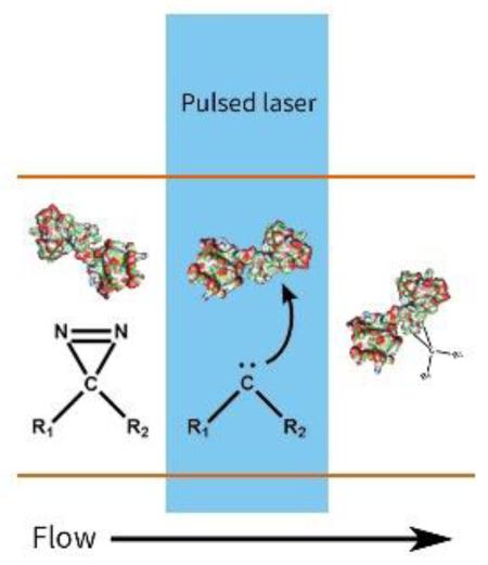
Protein footprinting combined with mass spectrometry provides a method to study protein structures and interactions. To improve further current protein footprinting methods, we adapted a fast photochemical oxidation of proteins (FPOP) platform to utilize carbenes as the footprinting reagent. A Nd-YAG laser provides 355 nm laser for carbene generation in situ from photoleucine as the carbene precursor in a flow system with calmodulin as the test protein. Reversed-phase liquid chromatography coupled with mass spectrometry is appropriate to analyze the modifications produced in this footprinting. By comparing the modification extent of apo and holo calmodulin on the peptide level, we can resolve different structural domains of the protein. Carbene footprinting in a flow system is a promising strategy to investigate protein structures.
Introduction
New sources of structural information are needed to understand protein structure and function in complex biological milieu. New opportunities based on mass spectrometry are emerging to learn about structure; probing solvent-accessible area [1], determining interfaces, and mapping epitopes are three of them. A common feature of these opportunities is that they can be studied by protein footprinting [2-4]. Furthermore, footprinting can follow fast folding in a dynamic process [5] and determine affinity as exemplified by PLIMSTEX [6] and SUPREX [7].
In modification-based footprinting, protein conformation is probed by reactions of reactive species in solution and amino-acid side chains to assess their solvent exposure. The reactive species can be deuterium that replaces amide hydrogen atoms on a protein backbone, as in hydrogen deuterium exchange [8]. Alternatives are reactive species that form irreversible covalent bonds to modify protein side chains. Some of these covalent-labeling reagents target specific amino acids [9, 10]. Their reactions with targets are often slow, raising concerns that the labelling perturbs conformation. On the other hand, many free radicals react rapidly, even on the microsecond time scale, and with more generality, making them broader based for probing solvent accessibility and determining binding interfaces.
The hydroxyl radical is now a widely-used footprinting reagent [11]. In its most reliable format, it is generated via synchrotron radiolysis [12] or FPOP [13, 14]. The lifetime of HO∙ in FPOP is in the micro-second range, sufficiently fast to capture even fast protein unfolding. Although hydroxyl radicals can react with almost all amino acid side chains, with rates from 1010 M−1s−1 to 107 M−1s−1 [11], Gln, Glu, Asp, Asn, Ala, and Gly are relatively “silent” in the FPOP platform under conditions where over oxidation of reactive residues is prevented.
Carbenes are an alternative to modify residues that are relatively unreactive with HO∙. A convenient means of generating carbene radicals is photolysis of diazirine precursors. In addition to complementing the reactivity of HO∙ and other radicals, carbenes in solution react on the nanosecond time scale as determined by their rapid quench with solvent H2O [15], even shorter than HO∙. Richards et al. [16] appears to be the first to study the properties of the simplest carbene, methylene, as a footprinting reagent for proteins in solution with mass spectrometry analysis. Delfino et al [17, 18] used 3H-methylene so that its concentration in solution can be monitored, and they demonstrated that methylene reactivity is a monitor of protein conformational transitions and protein interactions. In their later work, Delfino et al [19, 20] used mass spectrometry as the analytical method to achieve accurate detection of footprinting products and map solvent accessibility of proteins. Gas bubbling devices were used in their experiments to mix methylene gas with protein solutions, and reactions were initiated by a halogen/Hg lamp or a Hg/Xe arc source. Schriemer et al. [21, 22] used a more readily implemented carbene precursor, photoleucine, for protein footprinting with a pulsed 355 nm Nd-YAG laser to irradiate a sample droplet on glass. They demonstrated footprinting with a single laser shot but not in a flow system.
We report here the extension of the carbene footprinting strategy to a FPOP flow system. The principal advantage of the flow system is the ability to use many laser shots but with limited exposure per shot. After each laser shot, the induced footprinting reactions go to completion before the protein unfolds, because unfolding usually requires a timescale longer than microseconds. By confining the reactions within a tiny spot, we are able to use laser pulses with relatively low energy to achieve good modification yields. To test the effectiveness of carbene labelling, we used calmodulin as the model protein with photoleucine as the carbene precursor.
Methods
Bovine calmodulin was purchased from Ocean Biologics (Seattle, WA). Trypsin was obtained from Promega (Madison, WI). Photoleucine was from Thermo Fisher Scientific (Waltham, MA).
Apo and holo CaM samples were prepared by incubating 50 μM bovine calmodulin with 0.5 mM EGTA and 1 mM CaCl2, respectively. The protein solutions were diluted with tris buffer, and photoleucine was added to make a solution of 10 μM CaM (and 0.1 mM EGTA for apo, or 0.2 mM CaCl2 for holo conditions), 100 mM photoleucine, 10 mM tris, 100 mM KCl, pH = 7.5.
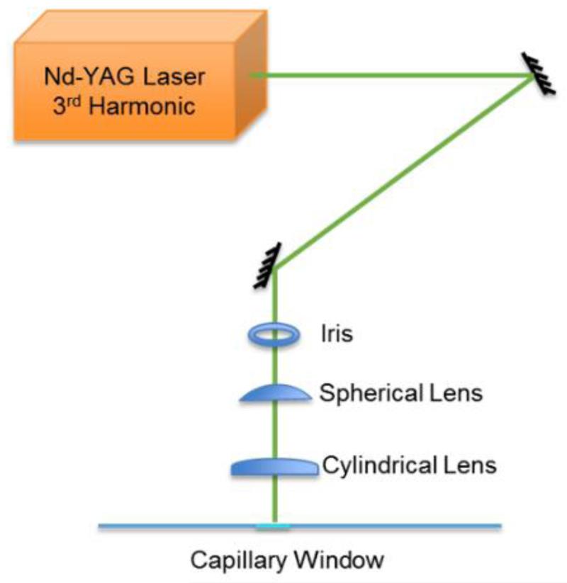
The platform is identical to that reported in a recent FPOP study. The sample was introduced to a 150-μm I.D. capillary by a syringe pump and passed a laser window where it was irradiated by a frequency tripled Nd-YAG laser (355 nm) (Quanta-Ray, Model DCR-2A, Mountain View, CA) (see Figure 1 for a diagram). A flow rate (~ 15-20 μL/min) was calculated to ensure a 25% exclusion volume, based on laser spot size (~ 2 mm) and laser frequency (~ 6 Hz). The average laser energy was 7.5 mJ/pulse. The 50 μL sample solution was subjected to 900 to 1200 laser shots in total.
Figure 1.
Schematic of a custom-built flow system for carbene footprinting on an FPOP platform. A pulsed laser beam at 355 nm (Nd-YAG laser) was focused on a transparent window in silica tubing of 150 μm inner diameter.
After labeling, an aliquot representing 0.3 μg of protein was analyzed by using a Bruker Maxis Q-TOF mass spectrometer under denaturing conditions to provide protein-level information. Another aliquot of 0.8 μg protein was incubated at 65 °C for 90 min with trypsin to digest the protein. The resulting peptides were separated by reversed-phase HPLC by an Eksigent NanoLC Ultra (Dublin, CA) and introduced to a Thermo LTQ-FT mass spectrometer (Waltham, MA) via nano-ESI. Peptide ions were fragmented by CID, and the m/z values of product ions were recorded. Unmodified and modified peptides were identified by Mascot (Matrix Science, Boston, MA) and Byonic (Protein Metrics, San Carlos, CA).
Results and discussions
An advantage of the FPOP platform is that it is versatile and can utilize other reactive species (SO4−•[23] and I∙ [24]) in addition to HO∙ for protein footprinting, providing opportunities to achieve new specificity and coverage. To test this prospect, we used our standard FPOP platform and submitted apo and holo CaM to laser photolysis of photoleucine to generate a carbene reagent. The only modifications of the platform are incorporation of a second laser (here a YAG) and a small change in the optical path to allow switching between the YAG and the standard KrF laser.
Significant photoleucine modifications (+115 Da) on apo and holo CaM occur upon laser irradiation in our flow system. Control experiments confirm that the modifications are not found without laser or without photoleucine present (data not shown). Overall, apo CaM shows higher modification extent than holo CaM (Fig. 2). Crystal structures [25] and solution NMR [26] studies show that upon calcium binding, the binding regions of CaM that bind Ca2+ become more protected, whereas the central linker becomes more solvent-accessible. In our experiment, the exposure of the linker region has less impact on the carbene modification extent at the global level than does the protection of the calcium binding regions.
Figure 2.
(a) Structure of photoleucine. (b) Mass spectrum of apo CaM following modifications with carbenes from photoleucine. (c) Deconvolution of spectrum b shows the mass shift between two adjacent peaks is consistent with modification by photoleucine. (d) Comparison of global modification extent on apo and holo CaM after photoleucine footprinting.
A small difficulty occurs when the 355 nm YAG beam irradiates the flowing solution (Figure 1). We observed bubbles in the silica tubing at the laser spot. The N2 production is largely unavoidable during carbene generation from a diazirine. Whereas reflection and diffraction by those bubbles may cause some concerns about the reproducibility of laser irradiation, the bubbles are always carried downstream with the solution flow (15-25 μL/min). By using a 25% exclusion volume, we insured that the solution in the laser window is always replenished and bubble-free for a new laser shot.
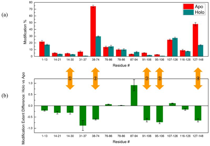
We calculated the modification extent of the tryptic peptides from mass spectral peak areas in extracted ion chromatograms of the unmodified vs. modified protein. We carefully examined each individual peak by comparing accurate masses and isotopic patterns with theoretical values and only used data that could be validated in this manner. The modification extent is calculated as [modified / (unmodified + modified] (Fig. 3a). To illustrate the differences between modification extent of apo and holo CaM, we calculated the [(holo-apo)/apo] ratio as shown in (Fig. 3b). All four peptides that contain Ca2+ binding sites show decreased modification upon Ca2+ binding. Peptides that correspond to the central linker region form F65 to V91 show higher modification extent for holo CaM than for apo CaM. This is expected from structural studies on CaM by crystallography [25], NMR [26] and FPOP [27]. It adds confidence that carbene footprinting can provide reliable information on protein conformational change.
Figure 3.
(a) Comparison of modification extent of peptides from apo and holo CaM. Calcium binding sites are marked by yellow arrows. On the crystal structure of holo Cam (PDB: 1CLL), two calcium binding domains are connected by an ɑ-helix from F65 to V91. (b) A comparison between apo and holo states calculated from ion intensities [(holo - apo)/apo].
MS/MS experiments indicate the most favorable amino acid residues for photoleucine are Asp and Glu. We also found modifications of Arg, Tyr, Ser and Thr with a false discovery rate < 10−4 (see supplemental information). We are reluctant to quantify modification extents on the residue level because we are not yet certain that the extracted ion chromatograms are free of co-eluting species that have different modification sites on the same peptide. Nevertheless, the identified reaction sites are consistent with the chemical property of carbenes that they react more favorably with active hydrogens than with C-H bonds. This phenomenon can also be explained by a favored distribution of photoleucine around charged residues and limited diffusion during the short lifetime of carbenes [22]. This special property of photoleucine and related carbene sources makes them good candidates to map metal-binding regions. Furthermore, the results suggest that some carbenes are specific footprinting reagents, unlike what may be anticipated given their high reactivity in organic synthesis.
Compared to methylene, photoleucine has the advantages of good solubility in aqueous solution and feasibility for quantification. Photoleucine is commercially available, whereas methylene is usually synthesized just before using owing to inconvenience of storage and transport. The polar groups on photoleucine may introduce bias, however, towards charged residues, especially carboxylate groups. Furthermore, the amine group on photoleucine introduces an extra charge on modified peptide, requiring more effort for peptide identification and data analysis. Unlike for OH radicals, the reaction rates between carbene radicals and amino acid side chains are not well-known. The structures of modified side chains also need to be investigated.
Conclusions
Carbenes, which have been traditionally used for organic synthesis and have high reactivity towards various X–H bonds and double bonds, are becoming of interest in protein footprinting. When used on the FPOP flow platform, additional method development can readily occur. Even though carbenes are highly reactive and might produce excessively complex footprints, the footprints are tractable. Moreover, carbenes can be conveniently generated by photolysis of diazirines, where the reactive site can be located in a controlled way and reactivity can be tailored, as demonstrated by Schriemer and coworkers [21, 22]. Using diazirines of various structures, we can increase the versatility of carbene footprinting to accomplish different goals (e.g., footprint membrane regions using membrane-seeking precursors). This approach is complementary to hydroxyl and other radical footprinting, especially for the protein regions that are FPOP “silent” and should conveniently enhance the scope of MS-based footprinting with an FPOP platform.
Supplementary Material
Acknowledgment
This work was supported by a grant (P41 GM103422) from the NIH NIGMS.
References
- 1.Lee B, Richards FM. The interpretation of protein structures: Estimation of static accessibility. J. Mol. Biol. 1971;55:379–400. doi: 10.1016/0022-2836(71)90324-x. [DOI] [PubMed] [Google Scholar]
- 2.Maleknia SD, Downard KM. Advances in radical probe mass spectrometry for protein footprinting in chemical biology applications. Chem. Soc. Rev. 2014;43:3244–3258. doi: 10.1039/c3cs60432b. [DOI] [PubMed] [Google Scholar]
- 3.Chen J, Rempel DL, Gau BC, Gross ML. Fast Photochemical Oxidation of Proteins and Mass Spectrometry Follow Submillisecond Protein Folding at the Amino-Acid Level. J. Am. Chem. Soc. 2012;134:18724–18731. doi: 10.1021/ja307606f. [DOI] [PMC free article] [PubMed] [Google Scholar]
- 4.Jones LM, B. Sperry J, A. Carroll J, Gross ML. Fast Photochemical Oxidation of Proteins for Epitope Mapping. Anal. Chem. 2011;83:7657–7661. doi: 10.1021/ac2007366. [DOI] [PMC free article] [PubMed] [Google Scholar]
- 5.Chen J, Rempel DL, Gross ML. Temperature Jump and Fast Photochemical Oxidation Probe Submillisecond Protein Folding. J. Am. Chem. Soc. 2010;132:15502–15504. doi: 10.1021/ja106518d. [DOI] [PMC free article] [PubMed] [Google Scholar]
- 6.Zhu MM, Chitta R, Gross ML. PLIMSTEX: a novel mass spectrometric method for the quantification of protein–ligand interactions in solution. Int. J. Mass Spectrom. 2005;240:213–220. [Google Scholar]
- 7.Ghaemmaghami S, Fitzgerald MC, Oas TG. A quantitative, high-throughput screen for protein stability. Proc. Natl. Acad. Sci. 2000;97:8296–8301. doi: 10.1073/pnas.140111397. [DOI] [PMC free article] [PubMed] [Google Scholar]
- 8.Englander SW, Kallenbach NR. Hydrogen exchange and structural dynamics of proteins and nucleic acids. Q. Rev. Biophys. 1983;16:521–655. doi: 10.1017/s0033583500005217. [DOI] [PubMed] [Google Scholar]
- 9.Mendoza VL, Vachet RW. Probing protein structure by amino acid-specific covalent labeling and mass spectrometry. Mass Spectrom. Rev. 2009;28:785–815. doi: 10.1002/mas.20203. [DOI] [PMC free article] [PubMed] [Google Scholar]
- 10.Zhang H, Wen J, Huang RY-C, Blankenship RE, Gross ML. Mass spectrometry-based carboxyl footprinting of proteins: Method evaluation. Int. J. Mass Spectrom. 2012;312:78–86. doi: 10.1016/j.ijms.2011.07.015. [DOI] [PMC free article] [PubMed] [Google Scholar]
- 11.Xu G, Chance MR. Hydroxyl Radical-Mediated Modification of Proteins as Probes for Structural Proteomics. Chem. Rev. 2007;107:3514–3543. doi: 10.1021/cr0682047. [DOI] [PubMed] [Google Scholar]
- 12.Kiselar JG, Maleknia SD, Sullivan M, Downard KM, Chance MR. Hydroxyl radical probe of protein surfaces using synchrotron X-ray radiolysis and mass spectrometry. Int. J. Radiat. Biol. 2002;78:101–114. doi: 10.1080/09553000110094805. [DOI] [PubMed] [Google Scholar]
- 13.Hambly DM, Gross ML. Laser Flash Photolysis of Hydrogen Peroxide to Oxidize Protein Solvent-Accessible Residues on the Microsecond Timescale. J. Am. Soc. Mass Spectrom. 2005;16:2057–2063. doi: 10.1016/j.jasms.2005.09.008. [DOI] [PubMed] [Google Scholar]
- 14.Gau BC, Sharp JS, Rempel DL, Gross ML. Fast Photochemical Oxidation of Protein Footprints Faster than Protein Unfolding. Anal. Chem. 2009;81:6563–6571. doi: 10.1021/ac901054w. [DOI] [PMC free article] [PubMed] [Google Scholar]
- 15.Matthew P, Modarelli D, Morgan S, White W, Mullins M, Celebi S, Toscano J. Lifetimes of alkyl and dialkyl carbenes in solution. Prog. React. Kinet. 1994;19:93–137. [Google Scholar]
- 16.Richards FM, Lamed R, Wynn R, Patel D, Olack G. Methylene as a possible universal footprinting reagent that will include hydrophobic surface areas: Overview and feasibility: Properties of diazirine as a precursor. Protein Sci. 2000;9:2506–2517. doi: 10.1110/ps.9.12.2506. [DOI] [PMC free article] [PubMed] [Google Scholar]
- 17.Craig PO, Ureta DB, Delfino JM. Probing protein conformation with a minimal photochemical reagent. Protein Sci. 2002;11:1353–1366. doi: 10.1110/ps.4710102. [DOI] [PMC free article] [PubMed] [Google Scholar]
- 18.Gómez GE, Cauerhff A, Craig PO, Goldbaum FA, Delfino JM. Exploring protein interfaces with a general photochemical reagent. Protein Sci. 2006;15:744–752. doi: 10.1110/ps.051960406. [DOI] [PMC free article] [PubMed] [Google Scholar]
- 19.Gómez GE, Mundo MR, Craig PO, Delfino JM. Probing Protein Surface with a Solvent Mimetic Carbene Coupled to Detection by Mass Spectrometry. J. Am. Soc. Mass Spectrom. 2012;23:30–42. doi: 10.1007/s13361-011-0266-x. [DOI] [PubMed] [Google Scholar]
- 20.Gómez GE, Monti JLE, Mundo MR, Delfino JM. Solvent Mimicry with Methylene Carbene to Probe Protein Topography. Anal. Chem. 2015 doi: 10.1021/acs.analchem.5b02724. [DOI] [PubMed] [Google Scholar]
- 21.Jumper CC, Schriemer DC. Mass Spectrometry of Laser-Initiated Carbene Reactions for Protein Topographic Analysis. Anal. Chem. 2011;83:2913–2920. doi: 10.1021/ac102655f. [DOI] [PubMed] [Google Scholar]
- 22.Jumper CC, Bomgarden R, Rogers J, Etienne C, Schriemer DC. High-Resolution Mapping of Carbene-Based Protein Footprints. Anal. Chem. 2012;84:4411–4418. doi: 10.1021/ac300120z. [DOI] [PubMed] [Google Scholar]
- 23.Gau BC, Chen H, Zhang Y, Gross ML. Sulfate Radical Anion as a New Reagent for Fast Photochemical Oxidation of Proteins. Anal. Chem. 2010;82:7821–7827. doi: 10.1021/ac101760y. [DOI] [PMC free article] [PubMed] [Google Scholar]
- 24.Chen J, Cui W, Giblin D, Gross ML. New Protein Footprinting: Fast Photochemical Iodination Combined with Top-Down and Bottom-Up Mass Spectrometry. J. Am. Soc. Mass Spectrom. 2012;23:1306–1318. doi: 10.1007/s13361-012-0403-1. [DOI] [PMC free article] [PubMed] [Google Scholar]
- 25.Chattopadhyaya R, Meador WE, Means AR, Quiocho FA. Calmodulin structure refined at 1.7 A resolution. J. Mol. Biol. 1992;228:1177–1192. doi: 10.1016/0022-2836(92)90324-d. [DOI] [PubMed] [Google Scholar]
- 26.Kuboniwa H, Tjandra N, Grzesiek S, Ren H, Klee CB, Bax A. Solution structure of calcium-free calmodulin. Nat.Struct.Biol. 1995;2:768–776. doi: 10.1038/nsb0995-768. [DOI] [PubMed] [Google Scholar]
- 27.Zhang H, Gau BC, Jones LM, Vidavsky I, Gross ML. Fast Photochemical Oxidation of Proteins for Comparing Structures of Protein–Ligand Complexes: The Calmodulin–Peptide Model System. Anal. Chem. 2011;83:311–318. doi: 10.1021/ac102426d. [DOI] [PMC free article] [PubMed] [Google Scholar]
Associated Data
This section collects any data citations, data availability statements, or supplementary materials included in this article.


