

Abstract
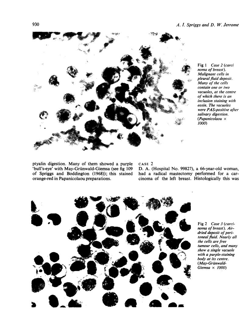
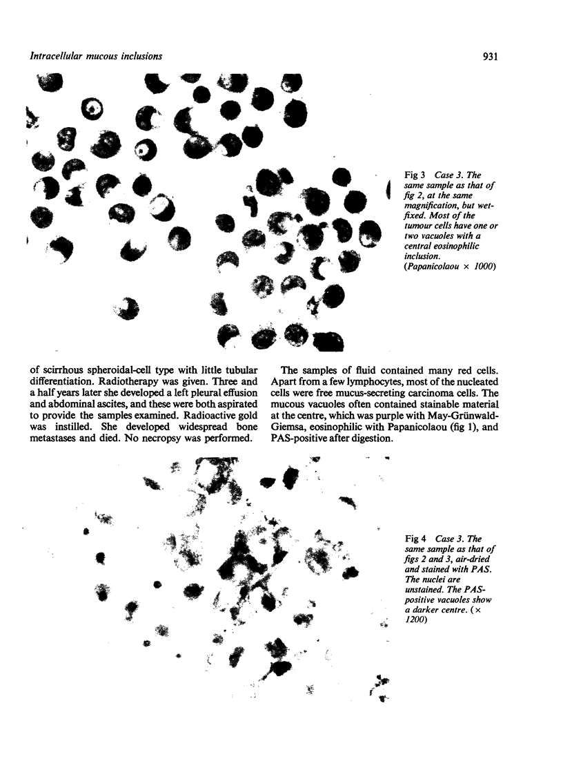
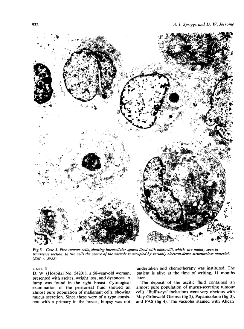
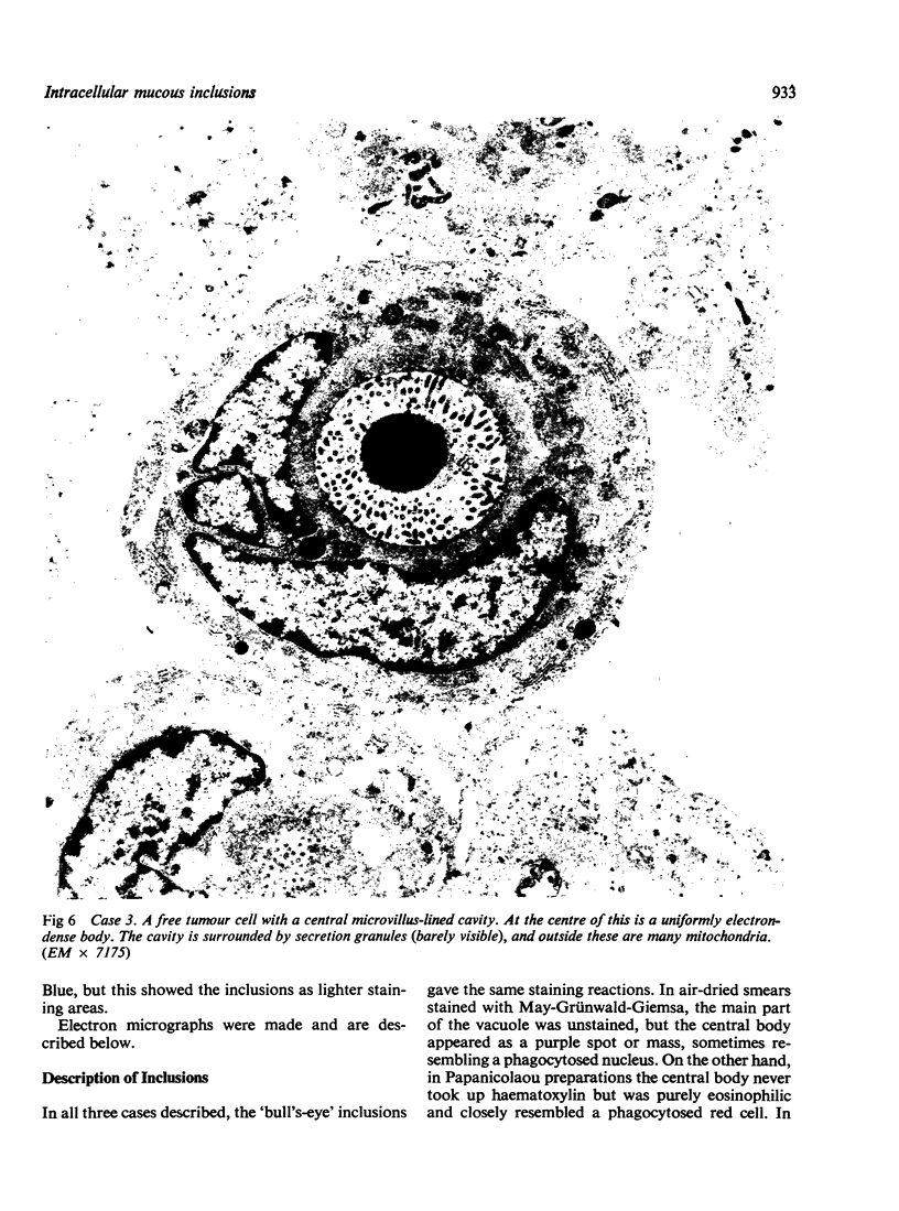
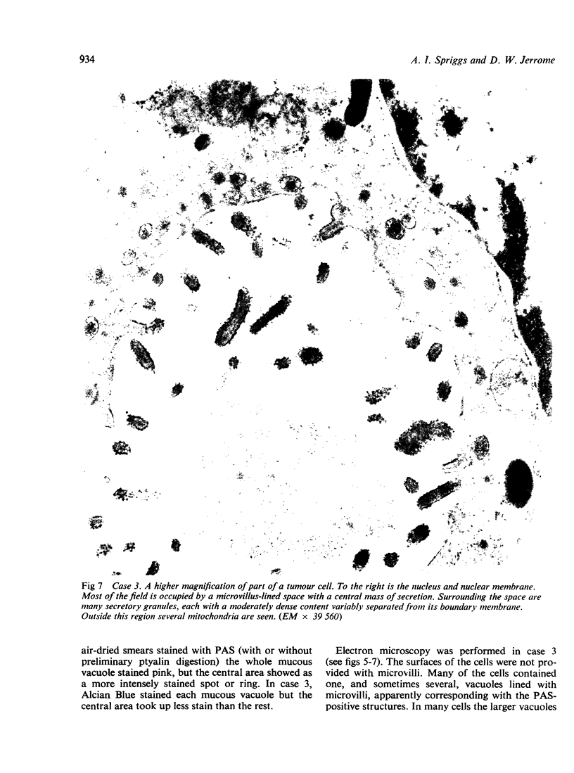
Three cases of carcinoma of the breast are described, in which similar tumour cells were found in pleural or peritoneal fluid. The cells were characterized by the frequent presence of 'bull's-eye' vacuoles, in which a central spot was deeply stained by Giemsa, eosin or periodic acid-Schiff (PAS). In one of these it was shown by electron microscopy that the vacuoles are lined by microvilli, and that their mucinous content is condensed in the central area, presumably as a result of failure to discharge the secretion.
Full text
PDF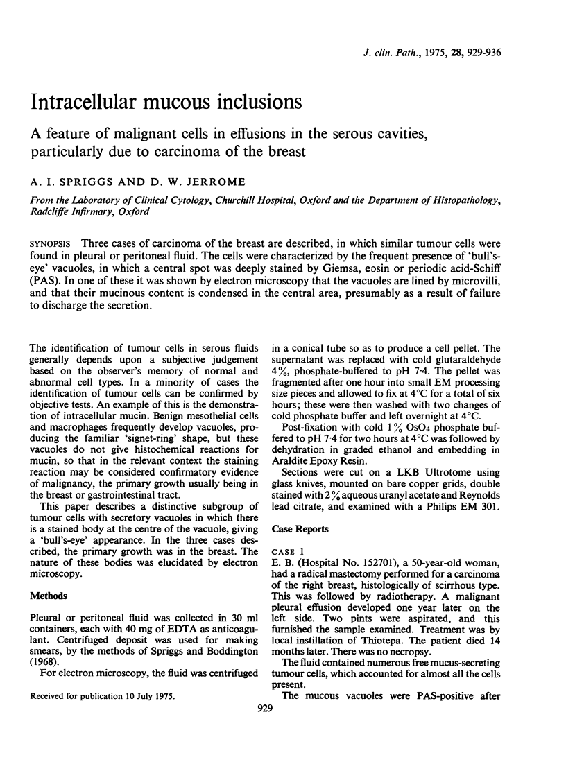
Images in this article
Selected References
These references are in PubMed. This may not be the complete list of references from this article.
- BARTON A. A. AN ELECTRON MICROSCOPE STUDY OF HUMAN BREAST CELLS IN FIBROADENOSIS AND CARCINOMA. Br J Cancer. 1964 Dec;18:682–685. doi: 10.1038/bjc.1964.78. [DOI] [PMC free article] [PubMed] [Google Scholar]
- Busch W., Merker H. J. Elektronenmikroskopische Untersuchungen an menschlichen Mammacarcinomen. Virchows Arch Pathol Anat Physiol Klin Med. 1967;344(4):356–371. [PubMed] [Google Scholar]
- Goldenberg V. E., Goldenberg N. S., Sommers S. C. Comparative ultrastructure of atypical ductal hyperplasia, intraductal carcinoma, and infiltrating ductal carcinoma of the breast. Cancer. 1969 Dec;24(6):1152–1169. doi: 10.1002/1097-0142(196912)24:6<1152::aid-cncr2820240614>3.0.co;2-5. [DOI] [PubMed] [Google Scholar]
- HAGUENAU F. Le cancer mammaire de la souris et de la femme; étude comparative au microscope électronique. Pathol Biol. 1959 May;7(9-10):989–1015. [PubMed] [Google Scholar]
- Murad T. M., Scharpelli D. G. The ultrastructure of medullary and scirrhous mammary duct carcinoma. Am J Pathol. 1967 Feb;50(2):335–360. [PMC free article] [PubMed] [Google Scholar]
- Ozzello L. Ultrastructure of the human mammary gland. Pathol Annu. 1971;6:1–59. [PubMed] [Google Scholar]
- Sagebiel R. W. Ultrastructural observations on epidermal cells in Paget's disease of the breast. Am J Pathol. 1969 Oct;57(1):49–64. [PMC free article] [PubMed] [Google Scholar]
- Sykes J. A., Recher L., Jernstrom P. H., Whitescarver J. Morphological investigation of human breast cancer. J Natl Cancer Inst. 1968 Feb;40(2):195–223. [PubMed] [Google Scholar]
- WELLINGS S. R., ROBERTS P. Electron microscopy of sclerosing adenosis and infiltrating duct carcinoma of the human mammary gland. J Natl Cancer Inst. 1963 Feb;30:269–287. [PubMed] [Google Scholar]