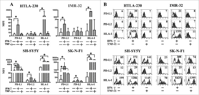
Figure 2.
PD-L1 and PD-L2 expression in INFγ− or TNF-α−treated NB cell lines. Panel A: cytofluorimetric analysis of the expression of PD-L1, PD-L2 and HLA-I in representative MYCNampl(HTLA-230, IMR-32) and non-MYCNampl (SH-SY5Y and SK-N-F1) cell lines cultured for 48 h either in the absence (white bars) or in the presence of IFNγ (gray bars) or TNF-α (striped bars). Mean of MFI and 95% confidence intervals are indicated. *p < 0 .05. Panel B: Representative cytofluorimetric analysis of PD-L1, PD-L2 and HLA-I expression in untreated or cytokine-treated NB cell lines. White profiles refer to cells incubated with isotype-matched controls. Values inside each histogram indicate the MFI.

