Fig. 5.
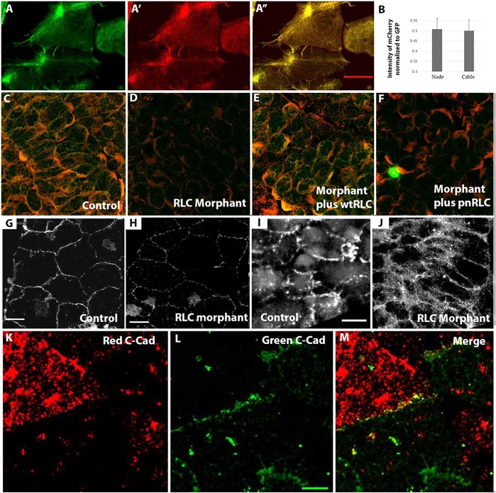
Colocalization and interdepencies of transcellular array components. (A,B) Imaging of F-actin with moe-GFP (A) and RLC with wtRLC-mCherry (A′) shows colocalization (A″) in both node-and-cable structures with the same relative concentrations in both (B; n=8 regions of interest). (C) Both pRLC and MHC localize to cell cortices in stage 13 explants using both MHC-IIB (red) and mono-phosphorylated RLC (green) (S19-P) antibodies. Levels of both are reduced in RLC morphants (D), and rescued by expression of wtRLC (E), whereas pnRLC expression rescues heavy chain IIB but not pRLC levels (F). At both stage 10 (G,H) and stage 12 (I,J), C-cadherin localization by immunostaining is perturbed in RLC morphant cells (H). Expression of red (K)- and green (L)-labeled C-cadherin in neighboring cells reveals that they colocalize at the membrane (yellow in M), as expected if they have a role in adhesion. Scale bars: 20 µm in A*,G-I and 10 µm in L.

