
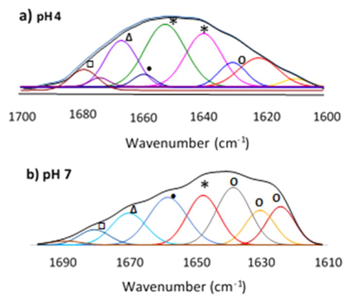
Figure 6.
ATR-FTIR spectra of 1 mg/mL Mfp3F in deuterated buffer at (a) pH 4 and (b) pH 7: (○) β-sheet, (*) random coil, (●) α-helix, (Δ) 310 helix, and (□) turns. Secondary derivatives are shown in the Supporting Information (S9).
Official websites use .gov
A
.gov website belongs to an official
government organization in the United States.
Secure .gov websites use HTTPS
A lock (
) or https:// means you've safely
connected to the .gov website. Share sensitive
information only on official, secure websites.
ATR-FTIR spectra of 1 mg/mL Mfp3F in deuterated buffer at (a) pH 4 and (b) pH 7: (○) β-sheet, (*) random coil, (●) α-helix, (Δ) 310 helix, and (□) turns. Secondary derivatives are shown in the Supporting Information (S9).