Fig. 2.
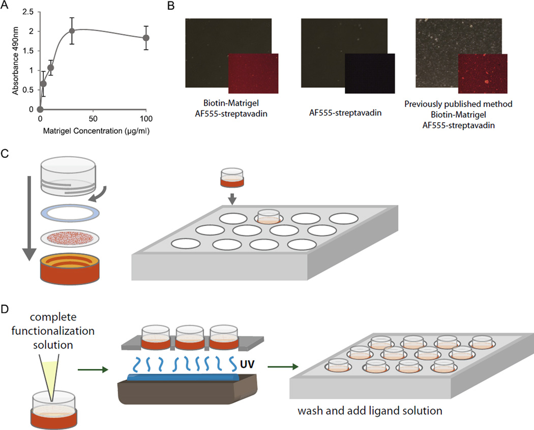
Functionalization of traction force gels compatible with multi-day culture of hESCs. (A) Relative PA gel surface bound biotin labeled Matrigel proteins as a function of total solution phase Matrigel concentration for the modified chemistry described herein. Biotin-labeled proteins were indirectly detected by streptavidin-horseradish peroxidase complex in an ELISA format using O-phenylenediamine-H2O2 as substrate with endpoint product measured by absorption at 490 nm. (B) Phase images of gel surfaces (large panels) and corresponding distribution of PA gel surface-bound biotin labeled Matrigel proteins detected with Alexa555 labeled streptavidin (insets). Leftmost two panels show specific (left panel; with Matrigel) and non-specific (middle panel; without Matrigel) streptavidin binding for the modified chemistry described herein compared to specific binding for the earlier chemistry described in [27] (right panel). Note the discrete co-polymeric deposits of N6 and bisacrylamide in this particular example of the latter compared to the more consistent uniform distribution obtained with the modified chemistry. (C) Traction force gels are re-inserted into cassettes and put into multi-well plates for functionalization. (D) Functionalization solution containing N6, Irgacure, and tetramethacrylate is added to gels before undergoing UV treatment and subsequent washes prior to plating cells.

