

Abstract
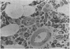
Preparation of human marrow sections has been studied systematically in order to facilitate accurate identification of marrow cells. Both of the methods developed involve embedding marrow cores in methyl methacrylate. In one, acrolein fixation is followed by staining of deplasticized sections with eosine-y followed by azure II; in the other, neutrophilic forms are identified by their esterase-specific reactivity in marrow fixed with neutral-buffered formalin. These preparations are suitable for quantitative studies of marrow cellularity.
Full text
PDF


Images in this article

Selected References
These references are in PubMed. This may not be the complete list of references from this article.
- BLOCK M., SMALLER V., BROWN J. An adaptation of the Maximow technique for preparation of sections of hematopoietic tissues. J Lab Clin Med. 1953 Jul;42(1):145–151. [PubMed] [Google Scholar]
- Burkhardt R. Präparative Voraussetzungen zur klinischen Histologie des menschlichen Knochenmarks. 1. Methodische Untersuchungen zur Acrylateinbettung grösserer lipidreicher Gewebsproben. Blut. 1966 Sep;13(6):337–357. doi: 10.1007/BF01633515. [DOI] [PubMed] [Google Scholar]
- CATHEY W. J. A PLASTIC EMBEDDING MEDIUM FOR THIN SECTIONING IN LIGHT MICROSCOPY. Stain Technol. 1963 Jul;38:213–216. doi: 10.3109/10520296309061180. [DOI] [PubMed] [Google Scholar]
- ELLIS L. D., JENSEN W. N., WESTERMAN M. P. NEEDLE BIOPSY OF BONE AND MARROW; AN EXPERIENCE WITH 1,445 BIOPSIES. Arch Intern Med. 1964 Aug;114:213–221. doi: 10.1001/archinte.1964.03860080063005. [DOI] [PubMed] [Google Scholar]
- Grann V., Pool J. L., Mayer K. Comparative study of bone marrow aspiration and biopsy in patients with neoplastic disease. Cancer. 1966 Dec;19(12):1898–1900. doi: 10.1002/1097-0142(196612)19:12<1898::aid-cncr2820191217>3.0.co;2-u. [DOI] [PubMed] [Google Scholar]
- Jamshidi K., Swaim W. R. Bone marrow biopsy with unaltered architecture: a new biopsy device. J Lab Clin Med. 1971 Feb;77(2):335–342. [PubMed] [Google Scholar]
- Jones S. E., Rosenberg S. A., Kaplan H. S. Non-Hodgkin's lymphomas. I. Bone marrow involvement. Cancer. 1972 Apr;29(4):954–960. doi: 10.1002/1097-0142(197204)29:4<954::aid-cncr2820290442>3.0.co;2-5. [DOI] [PubMed] [Google Scholar]
- Kadin M. E., Glatstein E., Dorfman R. F. Clinicopathologic studies of 117 untreated patients subjected to laparotomy for the staging of Hodgkin's disease. Cancer. 1971 Jun;27(6):1277–1294. doi: 10.1002/1097-0142(197106)27:6<1277::aid-cncr2820270602>3.0.co;2-t. [DOI] [PubMed] [Google Scholar]
- Mori M., Ito M., Fukui S. Decalcification for histochemical demonstration of hydrolytic and oxidative enzymes. Histochemie. 1965 Oct 1;5(3):185–195. doi: 10.1007/BF00306127. [DOI] [PubMed] [Google Scholar]
- Ruddell C. L. Embedding media for 1-2 micron sectioning. 2. Hydroxyethyl methacrylate combined with 2-butoxyethanol. Stain Technol. 1967 Sep;42(5):253–255. doi: 10.3109/10520296709115020. [DOI] [PubMed] [Google Scholar]
- Ruddell C. L. The demonstration of anions generated by the action of sodium hypochlorite (NaOCl) on tissue sections, including observations on the unmasking of carbonyl groups. Histochemie. 1969;19(4):319–339. doi: 10.1007/BF00279682. [DOI] [PubMed] [Google Scholar]
- Zambernard J., Block M., Vatter A., Trenner L. An adaptation of methacrylate embedding for routine histopathologic use. Blood. 1969 Mar;33(3):444–450. [PubMed] [Google Scholar]