Abstract
Unique electrodynamic response of graphene implies a manifestation of an unusual propagating and localised transverse-electric (TE) mode near the spectral onset of interband transitions. However, excitation and further detection of the TE mode supported by graphene is considered to be a challenge for it is extremely sensitive to excitation environment and phase matching condition adherence. Here for the first time, we experimentally prove an existence of the TE mode by its direct optical probing, demonstrating significant coupling to an incident wave in electrically doped multilayer graphene sheet at room temperature. We believe that proposed technique of careful phase matching and obtained access to graphene’s TE excitation would stimulate further studies of this unique phenomenon, and enable its potential employing in various fields of photonics as well as for characterization of graphene.
Graphene is an atomically thin crystal of carbon atoms with hexagonal lattice1 which exhibits extraordinary electrodynamic response, and has earned a close attention of optics and photonics communities2,3,4. One of the remarkable properties of graphene is its spectral transition5 between “metallic” (Drude conductivity) and “dielectric” (broadband and saturable absorbance6 of ~2.3%) electrodynamic response at energies near ħω ~ 2EF, where charge carriers’ Fermi energy EF in graphene can be efficiently controlled by doping5,7,8,9.
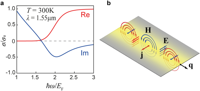
It was predicted10 that in the spectral transition region, the imaginary part of graphene conductivity (Im[σ] = σ″) changes sign from positive to negative, having its minimum at ħω = 2EF (Fig. 1a). The negative sign of σ″ has been associated with manifestation of transverse electric10,11 (TE) propagating electromagnetic mode in both single- and bi-layer12 graphene, contrary to conventional transverse magnetic (TM), i.e. plasmonic, modes in metals and graphene existing exclusively when σ″ >0. Yet, the main difficulty in detecting the TE mode is that its effective index is very close (Δn ~ 0.001) to that of propagating wave in a bulk material surrounding the graphene, which imposes a necessity of extremely precise phase matching with an excitation wave.
Figure 1. Spectral transition of graphene and the TE mode.
(a) Graphene sheet conductivity (red – real, blue – imaginary part) in the spectral transition region, obtained using the RPA at indicated parameters. (b) Schematic representation of graphene TE mode with wavevector q as an oscillation of surface current j under excitation by transverse electric field E.
In this article, for the first time eight years after its prediction, we demonstrate direct optical probing of the TE mode supported by graphene, employing modified Otto configuration that allows very precise phase matching of the incident wave to the TE excitation, and electrostatically doped multi-layer graphene stacks at room temperature, performed at the telecommunication wavelength λ0 = 1.55 μm.
The physical nature of TE excitations in graphene can be understood as magnetic dipole waves, i.e. self-sustained oscillations of current (Fig. 1b) with no spatial charge density perturbation, while conventional TM plasmons are an electric dipole waves. In analogy to a fundamental guided mode of a high-index dielectric slab waveguide13 when the slab thickness approaches zero, graphene TE mode is weakly bound but exhibits very low propagation loss10, and is extremely sensitive to the optical contrast between dielectrics sandwiching the graphene layer11 which may form the basis of extremely precise sensing of refractive index change in adjacent media11 with expected sensitivity exceeding 6.7 × 10⁻⁷ RIU. It was previously suggested that small effective index of the TE mode (close to that of the surrounding bulk10), neff = Re[q]/k0 where q is the modal wavenumber and k0 is that in free-space, makes it easily accessible to Otto excitation via evanescent field in attenuated total reflection (ATR) regime14,15,16 even at room temperature10, when doped graphene is sandwiched between two dielectric layers of same refractive index14.
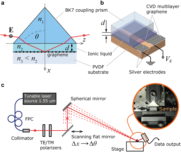
However, necessity of doping (i.e. special techniques of thereof) makes it difficult to provide symmetry between indices of materials above (n2) and below (n3) the graphene layer. Here we propose a TE mode excitation scheme where graphene is situated between dielectrics with asymmetric indices, n2 > n3, and placed at distance d from the coupling prism, n1 (Fig. 2a). In such system, in contrary to previously proposed ATR, there available a regime when incident wave experiences total internal reflection (TIR) at the n2|n3 interface where graphene layer is situated. By employing materials with low optical contrast between n2 and n3, TIR regime with close to 90° angle of incidence for the excitation wave is possible to achieve, which is strongly favourable for adherence of phase matching and efficient TE mode excitation.
Figure 2. Experimental setup.
(a) Modified Otto configuration with direct excitation of TE mode in multilayer graphene. (b) Multilayer graphene sample as inside the experimental setup, where graphene electrodes are electrostatically doped by the gate voltage Vg via the ionic liquid. (c) Experimental setup employing linear scan transition into the angular scan by a spherical mirror focused at the area where doped graphene is situated. Excitation wave’s polarization is controlled by a fibre polarization controller (FPC), and collimated beam diameter is 1 mm, providing an effective angular resolution of the system 0.05 deg.
Results
At first, we consider TE mode behaviour near the cutoff distance dcut, first described in our previous work14, and later confirmed by other research group16. The cutoff distance d = dcut denotes the condition of complete TE mode delocalisation in Otto configuration, i.e. when for n2 = n3 and d < dcut, the existence of localised at graphene TE mode is prohibited. When d > dcut, the solution of TE mode exists, but starts to be delocalised in the direction of n3 as d approaches to dcut. However, increasing of n2 > n3 makes delocalisation to occur in the opposite direction14. Through the balancing of this index asymmetry by a proper selection of distance d, a condition of propagating TE mode existence can be preserved.
In a stack of randomly oriented graphene layers (i.e., in a multilayer graphene with rotational faults17), an effective optical conductivity of the stack linearly increases proportional to the number of layers17,18,19,20 N (σeff = Nσ). Considering this, we employ a five-layer graphene stack to derive significantly more effective coupling to incident wave14. Graphene electrodes are deposited on a polyvinylidene fluoride resin (PVDF; n3(1.55 μm) ≈ 1.4045) with applied silver electrodes (Fig. 2b). Graphene is electrostatically doped by an applied bias voltage via top-gated configuration21 (essential for high doping values) with an ionic liquid of proper refractive index22 (n2(1.55 μm) ≈ 1.4070), such that desired index asymmetry condition n2 > n3 is satisfied. Employing of an ionic liquid instead of an ion gel23 allows scanning over the distance d between graphene and the coupling prism (BK7; n1(1.55 μm) ≈ 1.5006), approaching desired values of d ~ λ0, in order to obtain precise phase matching. To measure an angular reflectance, we employ a setup schematically shown in Fig. 2c, where linear shift of a scanning flat mirror is translated into a high-resolution angular scan by concave spherical mirror of large radius.
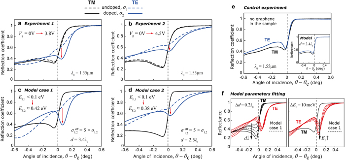
Experimentally measured angular reflectance for two different samples is shown in Fig. 3a,b, and is obtained for undoped (dashed lines) and doped (solid lines) 5-layer graphene stack under the bias voltage Vg = 3.8 V and 4.5 V respectively, for TM (black) and TE (blue) polarized incident wave at λ0 = 1.55 μm, and room temperature T = 300 K. While TM curves show no response to doping, TE reflectance of doped graphene exhibits a distinct dip in the vicinity of the critical angle θc and can be observed for both samples, indicating an excitation of a guided TE mode in the structure. It is important to note that in the absence of the graphene, reflectance profile indicates lack of any specific TE mode in the structure under applied experimental conditions, which was confirmed by the control experiment (Fig. 3e).
Figure 3. Angular reflectance signature of the TE mode.
(a) Experimentally measured angular reflectance of sample 1 with 5-layer graphene for TE (blue) and TM (black) incident waves, for undoped (dashed) and doped (solid) graphene under the bias voltage Vg = 3.8 V; at excitation wavelength 1.55 μm. (b) Same as in (a), measured for sample 2, when graphene is doped under the bias voltage Vg = 4.5 V. (c) Numerical reflectance data replicating experimental conditions for sample 1 (as in (a); model case 1), with fitting parameters d = 3.4λ0, n2 = 1.407, n2 – n3 = 0.0025, at indicated values of graphene conductivity; critical angle θc refers to the TIR at n2|n3 interface. (d) Numerical reflectance data replicating experimental conditions for sample 2 (as in (b); model case 2), with similar fitting parameters for n2,3 as in (c), and d = 2.5λ0, at indicated values of graphene conductivity. (e) Angular reflectance measured in the absence of graphene layer under experimental conditions same as in (a); inset – model numerical reflectance with no graphene for d = 3.4λ0. (f) Numerically demonstrated dependence of the reflectance profile on distance d (left) and graphene doping level (Fermi energy; right) for the basis of model parameters same as in (c) (dashed); arrows indicate change in reflectance profile according to denoted step change in parameters.
To confirm the experimental data, and to get an insight into the mode excitation dynamics, we use a numerical model of employed excitation scheme considering angular resolution Δθ ≈ 0.05 deg (same as experimental setup). We conduct model parameters fitting based on both TE and TM reflectance responses, providing roughly estimated tolerances of ±0.1λ0 for the d, and ±5 meV for the EF (Fig. 3f). In the model, sheet conductivity of single graphene layer is calculated using random phase approximation14,24,25,26 (RPA; see Supplementary Information) in the local limit (i.e. when q « kF; kF is the Fermi wavenumber), with assumed charge carriers’ mobility μ = 1 × 104 cm2(Vs)–1. In accordance with the RPA, fitted sheet conductivity is taken as σ1 = σu – i0.01σu for the undoped, and σ2 = 0.3σu – i0.4σu (model case 1) and σ2 = 0.7σu – i0.47σu (model case 2) for the doped graphene, corresponding to doping levels of EF ≈ 0.42 eV and 0.38 eV respectively; σu = e2/4ħ is the universal conductivity of graphene6. For the 5-layer graphene stack used in the experiment, its effective conductivity is taken as σeff1,2 = 5 × σ1,2. Additional notes on multilayer graphene doping can be found in Supplementary Information.
Numerical reflectance obtained for the values of σeff1,2, and d = 3.4λ0 (model case 1) and d = 2.5λ0 (model case 2) for both samples is shown in Fig. 3c,d, demonstrating the electrodynamic response in excellent agreement to that experimentally observed. Considering a uniqueness of reflectance profile for given values of d and σ, along with the estimated tolerances, it can be noted that fitted model parameters are very close to actual experimental conditions, thus confirming the doping of multilayer graphene coming up to negative values of σ″. Together with the reflectance minima located beyond the critical angle in both experimental and model studies, this indicates an excitation of TE mode in multilayer graphene.
It must be noted that an approximately 1 nm thick Debye layer of ions at the graphene/ionic liquid interface21 (see Supplementary Information) does not provide detectable interference with the measurements. Considering that the angular reflectance behavior for both TM and TE polarized light perfectly follows theoretical prediction of a model where TE mode is excited in a doped graphene stack (Fig. 3a–d), it is clear that the observed change of the TE reflectance under applied gate voltage is dominantly determined by a change of the optical conductivity in graphene.
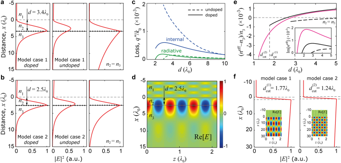
Further numerical investigation with a solver reveals a consistent presence of the TE eigenmode supported irrespectively of the structure variations, proving its existence solely due to the presence of multilayer graphene stack. Figure 4a,b demonstrate an electric field intensity |E|2 profile of this mode. Leftmost profiles correspond to reconstructed experimental conditions (Fig. 3c,d), and demonstrate the profile of the TE mode responsible for the observed reflectance dip at doped multilayer graphene (Fig. 3a,b). This doped graphene mode, although perturbed by the leaky term, is confined to graphene plane in contrary to the waveguide mode supported with presence of undoped lossy graphene in an asymmetric index (the midst in Fig. 4a,b). An unperturbed TE graphene mode is also supported by the structure with considered parameters, but only when n2 = n3, thus being tricky to be excited; its profile is the rightmost in Fig. 4a,b.
Figure 4. Transverse eigenmode of doped graphene.
(a) Obtained with a solver, electrical field intensity |E|2 profiles of TE eigenmodes in considered structure; left: TE eigenmode of doped graphene responsible for the experimentally observed TE reflectance dip, for the set of parameters of model case 1; middle: same as on the left, but for undoped graphene – a waveguide mode, with amplitude scaled according to theoretical coupling efficiency 59% of that for doped graphene mode; right: an unperturbed TE mode supported by doped graphene in the considered structure for model case 1 under assumption of n2 = n3. (b) Left: same as in (a), for the set of parameters of model case 2; middle: same as in (a), with amplitude scaled according to theoretical coupling efficiency 74% of that for doped graphene mode; right: same as in (a), for the set of parameters of model case 2. (c) Internal (blue) and radiative (green) losses of the TE eigenmode for the undoped (dashed) and doped (solid) graphene, for the set of parameters of model case 1. (d) Electric field spatial distribution of the TE eigenmode for the set of parameters of model case 2. (e) Dispersion of the TE eigenmode for the set of parameters of model case 1 (black), case 2 (pink), and case 1 with n2 = n3 (dashed) as a function of distance d; inset: cumulative losses of the TE eigenmode for all cases. (f) TE eigenmode’s electrical field intensity profile |E|2 shown at the indicated cutoff distances d = dcut, for model cases 1 and 2; insets: electrical field spatial distribution demonstrated for each cutoff case.
Graphene sheets in a multilayer stack are separated by approximately 0.3 nm distance20. Considering huge (about three orders of magnitude) difference between the size of the TE mode (Fig. 4a,b) and the actual thickness of the 5-layer graphene stack, experimentally detected TE mode is supported by a thin (i.e., with effectively zero thickness) layer of graphene with effective conductivity σeff proportional to the number of layers14,17,18,19,20. At the same time, the physical nature of the mode is that of graphene TE mode. Coupling between TE modes of two separated graphene layers is explicitly discussed in the authors’ previous work14.
Figure 4c displays the effect of doping on losses associated with the TE mode as a function of distance d. Due to reduction of internal losses (blue) in doped graphene (solid lines) at shorter d, their magnitude becomes comparable with that of radiative losses (green), providing significantly enhanced coupling to incident light13,14,27, and producing experimentally observed distinct reflectance dip. As one can note, cumulative losses of the excited mode are still very low, though higher than in case of n2 = n3 (see inset in Fig. 4e), giving its propagation length L = λP /(2πq″) ~ 1000 of plasmonic wavelengths.
TE mode’s electric field spatial distribution for the model case 2 is demonstrated at Fig. 4d, showing significant asymmetry along the x-z axes, revealing mode expansion by several λ0 into the bulk on both sides. Obtained with the solver dispersion of the TE mode for model case 1 (black), case 2 (pink), and case 1 with n2 = n3 (dashed) is demonstrated in Fig. 4e, with losses shown in the inset. Observed in Fig. 4e for model cases 1 and 2, the dispersion cutoff is associated with complete delocalization (leaking) of modes in the direction of n3, following prediction for the symmetrical (n2 = n3) configuration14. Modes’ delocalization is demonstrated in Fig. 4f where field’s intensity and spatial distribution are shown for distances d ≈ dcut for considered model cases. It is also clearly demonstrated (Fig. 4e) that cutoff distance in case when n2 = n3 is significantly larger comparing to that in employed experimental configuration, preventing efficient coupling to the TE mode.
To summarize, we conclude that exotic TE mode in graphene can be successfully excited in multilayer graphene at room temperature. Its first experimental observation is achieved employing 5-layer doped graphene in modified Otto configuration with very precise phase matching capability. Besides, we demonstrate successful doping of multilayer graphene up to significant values of EF using an ionic liquid, confirming predicted earlier increase of effective graphene conductivity17,18,19,20. We also believe that optical sensing of TE excitation in doped graphene can be used as a handy technique for characterisation of doping in multilayer graphene, and attract more attention to this unique phenomenon in the essential spectral transition domain.
Methods
Sample manufacturing and measurements
In this study we use PVDF film with thickness of 0.254 mm purchased from CS Hyde; silver electrodes were deposited manually using Pelco® colloidal silver liquid manufactured by Ted Pella, Inc. (product No. 16031), and cured at room temperature during 24 hours. 12.5 × 12.5 mm2 area five layer graphene was manufactured by chemical vapour deposition28 (CVD) and deposited layer by layer in acetone, and was purchased from Graphene Square, Inc. The ionic liquid 1-ethyl-3-methylimidazolium bis(trifluoromethylsulfonyl)imide was used as obtained from manufacturer, purchased from Sigma-Aldrich.
Infrared reflectance measurements were performed with a tunable diode laser at 1.55 μm wavelength and a power meter head with removed cover glass to avoid interference effects under varying angle. Gold-covered spherical mirror of 1250 mm radius was mounted at fixed stage while angular scan was conducted by a flat mirror on a moving linear stage (Fig. 2c), providing, together with collimated laser beam with diameter of 1 mm, an effective angular resolution of 0.05 deg, while mechanically limited angular resolution allowed by the setup is 3 × 10⁻⁵ deg. All measurements were performed under normal ambient conditions at 300 K.
Analytical model
In the numerical model, the excitation scheme shown in Fig. 2(a) is considered as a three-layered structure with an effectively semi-infinite top (coupling prism, n1) and bottom (PVDF substrate, n3) layers, where the plane x = 0 corresponds to the n1|n2 interface, and the multilayer graphene stack of effectively zero thickness occupies the x = d plane. TE-polarized plane wave is incident onto the n1|n2 interface at angle θ. The ansatz is made that the electric field in each medium m = 1,2,3 takes the form  , where,
 |
Substituting the ansatz into the Helmholtz equation  (where k0 = ω/c) we obtain
  , where q = n1k0sin(θ) is the projection of the incident wavevector onto the z-axis. Then, assuming the harmonic time dependence exp(–iωt) of the magnetic field H, substituting it into the Maxwell curl equation
  we apply electromagnetic boundary conditions
 at the n1|n2 and n2|n3 interfaces, noting that K(x = 0) = 0 and K(x = d) = σeff
E(x = d), thus obtaining the system of linear equations in complex amplitudes Am and Bm. Taking B3 = 0 (i.e., absence of a reflected wave in medium 3) and A1 = 1 (i.e., continuous incident wave), solving the system, we obtain the reflection coefficient of the incident wave R = |r|2 (r = B1/A1). The same system of linear equations in complex amplitudes is used in a solver to find eigenmodes solutions supported by the structure, solving the determined system in the absence of the incident wave A1 = 0.
Additional Information
How to cite this article: Menabde, S. G. et al. Direct Optical Probing of Transverse Electric Mode in Graphene. Sci. Rep. 6, 21523; doi: 10.1038/srep21523 (2016).
Supplementary Material
Acknowledgments
This work was supported by the National Research Foundation of Korea (NRF) under the Ministry of Science, ICT & Future Planning; the Global Frontier Program NRF-2014M3A6B3063708, and the Global Research Laboratory (GRL) Program K20815000003 (2008-00580), all funded by the South Korean government.
Footnotes
Author Contributions S.M. developed numerical model, proposed and performed experimental work. D.M. performed theoretical and numerical calculations, E.K. and C.L. assisted in samples manufacturing, and N.P. supervised the work, participated in discussion and in writing the manuscript.
References
- Geim A. K. Graphene: status and prospects. Science 324, 1530–1534 (2009). [DOI] [PubMed] [Google Scholar]
- García de Abajo F. J. Graphene plasmonics: Challenges and opportunities. ACS Photonics 1, 135–152 (2014). [Google Scholar]
- Bao Q. & Loh K. P. Graphene photonics, plasmonics, and broadband optoelectronic devices. ACS Nano 6, 3677–3694 (2012). [DOI] [PubMed] [Google Scholar]
- Koppens F. H. L., Chang D. E. & García de Abajo F. J. Graphene plasmonics: A platform for strong light-matter interactions. Nano Lett. 11, 3370–3377 (2011). [DOI] [PubMed] [Google Scholar]
- Wang F. et al. Gate-variable optical transitions in graphene. Science 320, 206–209 (2008). [DOI] [PubMed] [Google Scholar]
- Nair R. R. et al. Fine structure constant defines visual transparency of graphene. Science 320, 1308 (2008). [DOI] [PubMed] [Google Scholar]
- Mason D. R., Menabde S. G., Yu S. & Park N. Plasmonic excitations of 1D metal-dielectric interfaces in 2D systems: 1D surface plasmon polaritons. Scientific Reports 4, 4536 (2014). [DOI] [PMC free article] [PubMed] [Google Scholar]
- Kim J. et al. Electrical control of optical plasmon resonance with graphene. Nano Lett. 12, 5598–5602 (2012). [DOI] [PubMed] [Google Scholar]
- Thareja V. et al. Electrically Tunable Coherent Optical Absorption in Graphene with ion gel. Nano Lett. 15, 1570–1576 (2015). [DOI] [PubMed] [Google Scholar]
- Mikhailov S. A. & Ziegler K. New electromagnetic mode in graphene. Phys. Rev. Lett. 99, 016803 (2007). [DOI] [PubMed] [Google Scholar]
- Kotov O. V., Kol’chenko M. A. & Lozovik Y. E. Ultrahigh refractive index sensitivity of TE-polarized electromagnetic waves in graphene at the interface between two dielectric media. Opt. Express 21, 13533–13546 (2013). [DOI] [PubMed] [Google Scholar]
- Jablan M., Buljan H. & Soljačić M. Transverse electric plasmons in bilayer graphene. Opt. Express 19, 11236–11241 (2011). [DOI] [PubMed] [Google Scholar]
- Yeh P. Optical Waves in Layered Media Ch. 11 (John Wiley & Sons, New Jersey, 2005). [Google Scholar]
- Mason D. R., Menabde S. G. & Park N. Unusual Otto excitation dynamics and enhanced coupling of light to TE plasmons in graphene. Opt. Express 22, 847–858 (2014). [DOI] [PubMed] [Google Scholar]
- Bludov Y. V., Ferreira A., Peres N. M. R. & Vasilevskiy M. I. A primer on surface plasmon-polaritons in graphene. Int. J. Mod. Phys. B 27, 1341001 (2013). [DOI] [PubMed] [Google Scholar]
- Ramos-Mendieta F. Mid-infrared Otto excitation of transverse electric modes in doped graphene. J. Appl. Phys. 117, 133101 (2015). [Google Scholar]
- Hass J. et al. Why multilayer graphene on 4H-SiC(0001) behaves like a single sheet of graphene. Phys. Rev. Lett. 100, 125504 (2008). [DOI] [PubMed] [Google Scholar]
- Dawlaty J. M. et al. Measurement of the optical absorption spectra of epitaxial graphene from terahertz to visible. Appl. Phys. Lett. 93, 131905 (2008). [Google Scholar]
- Yan H. et al. Infrared spectroscopy of wafer-scale graphene. ACS Nano 5, 9854–9860 (2011). [DOI] [PubMed] [Google Scholar]
- Baek I. H. et al. Terahertz transmission and sheet conductivity of randomly stacked multi-layer graphene. Appl. Phys. Lett. 102, 191109 (2013). [Google Scholar]
- Das A. et al. Monitoring dopants by Raman scattering in an electrochemically top-gated graphene transistor. Nature Nanotech. 3, 210–215 (2008). [DOI] [PubMed] [Google Scholar]
- Seki S. et al. Comprehensive refractive index property for room-temperature ionic liquids. J. Chem. Eng. Data 57, 2211–2216 (2012). [Google Scholar]
- Chen C. F. et al. Controlling inelastic light scattering quantum pathways in graphene. Nature 471, 617–620 (2011). [DOI] [PubMed] [Google Scholar]
- Wunsch B., Stauber T., Sols F. & Guinea F. Dynamical polarization of graphene at finite doping. New J. Phys. 8, 318 (2006). [Google Scholar]
- Hwang E. H. & Das S. Dielectric function, screening, and plasmons in two-dimensional graphene. Phys. Rev. B 75, 205418 (2007). [Google Scholar]
- Falkovsky L. A. Optical properties of graphene. J. Phys.: Conference Series 129, 012004 (2008). [Google Scholar]
- Yeatman E. M. Resolution and sensitivity in surface plasmon microscopy and sensing. Biosensors and Bioelectronics, 11, 635–649 (1996). [Google Scholar]
- Li X. et al. Large-area synthesis of high-quality and uniform graphene films on copper foils. Science 342, 1312–1314 (2009). [DOI] [PubMed] [Google Scholar]
Associated Data
This section collects any data citations, data availability statements, or supplementary materials included in this article.


