Abstract
The purpose of this study was to examine normal vision and eye disease in relation to art. Ophthalmology cannot explain art, but vision is a tool for artists and its normal and abnormal characteristics may influence what an artist can do. The retina codes for contrast, and the impact of this is evident throughout art history from Asian brush painting, to Renaissance chiaroscuro, to Op Art. Art exists, and can portray day or night, only because of the way retina adjusts to light. Color processing is complex, but artists have exploited it to create shimmer (Seurat, Op Art), or to disconnect color from form (fauvists, expressionists, Andy Warhol). It is hazardous to diagnose eye disease from an artist's work, because artists have license to create as they wish. El Greco was not astigmatic; Monet was not myopic; Turner did not have cataracts. But when eye disease is documented, the effects can be analyzed. Color-blind artists limit their palette to ambers and blues, and avoid greens. Dense brown cataracts destroy color distinctions, and Monet's late canvases (before surgery) showed strange and intense uses of color. Degas had failing vision for 40 years, and his pastels grew coarser and coarser. He may have continued working because his blurred vision smoothed over the rough work. This paper can barely touch upon the complexity of either vision or art. However, it demonstrates some ways in which understanding vision and eye disease give insight into art, and thereby an appreciation of both art and ophthalmology.
Introduction
It is a great honor to be delivering the Keeler Lecture, which celebrates 25 years of extraordinary philanthropy and scholarship in our field of ophthalmology. The Keeler Company, on its fiftieth anniversary in 1967, began a program of small travel grants to UK ophthalmologists, called Keeler Awards for Clinical Studies. Then, a Scholarship program was established in 1990 to make a biennial award that is currently worth £30 000 and has helped to train a generation of academic and clinical ophthalmologists in the UK. I congratulate the Keeler Scholars and the Keeler Scholarship program on this anniversary.
I was asked to speak about the relationship of vision, eye disease, and art—and one might ask how this belonged at an ophthalmology congress as art is a cultural experience. But we manage patients from different cultures. And art is visual, as is our field of ophthalmology. The creation of art depends upon mechanisms of vision, as does our recognition of arzt as viewers, and artists are not immune to eye disease. Furthermore, the ways in which ocular abnormality may—or may not—alter paintings can be complex and intriguing.
I must emphasize, however, that art is not science. Ophthalmology will not ‘explain' art, which is an extraordinarily complex cultural phenomenon based on personal experience, historical context, political influence, economic factors, social pressures, and many more. These are not issues that knowledge about the retina, or about a cataract, can resolve. And yet ophthalmology has relevance insofar as vision is a tool for the artist and viewer, and understanding how this tool (normal or flawed) relates to the complex choices of an artist gives insight into art and how we see it.1, 2
Normal vision
Vision begins in the eye, but the eye is not a simple camera that transmits an image to the brain. The retina has roughly 120 000 000 rods and 6 000 000 cones, and yet we have only about 1 000 000 fibers in the optic nerve. There is no way that a fully pixelated image representing all of these photoreceptors can be transmitted through the optic nerve. Rather, the retina, using several layers of neurons, becomes an ingenious device for coding visual information into a simplified ‘language' that emphasizes only the most critical aspects of visual objects. In essence, we code the world. But while this code lets us be efficient in managing visual data, and allowing the brain to concentrate just on what is important for recognition, it also puts constraints on our interpretation of the world and occasionally leads to confusions and illusions as will be noted below. I will touch upon a few highlights of perception that are particularly interesting and important with respect to the creation and interpretation of art.
Contrast
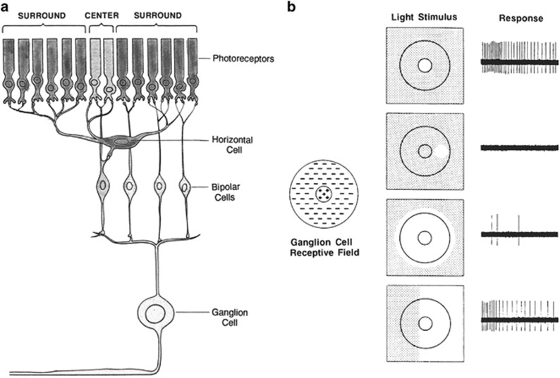
The most basic aspect of retinal coding is recognition of contrast. This begins at the level of the photoreceptor cells and the bipolar cells (that link the photoreceptors to ganglion cells), with help from the horizontal cells, through the development of ‘center-surround' receptive fields (Figure 1a).2 The receptive field of a cell is simply the portion of the world that the cell perceives. The receptive fields of the bipolar cells (and ganglion cells) are basically donut-shaped, with a center zone that receives direct excitatory input from one group of photoreceptors, and inhibitory input from a halo of photoreceptors that do not synapse directly with the given bipolar cell but make contact with processes of the horizontal cells. The horizontal cells feed into the central photoreceptor-bipolar synapses with inhibitory effect. The excitatory center and inhibitory surround roughly cancel each other, so that turning on a room light has little effect on the bipolar cell (Figure 1b). Thus, we are not visually sensitive to absolute levels of light, but primarily to contrast when an edge crosses the donut to stimulate the center and surround asymmetrically. An edge of light just covering the center, for example, gives strong excitation but only partial inhibition.
Figure 1.
Retinal organization for contrast. (a) A core of photoreceptors feed into a bipolar cell, while a surrounding ring of photoreceptors stimulates horizontal cells to inhibit the response. This creates a center-surround receptive field. (b) Edge recognition. A spot of light in the center or surround will excite or inhibit—but overall illumination (third example) balances the two and is hardly recognized. An edge (bottom example) illuminates the center and surround disparately, and is perceived well. Image details: (a, b) M F Marmor, after reference 2. Notes: Google: Google Art Project (free Access); MMA: Metropolitan Museum of Art, New York, NY, USA (free access); MFM: Michael F Marmor; NGA: National Gallery of Art, Washington, DC, USA (free access); Wiki: Wikimedia Commons and Foundation (free access).
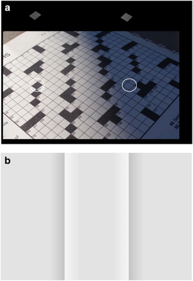
Insofar as the bipolar and ganglion cells are activated primarily by contrast and edges, these drive our recognition of form and our perception of brightness. For example, Figure 2a shows a crossword puzzle with a shadow crossing it on one side. A black square will always appears dark relative to nearby white squares, but in fact a black square in the light may be no darker than a white square in the shadow. The illusion in Figure 2b is very powerful to most people. Are the three zones identical? The middle zone clearly looks lighter, although if you hold a pencil or finger across the junction between the center and either side, you will see that these two regions are identical except at the boundary. A computer has made each of the outer zones get a little bit darker, and the central zone get a little bit lighter, towards the junction. This creates a sharp light-to-dark edge, which fools the retina and brain into thinking that everything beyond the junction is also light or dark.
Figure 2.
Contrast recognition. (a) We perceive squares properly as black or white whether in or out of the shadow, but the circled squares are actually of equal brightness (see above the puzzle). (b) The center region looks lighter than the sides because of the sharp light-dark junctions. If you cover one or another of the junctions, you will see that the cores of the regions are identical. Image details: (a) © M F Marmor. (b) M F Marmor, from reference 2.
Contrast is very much a part of art. For example, Figure 3a shows an Asian brush painting in which the artist has created effects very similar to the junctions in Figure 2b. There is a beautiful image of a dark mountain against the light sky—except that the core of the mountain, much like the lateral zones in Figure 2b, is actually the same brightness as the sky. The painter has understood intuitively that large swaths of white and black can be avoided by the judicious use of shading. Many other artists have used subtle shading to great effect, such as the twentieth century painter Georgia O'Keeffe.
Figure 3.
Contrast in art. (a) Ma Yuan (active ca. 1190–1225): Viewing Plum Blossoms by Moonlight (early thirteenth century). This Chinese brush painting creates light-dark junctions to give an illusion of dark rocks and mountains. (b) Caravaggio (Michelangelo Merisi) (1571–1610): Saint John the Baptist in the Wilderness (ca. 1604–5). This painting shows extreme chiaroscuro, with a bright figure on a nearly black background. (c) Georges Seurat (1859–1891): A Sunday on La Grande Jatte (detail) (1884). In this part of Seurat's grand canvas, the water brightness, and even the lawn brightness, has been varied to contrast with overlying objects. Image details: (a) Ink on silk (25 × 27 cm). Metropolitan Museum of Art, New York: gift of John M Crawford Jr, in honor of Alfreda Murck, 1986. (b) Oil on canvas (173 × 132 cm). Google Art Project, Nelson-Atkins Museum of Art, William Rockhill Nelson Trust. (c) Oil on canvas (208 × 308 cm). Google: Art Institute of Chicago.
Leonardo did not understand retinal circuitry, but he wrote in his notebooks that the way to emphasize a dark object is to put it next to a light one, and vice versa. He was wise enough in his own art not to carry the dictum to extremes, but later artists were not always so cautious. The use of high contrast, often showing the subject against a dense dark background (‘chiaroscuro'), was popular for a time in the Renaissance, following the lead of Caravaggio (Figure 3b). The French neo-Impressionist and pointillist Georges Seurat rather compulsively read books about contrast and color, and took this advice to heart in a different way. His famous work A Sunday on La Grande Jatte, is revered for its portrayal of an afternoon on the riverbank. However, careful inspection will show that Seurat has lightened the water where the bathers are in shadow, and darkened it where they are in the sun; even the lawn varies where light or dark figures cross the grass (Figure 3c). The overall impact of the painting remains glorious, but the water is a rather curious pond!
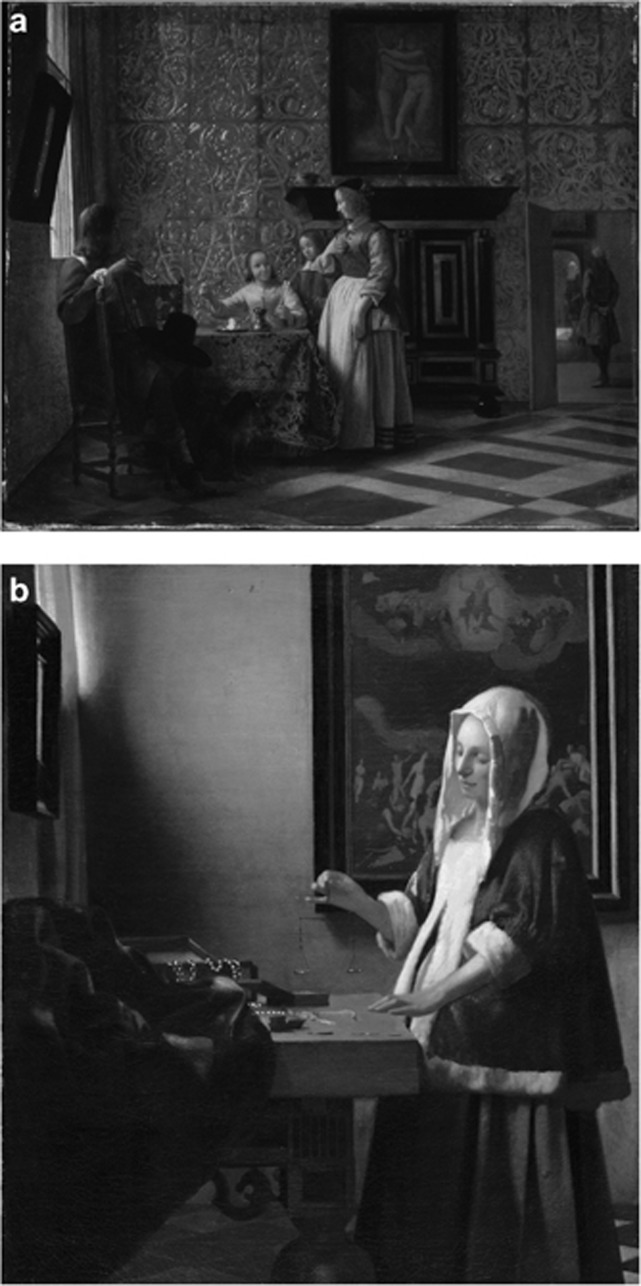
When contrast is used wisely, it can be very powerful in art, bringing life and excitement to a painting. Compare the handling of light by different seventeenth century painters. Indoor scenes by Pieter de Hooch were beautifully drawn (Figure 4a) but often lit rather evenly so that they rarely seem ‘brilliant'. Compare his painting with a similar scene by Vermeer (Figure 4b). Vermeer not only had an extraordinary eye for framing a poignant moment, but for highlighting the dialog of sunlight and shadow. Sunlight reflecting off the woman's blouse gives brilliance that is lacking in the de Hooch.
Figure 4.
Light and dark. (a) Pieter de Hooch (1629–1684): Leisure Time in an Elegant Setting (ca. 1663–65). This painting is beautifully composed, but the lighting is rather flat. (b) Johannes Vermeer (1632–1675): Woman Holding a Balance (c. 1664). This similar scene is much more dramatic because of the greater differences between light and dark. Image details: (a) Oil on canvas (40 × 36 cm). National Gallery of Art, Washington, DC: Widener Collection. (b) Oil on canvas (58 × 69 cm). Metropolitan Museum of Art, New York: Robert Lehman Collection, 1975.
Light and dark
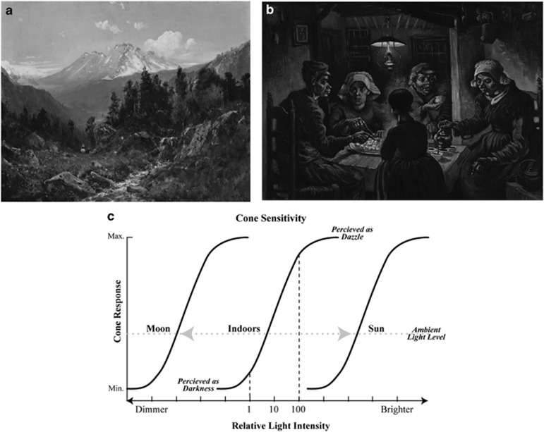
Outdoor sunlight may be a million times brighter than the light in a dim gallery where we view art. Thus, one may reasonably ask how art exists? How can we look at a painting in a gallery and see a bright, sunny day, when there is only indoor light emanating from the painting (Figure 5a)? Or how can we a recognize painting of a night scene, when the illumination entering our eyes is vastly greater (Figure 5b). The answer lies within the retina itself. The cone photoreceptors, which provide our vision in all but the dimmest light, actually have a very limited range of brightness sensitivity at any moment in time. There is only about a 100-fold spread of energy between a stimulus that fails to activate a cone, and one that maxes out the cone response. But the cones move this range of sensitivity (the stimulus response curve) up or down in accordance with the ambient (background) lighting as shown in Figure 5c.2, 3
Figure 5.
Picturing day and night. (a) William Keith (1838–1911) Mount Shasta (late nineteenth century). This shows a sunny scene, even when the light bouncing off the picture is much less bright. (b) Vincent van Gogh (1853–1890): The Potato Eaters (1885). An indoor scene would be evident even if the image were viewed outdoors. (c) Cone response curves show only about a 100-fold (2 log unit) response range, which adjusts according to ambient lighting. This lets us recognize the same objects in a dim room or the outdoor sun. Image details: (a) Oil on canvas (24 × 30 cm): Iris & B Gerald Cantor Center for Visual Arts at Stanford University, Stanford, CA, USA: Gift of Mrs. William Babcock. (b) Oil on canvas (82 × 114 cm). Google Art Project: Van Gogh Museum, Amsterdam. (c) M F Marmor, after reference 2.
This adjustment takes place within seconds and resets our recognition of white-to-black to correspond with the environment. For example, when you drive down the highway on a sunny day, the interior of a tunnel appears pitch black, but within seconds after entering, you adapt to the dimmer environment and can see the road again. As you have the same 100-fold range of white-to-black discrimination, whether indoors or outdoors, a painting that illustrates this range will appear realistic in either location. We judge the location of a scene by its content. And this accounts for the fascination of Magritte's famous painting that shows a bright sunny sky above a darkened row of houses illuminated by street lamps: our sense of light and dark is in conflict with different portions of the same scene.
Color
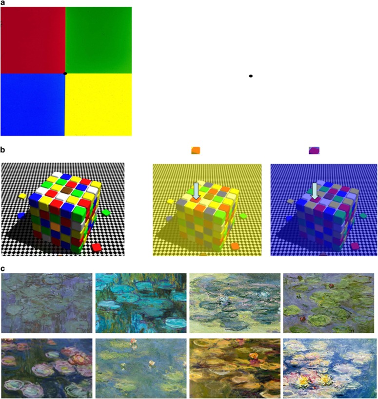
People recognize a wide array of colors, but color perception is actually processed in terms of contrast rather than absolute wavelength. We have red-, green-, and blue-sensitive cone pigments, but the bipolar cells have center-surround receptive fields that contrast red vs green and blue vs yellow. Thus, we do not so much perceive ‘red' as to recognize that something is redder than the area around it.2 For example, fix your gaze on the black dot on the left in Figure 6a for at least 30 s. When you shift your gaze to a white part of the page, you see the contrasting or complementary colors of red instead of green, and blue instead of yellow. Another example, devised by neuroscientist Dale Purves,4 is shown in Figure 6b. It shows three Rubik's cubes, one in normal lighting, one illuminated with yellow light and one illuminated with blue light. It appears that the colors on the cubes are relatively constant under all three conditions, and one has no trouble finding red and the green squares. However, if the red squares are isolated, apart from their relationship to other colors, you can see how different they are under yellow or blue illumination. Just as contrast and edge detection lets us recognize things under many different lighting conditions, color constancy lets us recognize the same faces or fruits in different places.
Figure 6.
Seeing colors. (a) Stare at the black dot amid the four primary colors for 30 s, then look at the dot on the right. You will see complementary colors that represent the color contrast mechanisms of the retina. (b) Color constancy (from Dale Purves): Rubik's cubes under white, yellow and blue illumination still look like a Rubik's cube. However, when the red squares are isolated (see above the arrows), the colors appear quite different. (c) Details from eight different Monet paintings of water lilies at his estate in Giverny. Image details: (a) M F Marmor, after reference 2. (b) Images devised by Dale Purves, MD: free access from http://www.purveslab.net/seeforyourself. (c) Detailed views of eight paintings entitled Waterlilies, all oil on canvas. In order: Google Art Project: Musee d'Orsay, Paris; Wikimedia Commons and Foundation (photograph Schlaier): Neue Pinothek, Munich, Germany; Google: Kawamura Memorial DIC Museum of Art, Sakura City, Japan; Google: National Gallery of Australia, Canberra, ACT, Australia; Wikimedia (photograph Bildum): National Museum of Western Art, Matsukata Collection, Tokyo, Japan; Google: Ohara Museum of Art, Kurashiki, Japan; National Gallery of Art, Washington, DC: The Walter H and Leonore Annenberg Collection, Gift of Walter H and Leonore Annenbrg, 1998, Bequest of Walter H Annenberg, 2002; Wikimedia (unknown photograph): Dallas Museum of Art.
Artists perceive colors in this same way, and both color contrast and color constancy will affect scenes being painted under different conditions such as the orange glow of twilight vs the blue noonday sky or a dim incandescent bulb. It is intriguing to look at the serial paintings of Monet, who often painted the same subject at different times of day or in different seasons. Figure 6c shows a variety of colors that he could apply to the same lily pads in his garden in Giverny. He did not write about his perceptions or intentions for these paintings, but we know he was an extraordinarily sensitive observer of light and color.
Equiluminance
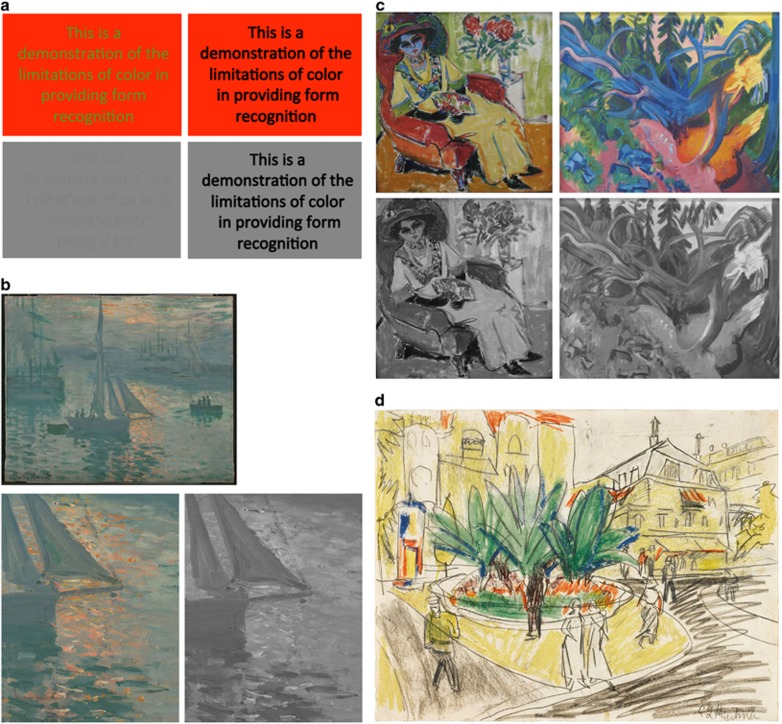
Although it may seem that we recognize many objects by color, the basic retinal circuitry for discriminating shape, form, movement and depth is driven solely by brightness (independent of color). Thus, to a large degree, this color sense is just superimposed upon our more basic discrimination of brightness and contrast. Objects that we ‘recognize' by color actually depend on brightness to recognize the shape and form.2, 5 Figure 7a shows how text may be difficult to read when printed in different colors that are similar in brightness. Students in an art school are taught that ‘value' (absolute brightness) is a critical component of color, in addition to hue (wavelength) and saturation (dilution with white). Even if art students do not understand the underlying physiology, they know that colors of equal brightness (ie ‘equiluminant' colors) cause problems.
Figure 7.
Color vs form. (a) Equiluminance. Green-on-red lettering is hard to read when the colors have equal brightness. The grey scale version shows how the lettering disappears. (b) Claude Monet (1840–1926): Sunrise (Marine) (1873). The water glistens in the red sun, because of equiluminance of the reflections and water. The grey scale detail shows how the red disappears. (c) Ernst Ludwig Kirchner (1880–1938): Seated Woman (Dodo) (1907) and Uprooted Tree (1922). Both of these paintings use unusual colors but the grey scale views show why only the woman looks realistic: the brightness cues in the forest are quite obscure. (d) Kirchner, The Trees in the Albertplatz in Dresden (1910–1911). This drawing shows a lovely group of trees in a square, surrounded by buildings with yellow facades. We are hardly concerned by the location the color fields because we recognize the objects by their black outlines. Image details: (a) © M F Marmor. (b) Oil on canvas (50 x 61 cm). Google Art Project: J Paul Getty Museum, Los Angeles, CA, USA. (c) Oil on canvas (112x114 cm). Seated Woman: Wikimedia Commons and Foundation (photograph Rufus 46): Pinothek der Moderne, Munich Germany; oil on canvas (100 × 120 cm). Uprooted Tree: Wikimedia (photograph Hajotthu): Sprengel Museum, Hanover, Germany. (d) Crayon and pencil on paper (27 × 35 cm). Wikimedia (photograph Sotheby's): sold by Sotheby's in 2012.
Artists from Seurat to modern Op Art painters, such as Bridget Riley and Richard Anuszkiewicz, have observed that relatively equiluminant dots or patterns become hard to distinguish, and create shimmer and vibrancy. Monet used this effect beautifully in Impression, Sunrise and Sunrise (Marine), where the red glow shimmers off the water because the red and grey have equal brightness (Figure 7b). Andy Warhol made use of this phenomenon in his series paintings, such as the portraits of Chairman Mao or Marilyn Monroe. In some paintings, faces with bizarre color were easy to recognize because the face was lighter than the hair; in other paintings, the brightness of face and hair was similar, creating an unstable image that is hard to interpret as a portrait. The French Fauvists and German Expressionists used colors wildly, to challenge our color expectations in art. This worked, as long as the colored objects showed normal luminance relationships. However, scenes would be difficult to interpret when brightness relationships did not match how the scene should appear in black and white. In the examples of Figure 7c, Kirchner's woman is easy to recognize despite unrealistic colors: however, the uprooted tree is very hard to distinguish despite the bright colors.
A secondary role for color in form perception is also illustrated by paintings in which colors spread across outlines or boundaries of objects. This is seen in some drawings and paintings by Picasso and Matisse, and strikingly in the paintings of Raoul Dufy. An example by Kirchner is shown in Figure 7d. At a quick glance, such paintings may seem realistic, but on a second look, it is obvious that the colors do not match the outlines. Why don't we notice immediately, or care? Because demarcated sharp outlines dominate our perception of form and our recognition of the objects. And as long as colors fall somewhere in the area, our brain happily associates them with the appropriate object.
Eye disease
We have seen how the complexities of normal vision influence the way we see contrast, form, and color, and thus are a part of the artist's toolbox as much as paintbrushes and pigments.
As ophthalmologists, we deal with dysfunction of these visual tools caused by age and disease, and it is reasonable to ask when and how this may affect how an artist works or an artist's style.1, 2, 6 There must be some point at which visual disability will put constraints on what an artist can do. It may modify the ability to see a scene, to put details or proper colors on a canvas, or to refine a work in the studio and recognize what needs to be improved. And yet artists with excellent vision may choose a style for their own aesthetic and other reasons, and paint photographically or abstractly, in greys or in color, and with precision or freedom. Thus, it is very difficult—indeed hazardous—to look at a work of art and make judgments about an artist's visual disability or eye disease. Only when eye disease or visual disability is clearly documented through historical or medical records, can one safely make correlations with what the artist says and does.
False judgments
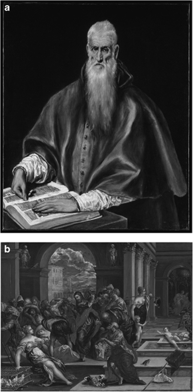
When people hear that I work with eye disease and art, they often say, ‘Oh, like El Greco and astigmatism'. I appreciate the interest, of course, but the assertion is wrong. El Greco did paint strikingly elongated figures (Figure 8a), and serious ophthalmologic articles have been written to show how the elongations can be corrected by viewing the paintings through astigmatic lenses. However, there are numerous counter-arguments.2, 7 El Greco typically painted religious figures with elongation, whereas secular figures and objects in the scenery were often painted without distortion. Although his figures were typically elongated in a vertical direction, hands that point horizontally are always long and thin (although the purported optical distortion would make them short and stubby). Furthermore, an astigmatic error causes visual blur rather than obvious distortion, as the optical effect is to create two blur circles on the retina. Finally, the whole concept falls apart if you picture El Greco painting an image on the canvas to match his model or subject. As he looks back and forth between canvas and subject, the painted image must be a realistic copy of the subject if the two are to look the same to him (no matter what distortion might exist within his eye). El Greco is often called a Mannerist because he was among a group of artists who took liberties with realism, and many of his early paintings were perfectly representational, before he developed his mature style (Figure 8b). The assertion that elongated works of art imply an astigmatic error becomes quite absurd with Modigliani paintings or Giacometti sculptures.
Figure 8.
El Greco's elongations. (a) El Greco (Domenikos Theotokopoulos) (1541–1614): St. Jerome as Scholar, ca. 1610. The saint is elongated vertically, but his left hand is horizontal; and the neither the book nor his tunic buttons seem distorted. (b) El Greco: Christ Cleansing the Temple (probably before 1570). This early painting is quite realistic. Image details: (a) Oil on canvas (108 × 89 cm). Metropolitan Museum of Art, New York: Robert Lehman Collection, 1975. (b) Oil on panel (65 × 83). National Gallery of Art, Washington, DC: Samuel H Kress Collection.
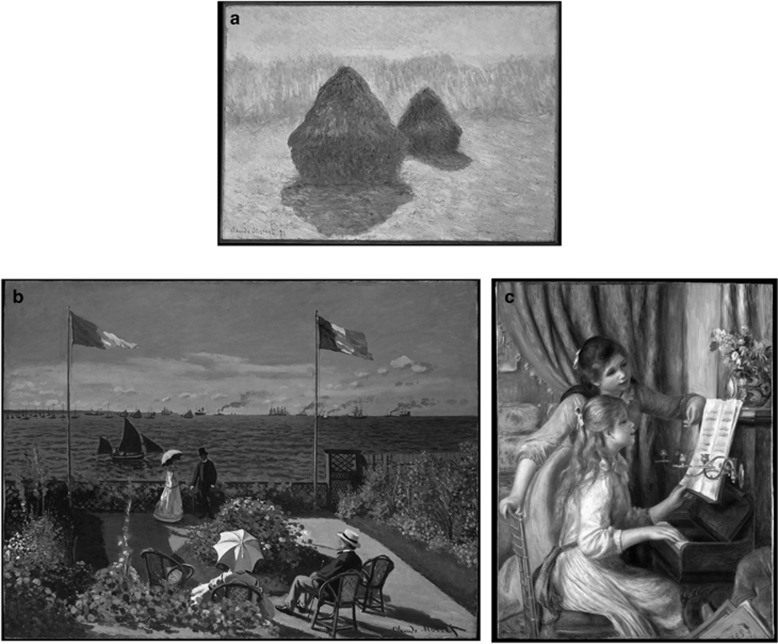
Another question that is occasionally asked is whether the impressionists like Monet (Figure 9a) simply had uncorrected myopia. This, too, has no basis in either ophthalmologic or artistic fact. Impressionism was a complex intellectual movement, in rebellion to the rigid stylistic expectations of the Paris Salon in the mid-nineteenth century. The painters sought freedom to do natural scenes, and to create emotions rather than historical or mythological scenes. Most of them were not myopic, and glasses were readily available for myopia. Monet could not have been severely myopic in his youth, as many of his early works are painted in exquisite detail and show far-off objects (Figure 9b). The impressionistic style was based on subject, color, and light rather than the presence or absence of detail, as shown in many works by Renoir, Degas, Pissarro, and others (Figure 9c).
Figure 9.
Impressionism. (a) Claude Monet: Haystacks (Effect of Snow and Sun) (1891). This is a study of light, not of blur. (b) Claude Monet: Garden at Sainte-Adresse (1867). This earlier work is painted realistically with precise details of both near and distant objects. (c) Auguste Renoir (1841–1919): Two Young Girls at the Piano (1892). This painting, done in the Impressionist period, shows precise details of hair, candlesticks, chair, etc. Image details: (a) Oil on canvas (65 x 92 cm). Metropolitan Museum of Art, New York: HO Havemeyer Collection, bequest of Mrs HO Havemeyer, 1929. (b) Oil on canvas (98 x 130 cm). Metropolitan Museum of Art, New York: purchase, with contributions and funds by friends of the Museum, 1967. (c) Oil on canvas (112 × 86 cm). Metropolitan Museum of Art, New York: Robert Lehman Collection, 1975.
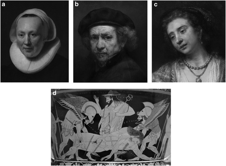
Does the broadening of style with age, which one sees in many painters such as Rembrandt (Figure 10a and b) and Titian, indicate eye disease? It is hard to deny that the 90-year-old Titian might have had some degree of cataract or possibly dry macular degeneration—but we have no historical documentation of failing vision. Rembrandt used impasto (thick application of paint) to create more expressive works in his last decades; but he could also limit the impasto where a subject was more delicate like a child or young woman (Figure 10c). Presbyopia should not have been an issue, as reading glasses were readily available (and Rembrandt painted members of his household using them).
Figure 10.
Presbyopia. Details of works by by Rembrandt van Rijn (1606–1669) and Euphronios (c. 540-470/480 BCE). (a) Portrait of a Woman (1633). This early painting is almost photographic. (b) Self-Portrait (1659). This painting shows his expressive late style, with heavy impasto (thick applications of paint). (c) Lucretia (1664). This painting was done 5 years later, but has little impasto, appropriate to the subject. (d) Portion of an Athenian red figure krater (c. 515 BCE). Image details: (a) Oil on wood (68 x 50 cm). Metropolitan Museum of Art, New York: bequest of Benjamin Altman, 1913. (b) Oil on canvas (85 × 66 cm). National Gallery of Art, Washington, DC: Andrew Mellon Collection. (c) Oil on canvas (120 × 101 cm). National Gallery of Art, Washington, DC: Andrew W Mellon Collection. (d) Krater (pottery) is 46 × 55 cm. Wikimedia Commons and Foundation (photograph J Ardiles): Villa Giulia, Rome, Italy.
However, presbyopia probably was an issue in ancient Greece and Rome, as there is no evidence that spectacles or reading lenses were known. An art professor at Stanford, Jody Maxmin, noted that one of the great Greek vase artists, Euphronius, stopped painting rather abruptly at about age 40, although he remained a workshop owner or potter for at least another 20 years.8 Good near vision would certainly have been needed to paint the delicate figures that decorated Greek vases (Figure 10d), and indeed, these were often drawn by boys or young men in the pottery workshops. Euphronios might well have found it impossible to paint as he became presbyopic. Another Greek vase painter, the Amasis painter, continued to paint throughout a career that spanned roughly 45 years (certainly well into his 50s)—and we might guess that he was myopic.
Color blindness
One might think that color blindness would be an obvious and evident ocular disorder in artists. However, there are very few well-known painters with documented color blindness, because young painters with color blindness shift their work towards other disciplines such as graphics or sculpture. Examples are Charles Meryon, the famous French etcher of Paris street scenes, and Paul Manship, an American sculptor: both discovered as young men that they mixed up colors badly.
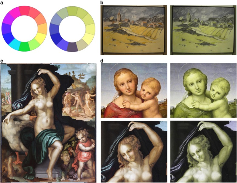
Color blindness (better termed color deficiency) afflicts roughly 8% of males in Western society, although most can resolve strong colors. Only about 1.5% of men have a total loss of red-green discrimination. Figure 11a simulates the effect of deuteranopia (the most common form of dichromacy) on perception of a color wheel. Basically, there are no distinctions of hue between red and green, and green becomes grey or white as it lies between blue and yellow and represents a mix of all colors (white light).
Figure 11.
Color-blindness. (a) Color circles, normal and deuteranopic. (b) Jens (Johannsen) (1934–2010) was deuteranopic: Fields of Wheat (1980s). The painting (left) shows reddish roofs, but is otherwise predominately amber and blue-grey. Deuteranopic simulation (right) makes little difference except for loss of the roofs. (c) Baccio Bandinelli (1493–1560) was color-blind: Leda and the Swan (ca. 1512). The painting is beautifully drawn, but Leda's flesh is pasty without highlights. (d) Details of paintings by Raphael (1483-1520), who had normal vision: The Small Cowper Madonna (ca. 1505); and Bandinelli: Leda and the Swan. Deuteranopic simulation destroys the warmth of Raphael‘s flesh tones, but Leda is hardly affected by the conversion. Image details: Deuteranopic simulations through Coblis: www.color-blind.com/coblis (a) M F Marmor. (b) Oil on canvas (∼ 80 × 100 cm). Image provided by the artist; simulation by M F Marmor. (c) Oil on panel (128 × 101). © Chancellery of Paris Universities. Used by permission. Simulation, M F Marmor. (d) Raphael: Oil on canvas (full painting 60 × 44 cm). National Gallery of Art, Washington, DC: Widener Collection. Simulations, M F Marmor.
Mild degrees of color deficiency would be exceedingly hard to detect in a painter's style or color choices, because colors are seen and paint tubes have labels. However, painters with strong or total red-green color blindness often use a characteristic palette of yellow or amber, contrasted with blue and grey (Figure 11b). Greens are assiduously avoided as they lack color and are hazardous to mix or use.9 Artists who use a plethora of green shades are unlikely to have major red-green color deficiency. For example, one critic speculated that the great English landscape painter Constable might have been color-deficient because of murky colors in some of his canvases.10 This seems highly unlikely, because his paintings are dominated by greens and subtle mixtures of earth tones. However, some color-blind painters do use a wide range of colors (usually without much color-mixing), as friends advise them about the color of grass, oranges, bricks, etc. and they have license to choose tubes of paint by name or by brightness.
A good example of the color-blind palette is the work of the twentieth century American painter Jens, who was known to have deuteranopia (Figure 11b).9 The Irish painter, Paul Henry, was most famous for paintings done in the Irish Isles, which are dominated by amber wheat, and blue-grey mountains in the distance, which matches the actual scenery. After his death, his ophthalmologist revealed that he was red-green color-blind. He occasionally added striking red highlights, such as a peasant dress, but these were typically done without mixing colors. An Australian artist, Clifton Pugh, had a protan deficit, and also painted with a limited palette, using slightly faded colors and avoiding greens.11
An unproven example is the Renaissance sculptor and painter, Baccio Bandinelli (1493–1560). He was famous as a sculptor in Florence, but never achieved recognition as a painter. The great historian of Renaissance artists, Giorgio Vasari, said repeatedly that although Baccio was a brilliant draughtsman, he could not handle colors! He said in one note,12 ‘Baccio's drawings were very beautiful, but in colors he executed them badly and without grace... he took into his service one who handled colors passing well.' The late Philippe Lanthony, a great scholar of color vision and art, speculated that Baccio might have been color blind,6 and evidence from paintings newly restored and attributed to Baccio supports this hypothesis.13 His Leda and the Swan shows the problem (Figure 11c). Despite a complex and well-drawn design, Leda's flesh is yellowish and pasty, indistinguishable from the color of the swan, and devoid of warmth. Compare this to a painting by Raphael, whose flesh tones are vibrant (Figure 11d). Simulating color blindness destroys the richness of the Raphael, but has little effect on Baccio's Leda. The story does not quite end here, because despite Baccio's fame, he was jealous, scheming and vindictive, and he repeatedly fought with his fellow artists. One cannot help but wonder whether color blindness contributed to this personality, as it must have been incredibly frustrating to make paintings that looked perfectly fine to him and yet were criticized by everyone else. No one understood why, of course, as color blindness would not be recognized as a congenital disorder for another 250 years (when the great English chemist John Dalton described his own affliction in 1794).
Cataracts
Cataracts were a major cause of visual disability in earlier eras, when cataract removal was difficult or not performed at all. Rosealba Carriera (1763–1757), the ‘divine Rosealba,' was famous for her pastel portraits. She was diagnosed with cataracts at age 51 in 1726, but continued to draw pastels until 1746.2 She finally consented to surgery and had couching procedures in each eye, with the second in 1749 described as showing unusual debris in the left eye. Thus, her cataracts may have been complicated or secondary. These left her essentially blind. It is noteworthy that Jacque Daviel had described cataract removal shortly before Carriera‘s second couching. However, a book by her surgeon (G. Reghellini) 15 years later questioned the Daviel procedure as causing too many failures.
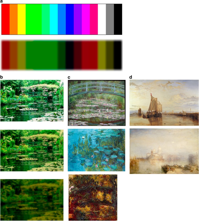
The typical aging cataract with nuclear sclerosis gets progressively more brown and dense over time. Thus, there is not only blurred vision, but less light, less contrast, and altered color perception. The diagram in Figure 12a shows a color spectrum modified to simulate the view through a 20/200 dense brunescent cataract. For an artist, the 20/200 blur may be less problematic than the striking loss of color discrimination: yellow and white can no longer be distinguished, blues are very dark, and distinctions between shades of green or shades of red are lost. This would be devastating to any artist whose work depended upon subtle color variations, such as Claude Monet.
Figure 12.
Cataract effects. (a) Color chart as it would appear through a dense brunescent cataract. Note the loss of color distinctions, disappearance of white, and darkening of blue. (b) Photographs of Monet's lily pond at Giverny showing how it would appear thorough moderate cataract (Monet 1915–17) and dense cataract (Monet 1922). (c) Monet paintings of the lily pond in these same years. Japanese Footbridge (1899) is quite representational. Waterlilies (1915) show strong flat fields of blue, perhaps seeking distinction from the leaves. Japanese Footbridge (1922) shows not only rough application of paint, but surprisingly strong orange color. (d) Joseph Mallord William Turner (1775–1851): The early Dort or Dordrecht: The Dort Packet-Boat from Rotterdam Becalmed (1818) is realistic in style, but suffused with yellow. The mature Dogana and Santa Maria della Salute, Venice (1843) is almost impressionistic, but still shows precise details near and far. Image details: (a) © M F Marmor. (b) Photograph of Giverny: © Elizabeth Murray, www.elizabethmurray.com. Used by permission. Simulations: Simulations, M F Marmor (From reference 14). (c) From top to bottom: 1899: Oil on canvas (81 × 102 cm). National Gallery of Art, Washington, DC: Gift of Victoria Nebeker Coberliy, in memory of her son John W Mudd, and Walter H and Leonore Annenberg. 1915: Oil on canvas (151 × 201 cm). Wikimedia Commons and Foundation (photograph Schlaier): Neue Pinothek, Munich, Germany. 1922: Oil on canvas (94 × 89). Google Art Project: Museum of Fine Arts, Houston, TX, USA. (d) From top to bottom: 1818: Oil on canvas (185 × 259 cm). Google: Yale Center for British Art, New Haven, CT, USA. 1843: Oil on canvas (62 × 93 cm). National Gallery of Art, Washington, DC: Given in memory of Governor Alvan T Fuller by the Fuller Foundation, Inc.
Monet was diagnosed with cataracts in 1912, and ophthalmologists recommended surgery in the worst eye. But he was apprehensive and refused. Over the next decade, his vision decreased progressively, finally reaching 20/200 in 1922 (documented by medical records). However, more distressing to him were the gradual alterations in color perception. By 1914, he was describing reds as dull and muddy. By 1918, he wrote that he was choosing colors by the labels on the tube. And yet he continued to paint until he acquiesced to surgery at the end of 1922 when he could no longer read or write, saying ‘I am almost blind.' One of Monet's close friends urging him to have surgery was George Clemenceau, the Prime Minister of France at the end of the First World War, and a physician by training. Clemenceau had motives beside medical advice, as he had negotiated from Monet the gift of his grand water lily paintings to the state, to be displayed in the Orangerie in Paris. As Monet's vision failed, Clemenceau feared that the works might not be finished.
It is instructive to look at a photograph of Monet's garden, using computer simulations to show the effects of his developing cataract.14 As his cataracts progressed (Figure 12b), his view of the garden became more yellowish and murky, blues became darker and indistinct, and colors were barely recognizable by 1922. It is important to emphasize that an artist with cataract may not paint these same yellow tones. Because a cataract is always present, vision adapts, and the sense of a yellowish world may be lost—however, color distortions do remain so that whites and yellows look alike, blues are hard to see, and color distinctions disappear. If a painter remembers how sky and water used to look, efforts to paint them that way can lead paradoxically to the use of brighter and stronger blues. Monet's paintings of the lily pond at Giverny reflected his perceptions (Figure 12c). Near 1900, the paintings were remarkably realistic, but paintings of the lily pond in 1915–17 often showed strong but flat blue fields of color for the water, unlike the varied and subtle colors of earlier years. This may have helped Monet to see distinction between water and leaves. By 1922, he could see little color beyond a murky green and many of his paintings showed strong colors ranging from bright orange to intense blue. Were these colors intentional by design, or do they reflect an attempt to get some feedback through his brunescent lens? These late paintings show dramatically the struggles of a great painter to continue working despite significant visual impairment.
One of Monet's reasons for refusing surgery was the fear that it would alter his color perception. He was right: it would have made it better. Monet's surgery in January of 1923 was a three-stage procedure with opening of the capsule, removal of the lens material, and an eventual posterior capsulotomy. He recovered excellent near vision and reasonable distance vision in the affected eye, but he complained bitterly about the world appearing too yellow or sometimes too blue. It took almost two years before he finally told friends that his color vision felt normal, and returned to work on the large canvases for the Orangerie. After his surgery, Monet destroyed many canvases (we have only what his family saved); and his painting (including the canvases for the Orangerie) returned to an earlier impressionist style. Thus, I doubt that Monet's abstract and strongly colored paintings done through dense cataracts were designed by aesthetic choice alone. We do not know whether he was trying to paint a memory, to paint what he saw, or to paint what he wanted us to see. We do know that his options were limited by failing vision.
Another impressionist who suffered from cataracts was Mary Cassatt, whose vision dropped towards 1913. Her pastels lost some detail, and she soon stopped painting, but one does not see obvious changes in color as with Monet. It is possible that these were subcapsular cataracts (she was diabetic), and sadly her surgery was complicated by uveitis and glaucoma, and despite multiple procedures, she ended up with minimal vision.
Richard Liebreich was a famous German ophthalmologist who worked for many years in England (and consulted on Claude Monet). He wrote an article in 1872 postulating that J. M. W. Turner, the great English painter of landscapes and the ocean, suffered from cataracts to account for his increasingly non-representational style and an intense use of yellow.15 However, Turner consciously described yellow as light and blue as darkness. His paintings were bathed in yellow light throughout his career (Figure 12d), and even at age 31, critics were complaining of his ‘jaundiced colors.' Even the most abstract of Turner's late works usually show some precise details. I know of no evidence that his vision was failing, and it is extremely unlikely that cataracts were an issue.
Macular disease
With most macular disease, the predominant symptom is a loss of visual acuity and contrast. Although color discrimination may be reduced, there is usually no strong or easily characterized deficit in color perception. It is easy to postulate macular disease when an artist's style broadens and becomes less precise, but stylistic changes can have so many other causes that it is only meaningful to discuss the effects of maculopathy in art where failing vision can be historically documented.
Georgia O'Keeffe, the great American painter of landscapes and natural objects, lived from 1887 to 1986. Medical records document that she was diagnosed with macular degeneration in 1964 at the age of 77, and also had a vein occlusion in the left eye in 1971. By 1972, her vision had fallen below 20/200 despite attempts at laser treatment, and towards the end of the 1970s she gave up painting altogether. Her rocks and landscapes prior to the development of macular degeneration were characterized by exquisite shading, delicacy of shadows, and fine details. Towards 1970, her canvases grew in size, and objects became hard-edged without the careful shading and shadowing that were so much a part of her mature work. She did not lose ability to see the natural shapes that fascinated her, but she lost the ability to paint them with subtlety.
The clearest example of the effects of macular disease upon a great artist is Edgar Degas (1834–1917). He had chronic and progressive visual loss over almost 50 years, which is well documented in his letters and other historical records. He discovered in his thirties that he could not fire a rifle with his right eye, and the vision in his left eye gradually decreased as well. As photographs show him walking by himself as an old man in the streets of Paris, we can surmise that he had primarily macular dysfunction, which affected his acuity but not his peripheral vision. He did not have age-related macular degeneration, given the young age at which his symptoms began. It could have been a dystrophy such as Stargardt disease, although there is no direct evidence for this. He had a first cousin with similar central visual loss as a young adult, but at best, they could have shared one recessive gene.
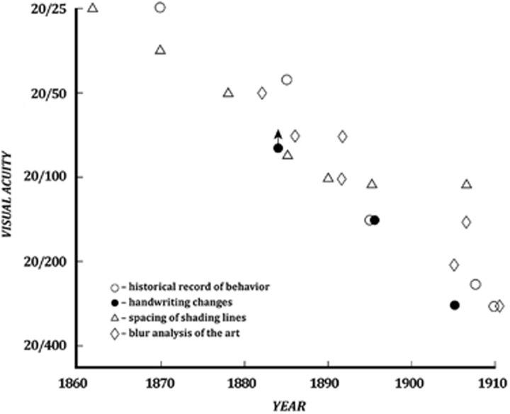
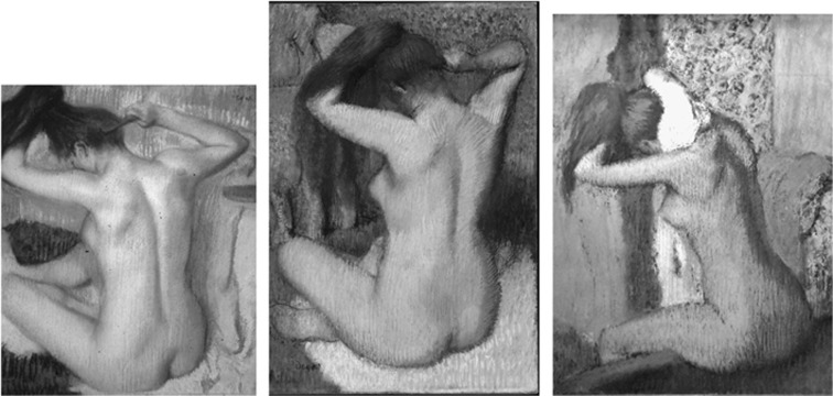
Whatever the cause, Degas' visual acuity fell progressively, reaching levels estimated to be below 20/400 by 1910 (Figure 13). And corresponding with this loss of vision, there was a progressive change in the precision of his paintings and pastels.14, 16 Over the years between 1880 and 1910, his depiction of pose and posture did not change, but his figures were less sharply outlined, details such as hair and facial features gradually disappeared, and subtleties such as shading and delineation of musculature were lost (Figures 14 and 15). It is important to emphasize that Degas drew rough working sketches throughout his career, so a lack of detail does not by itself prove poor vision. But finer works dominated early in his career, and had completely disappeared after the turn of the twentieth century.
Figure 13.
Degas' visual acuity. The chart shows different estimations, but all show a steady decline from excellent acuity in 1870 (in his best eye) to worse than 20/200 by 1905. Image details: M F Marmor, after reference 16.
Figure 14.
Changing detail in pastels by Edgar Degas (1834–1917). These images are scaled and cropped slightly to show Degas' technique over a 15-year period. Left: Woman Combing her Hair (ca. 1885) shows careful shading and musculature. Middle: Woman Combing her Hair (ca. 1888–90) has shading lines further apart and less precise. Right: After the bath (1899) has rather coarse outlining and shading. Image details: Left: Pastel on paper (52 × 51 cm). Wikimedia Commons and Foundation (photograph The Yorck Project): Hermitage Museum, St Petersburg, Russia. Middle: Pastel on paper (61 × 46 cm). Metropolitan Museum of Art, New York: Gift of Mr and Mrs Nate B Spingold, 1956. Right: Pastel on paper (62 × 65 cm). Wikimedia (photo by J-P Dalbéra): Musée d'Orsay, Paris, France.
Figure 15.
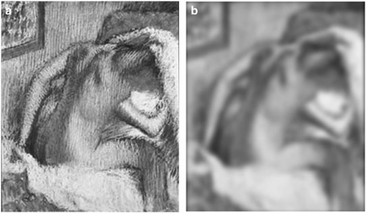
Simulation of Degas's view of his late work. (a) Woman Drying Her Hair (c. 1905) is very roughly drawn. (b) In Degas view, with 20/300 visual acuity at the time, the shading appears smoothed and much more refined. Image details: (a) Pastel on paper (71 × 63 cm). Norton Simon Art Foundation, Norton Simon Museum, Pasadena, CA, USA. (b) Simulation, M F Marmor, after reference 2.
Some of Degas' friends and critics advised him to stop working, as his pastels lost refinement, but Degas doggedly continued, still seeking the perfect pose. As with Monet, one may ask why a great artist would persist in the face of degrading vision. Mary Cassatt and Georgia O'Keeffe gave up their art when they could no longer maintain their mature style. Part of Degas' decision may have resulted from the way his visual loss affected his own perception of his pastels.14, 16 Figure 15 shows a simulated view of Degas' late Women Drying her Hair as it would have appeared to him with failing vision. His progressive visual loss limited his technique, but blurred vision also smoothed out coarse outlining and shadowing. In his own view, his later works still had a degree of shading and refinement. Degas was a consummate artist, and he must have realized that others would see roughness in the late pastels. Yet to him, the works were not so rough, and he could continue to refine the female body.
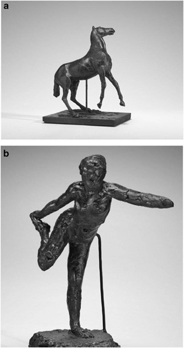
Some have suggested that Degas took up sculpture late in life to compensate for his loss of vision, as it was a ‘tactile art.' In reality, Degas worked with sculpture since the early 1870s, modeling horses and dancers out of wax or clay (all of the bronze casting was posthumous), mostly as studies of posture and form for his paintings and pastels. Like his pastels, his sculptures show a progression from a careful construction of details (face, hand, hair, etc.) in some works from the 1870s and 1880s to none at all in later years (Figure 16). Most of Degas' figures were quite tiny (typically12”–18” high) so that these details were well below tactile resolution and their construction was in fact visual with the use of fine tools. None of Degas‘ sculptures were sold or dated during his lifetime, and simulated images (showing what he could or could not see as his vision failed) have helped art historians to judge when some of these works were created.17
Figure 16.
Degas sculptures over the years. (a) Horse Rearing (1880s) is tiny (12 inches tall) but beautifully finished with fine details of the face, hooves, etc. (b) Dancer Holding Her Right Foot in Her Right Hand (ca. 1900–1911) is 18 inches tall, but roughly finished without any details at all. Image details: (a) Beeswax over metal armature (31 × 20 × 27 cm). National Gallery of Art, Washington, DC: Collection of Mr. and Mrs. Paul Mellon. (b) Beeswax over metal armature (50 × 24 × 38 cm). National Gallery of Art, Washington, DC: Collection of Mr and Mrs Paul Mellon.
Conclusions
This brief excursion only touched on a few aspects of normal vision and eye disease as they relate to art, and more information is available from other sources.1, 2, 5, 6, 18 Neither visual science nor ophthalmology can explain art or what artists have done creatively, but I hope my examples have shown where they give insight. Mechanisms of vision are relevant to the techniques of artists, and eye disease adds new challenges to the task of creating art. Ophthalmology may not explain art, but it can help us to appreciate art in new ways. And perhaps in some respect, art can help us to better appreciate ophthalmology.
Acknowledgments
All modern consideration of ophthalmology and art owes a debt to British ophthalmologist Patrick Trevor-Roper whose seminal book1 was originally published in 1970. I am grateful to Richard Keeler for proposing this topic for the Keeler Lecture, and to my colleague and co-author2 James G Ravin who has expanded my knowledge about eye disease in artists. Finally, as an academician writing about art, I am grateful to museums and organizations that are now making images of great art available as free access for scholarly publication: Google Art Project (and contributing museums), Metropolitan Museum of Art (New York), National Gallery of Art (Washington, DC), and Wikimedia Commons and Foundation (and contributing museums).
The author declares no conflict of interest.
References
- 1Trevor-Roper P. The World Through Blunted Sight. Allen Lane, The Penguin Press: London, UK, 1988. [Google Scholar]
- 2Marmor MF, Ravin JG. The Artist's Eyes. Abrams: New York, NY, USA, 2009. [Google Scholar]
- 3Normann RA, Perlman I. The effects of background illumination on the photoresponses of red and green cones. J Physiol 1979; 286: 491–507. [DOI] [PMC free article] [PubMed] [Google Scholar]
- 4Purves D, Lotto RB. Why We See What We Do Redux. Sinauer Associates: Sunderland, Massachusetts, 2011. [Google Scholar]
- 5Livingstone M. Vision and Art: the Biology of Seeing. Abrams: New York, NY, USA,, 2002. [Google Scholar]
- 6Lanthony P. Les Yeux des Peintres. L'Age d'Homme: Lausanne, Switzerland, 1999. [Google Scholar]
- 7Simunovic MP. “The El Greco fallacy” fallacy. JAMA Ophthalmol 2014; 132: 491–494. [DOI] [PubMed] [Google Scholar]
- 8Maxmin J. Euphronios epoiesen: portrait of the artist as a presbyopic potter. Greece & Rome 1974; 21: 178–180. [Google Scholar]
- 9Marmor MF, Lanthony P. The dilemma of color deficiency and art. Surv Ophthalmol 2001; 45: 407–415. [DOI] [PubMed] [Google Scholar]
- 10Law BM. Constable and Color. Paper read at Canadian Ophthalmol Congress: Banff, Alberta, 1957. [Google Scholar]
- 11Cole BL, Harris RW. Colour blindness does not preclude fame as an artist: celebrated Australian artist Clifton Pugh was a protanope. Clin Exp Optom 2009; 92: 421–428. [DOI] [PubMed] [Google Scholar]
- 12Vasari. Lives of the Painters, Sculptors and Architects, 1568 Transl: G de Vere, 1912. Everyman's Library, Alfred A, Vol 2. Knopf: New York, NY, USA, 1996, pp 277–278. [Google Scholar]
- 13Marmor MF. Bandinelli era daltonico?. In: Heikamp D, Strozzi BP (eds) Baccio Bandinelli: Scultore e Maestro (1493-1560). Giunti Editore: Florence, Italy, 2014. [Google Scholar]
- 14Marmor MF. Ophthalmology and art: simulation of Monet's cataracts and Degas' retinal disease. Arch Ophthalmol 2006; 124: 1764–1769. [DOI] [PubMed] [Google Scholar]
- 15Liebreich R. Turner and Mulready. On the effect of certain faults of vision on painting, with especial reference to their works. Proc Royal Inst Great Britain 1872; 6: 450–463. [Google Scholar]
- 16Marmor MF. Degas Through His Own Eyes: Visual Disability and the Late Style of Degas. Somogy Editions d'Art: Paris, France, 2002. [Google Scholar]
- 17Marmor MF. Simulating Degas' vision: implications for dating his sculpture. Sculpture Journal 2013; 22: 96–108. [Google Scholar]
- 18Lanthony P. Art and Ophthalmology. Wayenborgh Publications: Paraguay, 2009. [Google Scholar]


