Abstract
Emphysematous gastritis is a severe and rare form of gastritis with characteristic findings of intramural gas in the stomach. It is an acute life-threatening condition resulting from gas-producing microorganisms invading the stomach wall. Early diagnosis and initiation of treatment with bowel rest, hydration, and intravenous broad-spectrum antibiotics is imperative for an effective outcome. Surgical intervention is reserved for perforations, peritonitis, strictures, and uncontrolled disseminated sepsis. We present a case of an 82-year-old female with prior history of colon and uterine cancer on remission treated with surgeries who presented with bilious vomiting, abdominal discomfort, and nausea. She was tachycardic and had a diffusely tender abdomen with rebound on examination. Her laboratory results including blood count, serum chemistry, and coagulation studies were normal. She was diagnosed with emphysematous gastritis based on the characteristic radiographic findings of intramural stomach gas and also the presence of gas in the portal venous system. It is important to differentiate emphysematous gastritis from gastric emphysema because of the difference in management and prognosis, as emphysematous gastritis has a worse outcome and requires aggressive management. Despite an anticipated poor prognosis due to the known grave outcomes of emphysematous gastritis, our patient was successfully managed with conservative treatment. We concluded that she developed emphysematous gastritis probably secondary to immunosuppression and possible mucosal tears from multiple bouts of vomiting. She had a stable hospital course and resolution with medical management most likely due to early diagnosis and initiation of appropriate treatment.
Keywords: gastric emphysema, gastritis, portal venous air, stomach wall air
Emphysematous gastritis is a severe and rare form of gastritis with characteristic findings of intramural gas in the stomach. It is an acute life-threatening condition resulting from gas-producing microorganism invading the stomach wall. Early diagnosis and initiation of treatment with bowel rest, hydration, and intravenous broad-spectrum antibiotics is imperative for an effective outcome (1–3). Surgical intervention is reserved for perforations, peritonitis, strictures, and uncontrolled disseminated sepsis (2, 3).
Case report
We present a case of an 82-year-old female, an immigrant from Brazil, with past medical history of atrial fibrillation, colon cancer, and uterine cancer status post abdominal surgeries on remission who presented to our emergency room with severe abdominal discomfort, nausea, and 7–8 episodes of bilious vomiting without hematemesis. She did not have any constipation or diarrhea, and her last bowel movement was a day prior to presentation. She lived with her daughter and was bedridden due to advanced Alzheimer's disease.
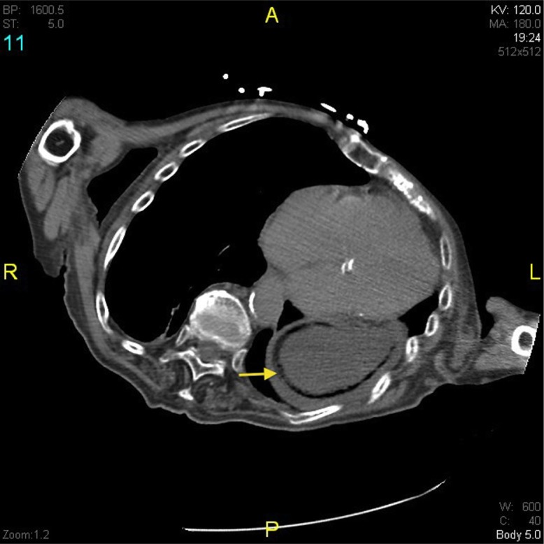
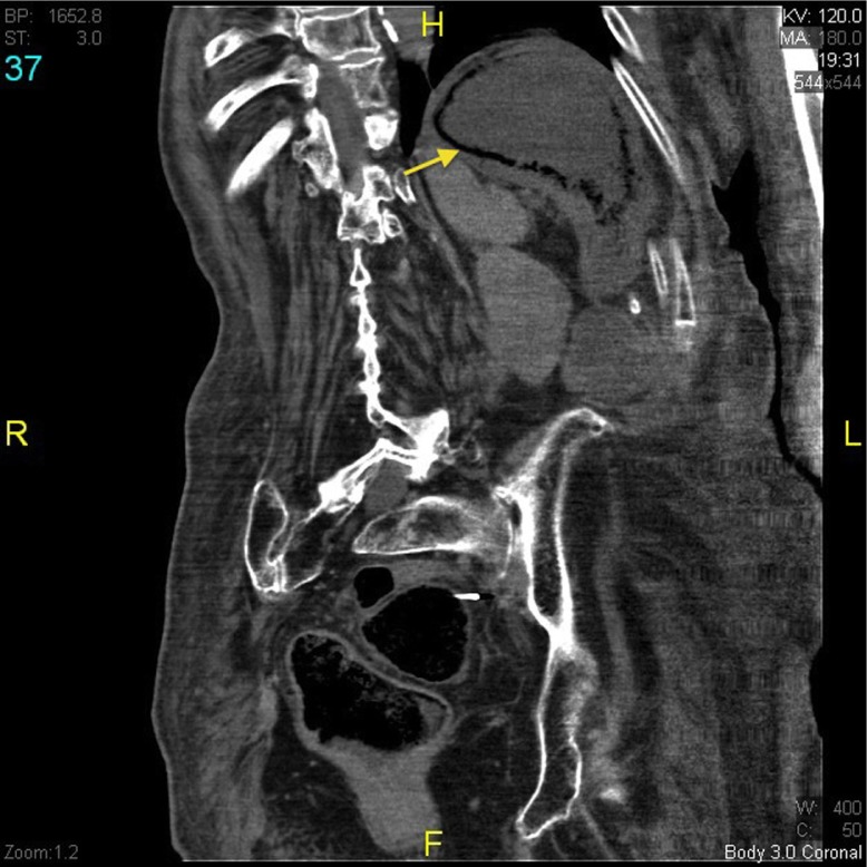
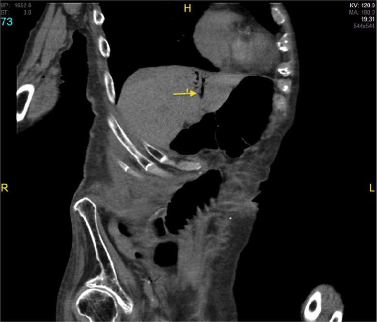
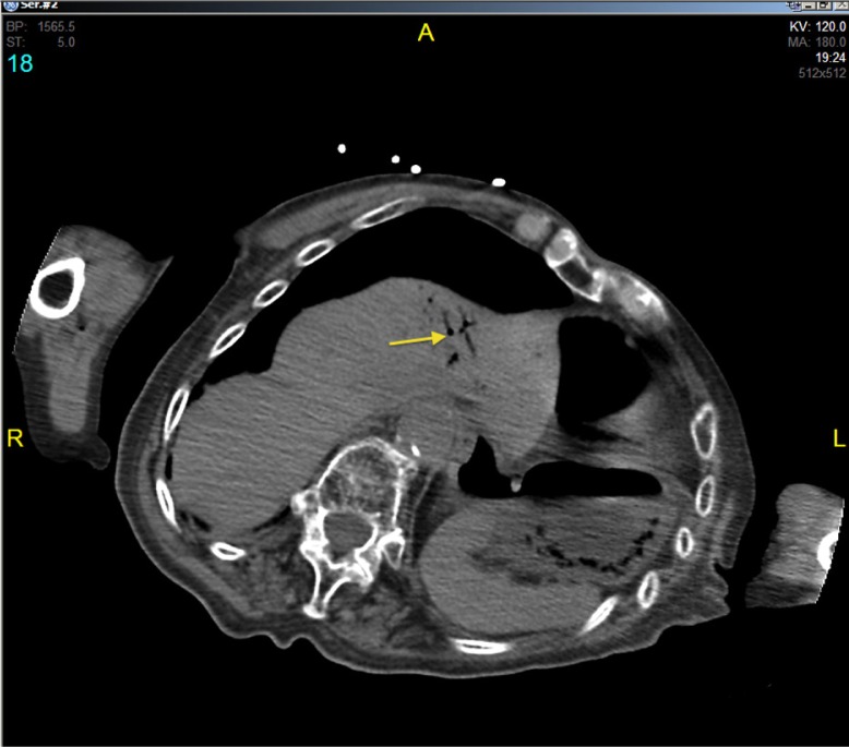
She was saturating 93% in room air and her temperature was 97.8°F. Physical examination revealed a diffusely tender abdomen with rebound but no guarding or rigidity was present. She had a hemoglobin level of 13 g/dL, a white blood cell count of 10,000 per microliter, and a platelet count of 80,000 per microliter. She had a lactate level of 1.8 mmol/L, and her lipase level was 29 U/L. Abdominal X-ray showed centrally dilated small bowel with large amount of stool within the rectum. Faecal occult test was positive. Computed tomography (CT) scan of the abdomen showed circumferential intramural air within the proximal–mid stomach (Figs. 1 and 2) and intrahepatic portal venous air within the left lobe of the liver (Figs. 3 and 4) consistent with emphysematous gastritis with no evidence of small bowel obstruction.
Fig. 1.
CT abdomen showing intramural air (yellow arrow) within the proximal–mid stomach consistent with emphysematous gastritis.
Fig. 2.
CT abdomen/coronal view showing intramural air (yellow arrow) within the proximal–mid stomach consistent with emphysematous gastritis.
Fig. 3.
CT abdomen/coronal view showing portal venous air (yellow arrow) within the left lobe of the liver.
Fig. 4.
CT abdomen showing portal venous air (yellow arrow) within the left lobe of the liver.
She was treated with intravenous hydration, intravenous vancomycin, cefepime, and metronidazole and kept NPO (nil per os). She improved clinically only with intravenous hydration and broad-spectrum antibiotics.
Discussion
Air in the stomach wall is a rare finding. Two conditions are included in the differential diagnosis of intramural gas in stomach: emphysematous gastritis and gastric emphysema (1).
Emphysematous gastritis, first described in 1889, is a rare form of phlegmonous gastritis caused by gas-forming organisms invading the stomach wall through a mucosal defect. The mortality rate is reported as high as 62% (2, 3). Recent systematic review suggested a mortality rate of 55% for emphysematous gastritis and 29% for gastric emphysema (4). The most common predisposing factors for emphysematous gastritis include ingestion of caustic substances, alcohol abuse, abdominal surgery, diabetes, immunosuppression, and phytobezoars (2, 3). Abundant blood supply, effective mucosal barrier, and an acidic PH make the stomach a very uncommon site for this finding (5). Organisms commonly involved are Streptococcus spp., Escherichia coli, Enterobacter spp., Clostridium spp., Pseudomonas aeruginosa, Staphylococcus aureus (2, 3), Sarcina spp. (6), Candida spp., and Mucor spp. (7). Previous reports have suggested a high mortality rate in cases with the finding of intramural stomach gas in association with hepatic portal venous gas (8, 9).
Gastric emphysema is a more benign condition in which air usually reaches within the wall from outside (1). The predisposing factors for gastric emphysema are mucosal disruption due to procedures like endoscopy (10) or nasogastric tube placement (11), increased intraluminal pressure secondary to pyloric or enteral stenosis (12, 13), gastric paresis and forceful vomiting, and chest wall injuries.
It is important to differentiate emphysematous gastritis from gastric emphysema mostly because of the difference in management and prognosis. Emphysematous gastritis usually presents in a sick, hemodynamically unstable patient and has increased mortality (1–5). It might also have an increased association of radiological finding of gas in the portal venous system in addition to the intramural stomach gas (5). Treatment is usually aggressive with antibiotics and surgical intervention. In contrast, patients with gastric emphysema generally present with milder symptoms of dyspepsia and epigastric discomfort or may be asymptomatic. They are usually managed with conservative management and observation (1–5). Two types of radiological pattern have been used to describe intramural stomach air. A cystic mottled pattern is usually associated with emphysematous gastritis, while linear lucency along the greater curvature of the stomach is present in gastric emphysema. Radiologists use terminologies like linear, curvilinear, or cystic to describe non-gangrenous changes, whereas bubbly signifies gangrenous gastritis (14, 15).
Our patient had prior history of multiple cancers and advanced Alzheimer's disease and was bed bound. We concluded that she developed emphysematous gastritis secondary to her immunosuppressed status and possible mucosal tears in the stomach from multiple bouts of vomiting. Emphysematous gastritis was diagnosed based on the characteristic radiographic signs and a good response to antibiotics. The radiological finding of intramural stomach gas and portal venous air predicted a poor prognosis. However, she had a stable hospital course and resolution with medical management, likely due to early diagnosis and initiation of appropriate treatment.
Conflict of interest and funding
The authors have not received any funding or benefits from industry or elsewhere to conduct this study.
References
- 1.Naqvi ZG, Shahzad N, Alvi AR, Effendi S. BMJ Case Rep 2013. 2013. Intramural gas in stomach along with acute calculus cholecystitis: an unusual association. bcr2012007757. [DOI] [PMC free article] [PubMed] [Google Scholar]
- 2.Iannuzzi J, Watson T, Litle V. Emphysematous gastritis: a young diabetic's recovery. Int J Surg Case Rep. 2012;3(4):125–7. doi: 10.1016/j.ijscr.2011.12.002. [DOI] [PMC free article] [PubMed] [Google Scholar]
- 3.Yalamanchili M, Cady W. Emphysematous gastritis in a hemodialysis patient. South Med J. 2003;96:84–8. doi: 10.1097/01.SMJ.0000048085.35271.75. [DOI] [PubMed] [Google Scholar]
- 4.Matsushima K, Won EJ, Tangel MR, Enomoto LM, Avella DM, Soybel DI. Emphysematous gastritis and gastric emphysema: similar radiographic findings, distinct clinical entities. World J Surg. 2015;39(4):1008–17. doi: 10.1007/s00268-014-2882-7. doi: http://dx.doi.org/10.1007/s00268-014-2882-7. [DOI] [PubMed] [Google Scholar]
- 5.Paul M, John S, Menon M, Golewale N, Weiss S, Murthy U. Successful medical management of emphysematous gastritis with concomitant portal venous air: a case report. J Med Case Rep. 2010;4:140. doi: 10.1186/1752-1947-4-140. [DOI] [PMC free article] [PubMed] [Google Scholar]
- 6.Laass MW, Pargac N, Fischer R, Bernhardt H, Knoke M, Henker J. Emphysematous gastritis caused by Sarcina ventriculi. Gastrointest Endosc. 2010;72(5):1101–3. doi: 10.1016/j.gie.2010.02.021. [DOI] [PubMed] [Google Scholar]
- 7.Jung JH, Choi HJ, Yoo J, Kang SJ, Lee KY. Emphysematous gastritis associated with invasive gastric mucormycosis: a case report. J Korean Med Sci. 2007;22:923–7. doi: 10.3346/jkms.2007.22.5.923. [DOI] [PMC free article] [PubMed] [Google Scholar]
- 8.Arezzo A, Famiglietti F, Garabello D, Morino M. Complete resolution of emphysematous gastritis after conservative management. Clin Gastroenterol Hepatol. 2011;9(4):e30. doi: 10.1016/j.cgh.2010.10.024. [DOI] [PubMed] [Google Scholar]
- 9.Abboud B, El Hachem J, Yazbeck T, Doumit C. Hepatic portal venous gas: physiopathology, etiology, prognosis and treatment. World J Gastroenterol. 2009;15(29):3585–90. doi: 10.3748/wjg.15.3585. [DOI] [PMC free article] [PubMed] [Google Scholar]
- 10.Hyun YS, Han DS, Lee HL, Bae JH, Eun CS. Gastric emphysema after endoscopic submucosal dissection. Endoscopy. 2011;43(Suppl 2 UCTN):E83–4. doi: 10.1055/s-0030-1255805. [DOI] [PubMed] [Google Scholar]
- 11.Zenooz NA, Robbin MR, Perez V. Gastric pneumatosis following nasogastric tube placement: a case report with literature review. Emerg Radiol. 2007;13(4):205–7. doi: 10.1007/s10140-006-0544-2. [DOI] [PubMed] [Google Scholar]
- 12.Lim RK, McKillop S, Karanicolas PJ, Scott L. Massive gastric pneumatosis from pyloric stenosis. CMAJ. 2010;182(5):E227. doi: 10.1503/cmaj.090200. [DOI] [PMC free article] [PubMed] [Google Scholar]
- 13.Gutiérrez Andreu M, Otero Perpiñá B, Muñoz C, De La Cruz Vigo F. Gastric pneumatosis secondary to a pyloric stenosis of peptic origin. Cir Esp. 2011;89(3):188. doi: 10.1016/j.ciresp.2009.09.010. [DOI] [PubMed] [Google Scholar]
- 14.McLaughlin SA, Nguyen JH. Conservative management of nongangrenous esophageal and gastric pneumatosis. Am Surg. 2007;73(9):862–4. [PubMed] [Google Scholar]
- 15.Soon MS, Yen HH, Soon A, Lin OS. Endoscopic ultrasonographic appearance of gastric emphysema. World J Gastroenterol. 2005;11:1719–21. doi: 10.3748/wjg.v11.i11.1719. [DOI] [PMC free article] [PubMed] [Google Scholar]


