Abstract
Median canaliform dystrophy of Heller is a rare entity characterized by a midline or a paramedian ridge or split and canal formation in nail plate of one or both the thumb nails. It is an acquired condition resulting from a temporary defect in the matrix that interferes with nail formation. Habitual picking of the nail base may be responsible for some cases. Histopathology classically shows parakeratosis, accumulation of melanin within and between the nail bed keratinocytes. Treatment of median nail dystrophy includes injectable triamcinalone acetonide, topical 0.1% tacrolimus, and tazarotene 0.05%, which is many a times challenging for a dermatologist. Psychiatric opinion should be taken when associated with the depressive, obsessive-compulsive, or impulse-control disorder. We report a case of 19-year-old male diagnosed as median nail dystrophy.
Keywords: Dystrophy, median nail dystrophy, nail matrix
Introduction
What was known?
Median canaliform dystrophy of Heller is a rare entity characterized by a midline or a paramedian ridge in nail plate, usually an acquired condition resulting from a temporary defect in the nail matrix that interferes with nail formation.
Median canaliform dystrophy (MCD) of Heller is a rare entity characterized by a midline or a paramedian ridge or split and canal formation in nail plate of one or both the thumb nails. The first case was recorded by Heller in 1928.[1] There is no sex predilection. Mean age of occurrence is 25.72 years. The condition is diagnosed based on its clinical features.[2] Its etiology is unknown, but it has been suggested that MCD is the result of a temporary defect in the nail matrix, following dyskeratinization or focal infection, or due to self-inflicted trauma to the nail or nail bed. The main condition from which it needs to be differentiated is habit tic deformity. Spontaneous remission is often seen after a period of months to years, but the condition can be recurrent. Avoidance of repetitive nail trauma can be achieved through behavioral counseling. Here, we report a case of 19-year-old male with a habit of biting the thumb nails while in stress diagnosed as median nail dystrophy.
Case Report
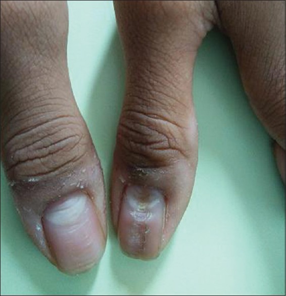
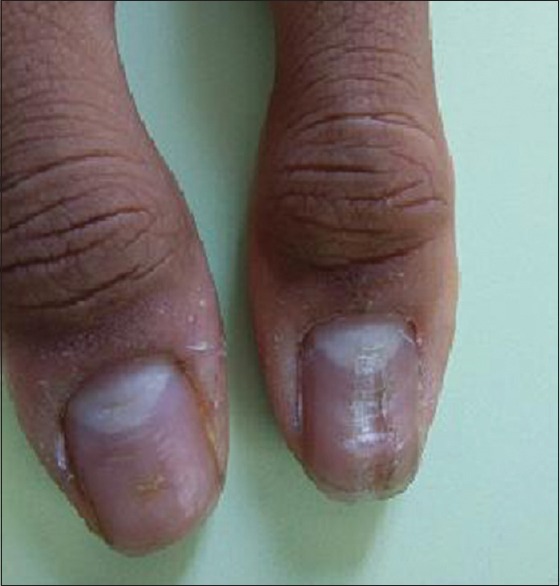
A 19-year-old male medical student attended skin outpatient department with complaints of lesions over both thumb nails since 4 months. History of biting of thumb nails during stress was present. No history of use of oral retinoids or other medications, or history of contact with irritants or allergens was present. He denied any nail disorders or psychiatric disorder in the family. On examination, single median longitudinal groove with transverse furrows arising from a median split on either side in a fir tree pattern present over both the thumb nails, more over, right thumb nail [Figure 1]. The median groove extended from the proximal nail fold up to the distal nail edge. Lunula was seemed to be enlarged in size. Exfoliation was present over lateral nail folds. Rest other finger and toe nails were normal. No skin lesions present elsewhere. Systemic examination was unremarkable. Diagnosis of median nail dystrophy was made on a clinical basis. Histopathology was not done for obvious reasons as there is no additional advantage in treatment and patient was put on 0.1% tacrolimus ointment topically at night, with the advice not to bite nails. Psychiatric consultation was sought; counselling was offered to the patient. Patient returned after 6 weeks, with visible improvement in the proximal part of the nail [Figure 2] and is still in follow-up.
Figure 1.

Single median longitudinal groove in a fir tree pattern over both thumb nails
Figure 2.

Follow-up after 6 weeks
Discussion
MCD also known as solenonychia or dystrophia unguis mediana canaliformis or nevus striatus unguis[3] presents with small cracks or fissures that extend laterally from the central canal or split toward the nail edge giving the appearance of an inverted fir tree or Christmas tree. The condition is usually symmetrical and most often affects the thumbs, although other fingers or toes may be involved.[2] Thickening of the proximal nail fold, enlargement, and redness of the lunula may occur.[3]
It is an acquired condition, but familial clustering of cases are reported by Sweeney et al. in 2005.[2] Presumably, the condition results from a temporary defect in the matrix that interferes with nail formation.[4] Trauma has been implicated as a causative factor.[4,5] Habitual picking of the nail base may be responsible for some cases as seen in our case. A few cases have been attributed to oral retinoid use also.[6] Intentional trauma in the form of pushing back of cuticle and proximal nail fold (habitual tic) is hypothesized in its pathogenesis.[7] However, subungual skin tumors such as glomus tumors,[8] myxoid tumors, have also been described to cause longitudinal grooving, lifting of the nail plate from the bed resulting in a tube-like structure (solenos) distal to it. The absence of keratinocytic adhesions within nail matrix with dyskeratosis is responsible for the formation of longitudinal grove with splitting of nail plate due to weaker tensile strength.[4] Histopathology classically shows parakeratosis, accumulation of melanin within and between the nail bed keratinocytes, which was not done in our case.
Habit tic deformity, digital mucous cyst (synovial cyst), lichen striatus, nail-patella syndrome, pterygium, Raynaud disease, and trachyonychia are other conditions in which a longitudinal nail defect have been described.[7] Habit tic deformity is usually present in one or both thumbnails and results in the alteration of normal nail growth. It is caused by the constant or habitual rubbing of the thumb's proximal nail fold by the tip of the second digit. The subsequent damage to the nail matrix produces transverse ridges along the central nail plate depression instead of a longitudinal groove with lateral projections as in median canaliform nail dystrophy. The depth of the central nail plate canal depends on the intensity of the inflicted trauma by the index finger to the matrix of the thumb nail.
Treatment of median nail dystrophy is many times challenging for a dermatologist as no therapy has been shown to be consistently successful. Stressful condition leading to depression can be the cause of this deformity as was seen in our case. An opinion of a psychiatrist should be sought, if the patient has the depressive, obsessive–compulsive, or impulse-control disorder. Injecting triamcinolone acetonide into the dystrophic nail is one option but is difficult to tolerate and has numerous adverse effects. Recently reported treatment is the topical application of 0.1% tacrolimus ointment once daily without occlusion as also seen in our case.[4] Calcineurin inhibitors are an effective treatment due to their interference with the inflammatory component. Topical tazarotene 0.05% ointment known to normalize the process of keratinization has been used by Madke et al.[9] Keeping the nail length short and buffing the surface of the nail can prevent the edge of the nail plate from catching on clothing and other objects. Covering the nail plate with tape or a nail wrap also can aid in it.
Financial support and sponsorship
Nil.
Conflicts of interest
There are no conflicts of interest.
What is new?
Stressful condition leading to depression can be the cause of median nail deformity. Not only in habit tic deformity but median nail dystrophy can be due to stressful condition. Hence, an opinion of a psychiatrist should be sought, if the patient has the obsessive–compulsive or impulse-control disorder.
References
- 1.Beck M, Wilkinson S. Disorders of nails: Medican canaliform dystrophy. In: Burns T, Breathnach S, Cox N, Griffiths C, editors. Rook's Textbook of Dermatology. 7th ed. Oxford: Blackwell Science; 2004. pp. 54–5. [Google Scholar]
- 2.Sweeney SA, Cohen PR, Schulze KE, Nelson BR. Familial median canaliform nail dystrophy. Cutis. 2005;75:161–5. [PubMed] [Google Scholar]
- 3.Wu CY, Chen GS, Lin HL. Median canaliform dystrophy of Heller with associated swan neck deformity. J Eur Acad Dermatol Venereol. 2009;23:1102–3. doi: 10.1111/j.1468-3083.2009.03104.x. [DOI] [PubMed] [Google Scholar]
- 4.Kim BY, Jin SP, Won CH, Cho S. Treatment of median canaliform nail dystrophy with topical 0.1% tacrolimus ointment. J Dermatol. 2010;37:573–4. doi: 10.1111/j.1346-8138.2009.00769.x. [DOI] [PubMed] [Google Scholar]
- 5.Olszewska M, Wu JZ, Slowinska M, Rudnicka L. The ‘PDA nail’: Traumatic nail dystrophy in habitual users of personal digital assistants. Am J Clin Dermatol. 2009;10:193–6. doi: 10.2165/00128071-200910030-00006. [DOI] [PubMed] [Google Scholar]
- 6.Dharmagunawardena B, Charles-Holmes R. Median canaliform dystrophy following isotretinoin therapy. Br J Dermatol. 1997;137:658–9. doi: 10.1111/j.1365-2133.1997.tb03815.x. [DOI] [PubMed] [Google Scholar]
- 7.Griego RD, Orengo IF, Scher RK. Median nail dystrophy and habit tic deformity: Are they different forms of the same disorder? Int J Dermatol. 1995;34:799–800. doi: 10.1111/j.1365-4362.1995.tb04402.x. [DOI] [PubMed] [Google Scholar]
- 8.Verma SB. Glomus tumor-induced longitudinal splitting of nail mimicking median canaliform dystrophy. Indian J Dermatol Venereol Leprol. 2008;74:257–9. doi: 10.4103/0378-6323.41375. [DOI] [PubMed] [Google Scholar]
- 9.Madke B, Gadkari R, Nayak C. Median canaliform dystrophy of Heller. Indian Dermatol Online J. 2012;3:224–5. doi: 10.4103/2229-5178.101832. [DOI] [PMC free article] [PubMed] [Google Scholar]


