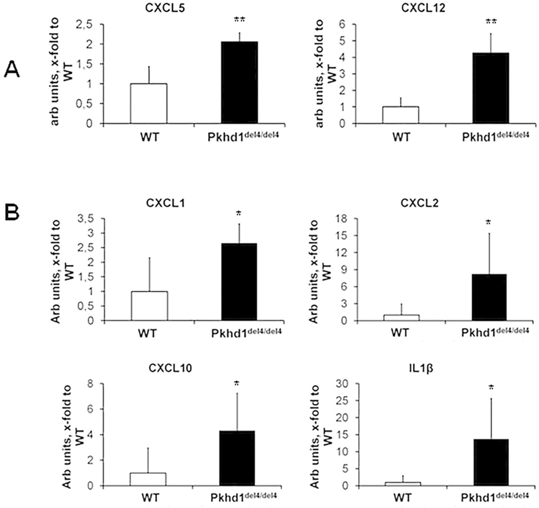
Figure 5. Gene expression analysis of biliary K19 positive structures isolated from Pkhd1del4/del4 and WT mice by LCM shows an increase in CXCL5, CXCL12, CXCL1, CXCL2, CXCL10 and IL1β.
To identify the soluble factors mediating macrophage recruitment by Pkhd1del4/del4 cholangiocytes, gene expression of a number of cytokines previously shown to be secreted by cholangiocytes in culture, was assessed in mRNA selectively captured from epithelial cells lining biliary cysts of Pkhd1del4/del4 and normal ducts of WT mice at 1 and 3 months of age by LCM. A. LIX/CXCL5 and SDF1/CXCL12 were already significantly increased in biliary cysts of Pkhd1del4/del4 mice at 1 month. B. Also KC/CXCL1, MIP-2/CXCL2, IP- 10/CXCL10, SDF1/CXCL12 and IL-1β became significantly increased at 3 months of age (n=4 for WT, n=5 for Pkhd1del4/del4 for both ages). *p<0.05 vs WT, **p<0.01 vs WT.

