

Abstract
Background:
Reconstruction of the anterior cruciate ligament (ACL) involves use of semintendinosis and gracilis tendons graft that is transplanted into bone tunnels at the femoral and tibial insertion sites and the sites and the bone tendon interface is a weak link in the early healing period due to slow rate of healing. We hypothesized that an addition of bone growth factor like Sadat-Habdan mesenchymal stimulating peptide (SHMSP) could enhance bone tendon healing rate so that re-rupture of the tendon does not take place.
Methodology:
Twenty skeletally mature rabbits underwent ACL reconstruction of the right knee. In 10 of the rabbits at the site of the tendon-graft 5 mg/kg body weight of SHMSP was put in the bone tunnel. In 10 other animals, nothing was added. At eight and 12 weeks 5 animals from each group were sacrificed. The tendon-graft site was harvested and sent for histopathological examination to assess the healing at the tendon-bone graft to the tibial tunnel.
Results:
There were no deaths in both the groups. One rabbit of the control group developed an infection. In all the animals of the study group from 4 weeks onward showed bone formation, wherein the control group only granulation tissue was observed. By 8 weeks in the study group, the canal was totally obliterated with the new bone formation which extended onto the periosteal area. In the control, there was minimal change in the formation of the new bone formation.
Conclusion:
Addition of a growth factor like SHMSP would enhance the osteo-integration of the tendon-graft in the bony tunnel after ACL reconstruction in vivo.
Keywords: Anterior cruciate ligament, healing growth factor, Sadat-Habdan mesenchymal stimulating peptide
INTRODUCTION
The anterior cruciate ligament (ACL) is one of major knee ligaments and is critical to knee stability. Injury to the ACL can be a debilitating musculoskeletal injury seen most often in athletes. The incidence of ACL injuries is currently estimated at approximately 200,000 in USA annually, with 100,000 ACL reconstructions performed each year.[1,2] In general, the incidence of ACL injury is higher in people who participate in high-risk sports, such as basketball, football, skiing, and soccer.[3,4,5]
The goal of the ACL reconstruction surgery is to prevent instability and restore the function of the torn ligament, creating a stable knee so that the young can go back to the sporting activities. ACL reconstruction is uasually performed using either the patellar bone tendon and semitendinosis and gracilis tendons, and both are not free from complications. The most common is the graft failure and stretching of the graft due to delay in the tendon-bone healing and tendon-bone incorporation of a tendon-graft within the bone tunnel. Improvement of graft healing to bone is crucial to facilitate early and aggressive rehabilitation and a rapid return to full activity. To counteract this bone morphogenetic factors have been used with good results to improve the bone in growth in the tendon.[6,7] Anoka et al. (2012)[8] reported a potential role of growth factors and bio-scaffolds for improving healing and mechanical integrity of the ACL injury that is reconstructed with a tendon-graft. It was shown that the use of a collagen-platelet-rich plasma scaffold stimulated healing of a defect in the canine ACL.[9,10]
Many other growth factors have been used in the early and better bone ingrowth at the site of ACL reconstruction with bone tendon-graft and tendon-graft.[11,12,13,14] Sadat-Habdan msenchymal stimulating peptide (SHMSP) was discovered at University of Dammam, Dammam and King Fahd Hospital of the University, AlKhobar and was patented in 2008 (United States Patency and Trade Office, US 7,399,826, B1 given on July 15, 2008). SHMP is a 13 amino acids with a molecular weight of 1460 KD, which is now available in the synthesized form. It was shown to stimulate bone growth and accelerate the healing of the fracture.[15,16] It was also shown to stimulate angiogenesis in a fracture module of healthy rabbits.[17] A recent study showed that when topically applied there was early and better healing in diabetic animals.[18]
The objective of this study is to assess the efficacy of a bone growth factor (SHMSP) in the rate of healing of bone tendon interface and osteo-integration of the tendon at the tunnel.
METHODOLOGY
The study was be carried out 20 skeletally mature male New Zealand rabbits. Rabbits were procured and were left in the animal house for 2 weeks for acclimatization to the surrounding. Under ketamine 50 ml/kg weight and xylazine 35 ml/kg weight animals were anesthetized. A 3 cm long lateral half of tendoachilles tendon of the left side was harvested and a bone tunnel was made in the region of ACL and a 1/0 ethilon suture was passed through the end of the harvested tendon [Figure 1]. The tendon was place in the amorphous powder of the SHMSP as a growth factor at a dose of 5 ml/kg body weight A 2.5 mm drill hole was made at the ACL going between the tibia and the femur. The tendon was passed through the bony tunnel made and secured to each end of the tunnel with a 2/0 dexon [Figures 2 and 3]. In the control group, the procedure was repeated without the addition of the SHMSP. Both groups of animals were kept in the similar circumstances and monitored on a regular basis. After 4 weeks, 5 animals from each group were euthanized and 8 weeks the rest of the animals were euthanized. The lower limb was disarticulated at the hip joint and stored in 2% formalin at a temperature of 4°C and before histopathological analysis was done.
Figure 1.
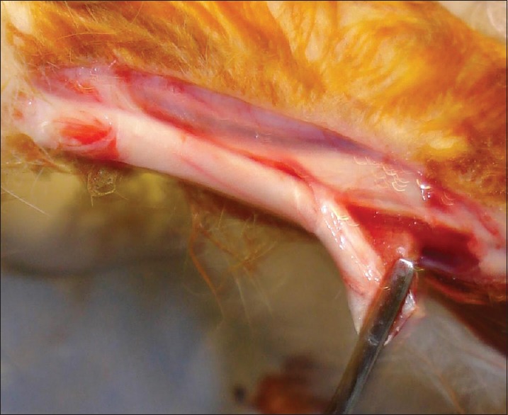
Harvesting of the tendon
Figure 2.
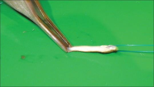
Harvesting and drilling of the tendon
Figure 3.
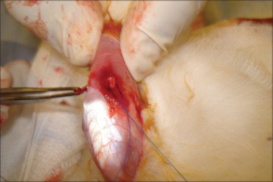
Positioning of the tendon
The two groups were compared specifically for the bone in growth in the drill hole made at the tibial end through, which the tendon-graft was passed. The study was approved by the Institutional Review Board of the University of Dammam and funded by the Deanship of Scientific Research of University of Dammam, Saudi Arabia.
RESULTS
There were no deaths in both the groups. One rabbit of the control group developed an infection. Figures 1–4 shows harvesting of the tendon, drilling, and position of the graft. In all the animals of the study group at 4 weeks showed, newly formed osteoid was observed at places early of the bone formation encroaching the tunnel, where in the control group tunnel was filled with the granulation tissue [Figures 5 and 6].
Figure 4.
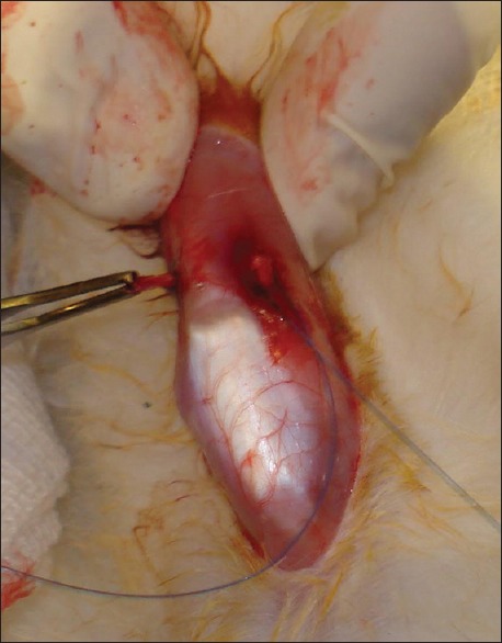
Tendon in position
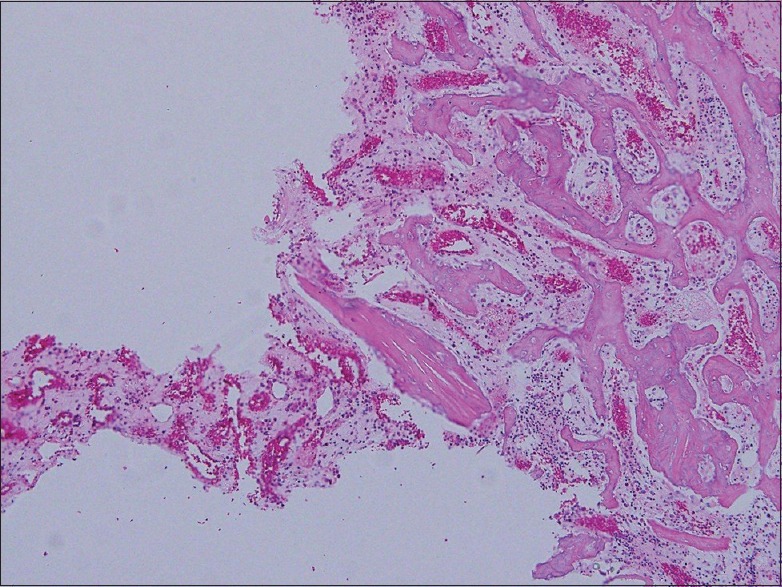
Figure 5.
Photomicrograph of control group at 4 weeks showing fibro-collagenous tissue (fibrosis); (H and E, ×40)
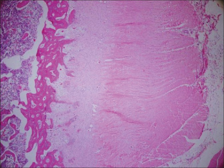
Figure 6.
Photomicrograph of study group at 4 weeks showing early bony exostosis in the tunnel; (H and E, ×40)
By 8 weeks in the study group, the canal was totally obliterated with increased mineralization of the new bone and at places seen extending onto the periosteal surface. In the control, there was minimal change in the formation of the new bone formation but there was more granulation tissue leading to form the connective tissue [Figures 7 and 8].
Figure 7.
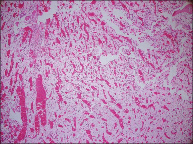
Photomicrograph of control group at 8 weeks showing the whole tunnel is filled with granulation tissue with no signs of any new bone formation; (H and E, ×40)
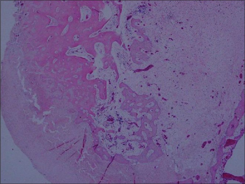
Figure 8.
Photomicrograph of study group at 8 weeks showing abundant new bony formation and little granulation tissue in the tunnel; (H and E, ×40)
DISCUSSION
Our study showed that in animals in which SHMSP was used to augment healing of the tendon-graft in the osseous tunnel there was exuberant bone formation, which was higher and more organized when compared to the control group of animals. The changes were subtle initially but 8 weeks the difference was more pronounced and appreciable. The histological specimens showed increased trabecular bone close to the grafted tendons as early as 4 weeks after implantation.
Different methods and growth factors were used to improve healing of the bone tendon interface. Rodeo et al.[19] found that improved bone formation around a tendon-graft using recombinant human bone morphogenetic protein-2 (BMP-2) in an extra-articular bone tunnel in a dog model. Similarly, Nicklin et al.[20] showed that exogenous osteogenic protein-1 results in improved bone formation at the tendon-bone interface in a sheep model. Recently, Pan et al.[21] injected fibrin sealant (IFS) combined with BMP after ACL reconstruction showed that the rate of new bone formation of IFS-BMP composite was significantly and achieved a more prolonged osteogenic effect. Lim et al.[14] coated mesenchymal stem cells to the tendon-graft thereby enhancing the of tendon-graft osteo-integration. In our study, we coated the tendon with the SHMSP to stimulate the osteo-integration in the tunnel with satisfactory results. In this study, we used SHMSP a small polypeptide, which was reported as an angiogenesis factor and showed results comparable to other growth factors.
The healing pattern between the tendon and the drill hole in the bone through which the tendon passes is not clearly understood but one this is certain that it takes many months probably to heal and incorporate and till that time certain activities are to be curtailed postsurgery.[22,23] For a young athlete to be away from sporting activities is quite difficult and early activity to jeopardize the repair causing up to 10,000 revision yearly of ACL reconstruction in the USA alone.[24] If a osteo-integration of the bone – tendon occurs early then recurrent injuries could be reduced. There is still no clinical evidence regarding the use of growth factors, due to the fact that dosage of these factors still remain undeterminable as most of the half-life of growth factors is too short to stimulate the healing for weeks. With regard to SHMSP which was used on a daily basis and a single dose proved the effect to be similar in fracture healing.[15,16]
We believe that our study has some limitations and the one which stands out that we did not perform bio-mechanical tests to assess the strength of the healing which was so convincing histologically and secondly a small sample of 10 animals on each side of the study arm.
CONCLUSION
Our study shows that local application of SHMSP on the tendon-graft and instilling the growth factor in the tunnel itself enhanced the oseto-integration of the tendon into the bony tunnel created. We believe there is opportunity to convert this animal-based study into a clinical study when the safety of SHMSP is established.
Financial support and sponsorship
Nil.
Conflicts of interest
There are no conflicts of interest.
Acknowledgments
The authors sincerely thank the Deanship of Scientific Research, the University of Dammam for supporting this study with a grant no. 2012088, without this the study could not be completed.
REFERENCES
- 1.Miyasaka KC, Daniel DM, Stone ML. The incidence of knee ligament injuries in the general population. Am J Knee Surg. 1991;4:43–8. [Google Scholar]
- 2.Brown CH, Jr, Carson EW. Revision anterior cruciate ligament surgery. Clin Sports Med. 1999;18:109–71. doi: 10.1016/s0278-5919(05)70133-2. [DOI] [PubMed] [Google Scholar]
- 3.Arendt E, Dick R. Knee injury patterns among men and women in collegiate basketball and soccer. NCAA data and review of literature. Am J Sports Med. 1995;23:694–701. doi: 10.1177/036354659502300611. [DOI] [PubMed] [Google Scholar]
- 4.Griffin LY, Agel J, Albohm MJ, Arendt EA, Dick RW, Garrett WE, et al. Noncontact anterior cruciate ligament injuries: Risk factors and prevention strategies. J Am Acad Orthop Surg. 2000;8:141–50. doi: 10.5435/00124635-200005000-00001. [DOI] [PubMed] [Google Scholar]
- 5.Viola RW, Steadman JR, Mair SD, Briggs KK, Sterett WI. Anterior cruciate ligament injury incidence among male and female professional alpine skiers. Am J Sports Med. 1999;27:792–5. doi: 10.1177/03635465990270061701. [DOI] [PubMed] [Google Scholar]
- 6.Martinek V, Latterman C, Usas A, Abramowitch S, Woo SL, Fu FH, et al. Enhancement of tendon-bone integration of anterior cruciate ligament grafts with bone morphogenetic protein-2 gene transfer: A histological and biomechanical study. J Bone Joint Surg Am. 2002;84-A:1123–31. doi: 10.2106/00004623-200207000-00005. [DOI] [PubMed] [Google Scholar]
- 7.Hashimoto Y, Yoshida G, Toyoda H, Takaoka K. Generation of tendon-to-bone interface “enthesis” with use of recombinant BMP-2 in a rabbit model. J Orthop Res. 2007;25:1415–24. doi: 10.1002/jor.20447. [DOI] [PubMed] [Google Scholar]
- 8.Anoka N, Nyland J, McGinnis M, Lee D, Doral MN, Caborn DN. Consideration of growth factors and bio-scaffolds for treatment of combined grade II MCL and ACL injury. Knee Surg Sports Traumatol Arthrosc. 2012;20:878–88. doi: 10.1007/s00167-011-1641-7. [DOI] [PubMed] [Google Scholar]
- 9.Murray MM, Spindler KP, Abreu E, Muller JA, Nedder A, Kelly M, et al. Collagen-platelet rich plasma hydrogel enhances primary repair of the porcine anterior cruciate ligament. J Orthop Res. 2007;25:81–91. doi: 10.1002/jor.20282. [DOI] [PubMed] [Google Scholar]
- 10.Murray MM, Spindler KP, Ballard P, Welch TP, Zurakowski D, Nanney LB. Enhanced histologic repair in a central wound in the anterior cruciate ligament with a collagen-platelet-rich plasma scaffold. J Orthop Res. 2007;25:1007–17. doi: 10.1002/jor.20367. [DOI] [PubMed] [Google Scholar]
- 11.Karaoglu S, Celik C, Korkusuz P. The effects of bone marrow or periosteum on tendon-to-bone tunnel healing in a rabbit model. Knee Surg Sports Traumatol Arthrosc. 2009;17:170–8. doi: 10.1007/s00167-008-0646-3. [DOI] [PubMed] [Google Scholar]
- 12.Ju YJ, Muneta T, Yoshimura H, Koga H, Sekiya I. Synovial mesenchymal stem cells accelerate early remodeling of tendon-bone healing. Cell Tissue Res. 2008;332:469–78. doi: 10.1007/s00441-008-0610-z. [DOI] [PubMed] [Google Scholar]
- 13.Yamazaki S, Yasuda K, Tomita F, Tohyama H, Minami A. The effect of transforming growth factor-beta1 on intraosseous healing of flexor tendon autograft replacement of anterior cruciate ligament in dogs. Arthroscopy. 2005;21:1034–41. doi: 10.1016/j.arthro.2005.05.011. [DOI] [PubMed] [Google Scholar]
- 14.Lim JK, Hui J, Li L, Thambyah A, Goh J, Lee EH. Enhancement of tendon graft osteointegration using mesenchymal stem cells in a rabbit model of anterior cruciate ligament reconstruction. Arthroscopy. 2004;20:899–910. doi: 10.1016/j.arthro.2004.06.035. [DOI] [PubMed] [Google Scholar]
- 15.Sadat-Ali M, Al-Habdan I. Enhancement of Fracture Healing by a New Peptide. European Orthopaedic Research Society Meeting, Helsinki 4-7th June. Helsinki, Finland, Book of Abstracts. 2003:44. [Google Scholar]
- 16.Sadat-Ali M, Sadat-Ali M, Al-Habdan I. Osteogenic activity of Sadat-Habdan mesenchymal stimulating peptide in diaphyseal segmental defects. Saudi Med J. 2008;29:464–6. [PubMed] [Google Scholar]
- 17.Sadat-Ali M, Al-Habdan I, Shawarby MA. Angiogenesis: A new factor on the block. Angiology. 2005;14:87–91. [Google Scholar]
- 18.Al-Elq AH, Sadat-Ali M, Elsharawy M, Al-Habdan I, Al-Aqeel FO, Naim MM. Topical application of Sadat-Habdan mesenchymal stimulating peptide (SHMSP) accelerates wound healing in diabetic rabbits. Exp Diabetes Res. 2012;2012:531961. doi: 10.1155/2012/531961. [DOI] [PMC free article] [PubMed] [Google Scholar]
- 19.Rodeo SA, Suzuki K, Deng XH, Wozney J, Warren RF. Use of recombinant human bone morphogenetic protein-2 to enhance tendon healing in a bone tunnel. Am J Sports Med. 1999;27:476–88. doi: 10.1177/03635465990270041201. [DOI] [PubMed] [Google Scholar]
- 20.Nicklin S, Morris H, Yu Y, Harrison J, Walsh WR. OP-1 augmentation of tendon-bone healing in an ovine ACL reconstruction. Orthop Trans. 2000;25:155. [Google Scholar]
- 21.Pan W, Wei Y, Zhou L, Li D. Comparative in vivo study of injectable biomaterials combined with BMP for enhancing tendon graft osteointegration for anterior cruciate ligament reconstruction. J Orthop Res. 2011;29:1015–21. doi: 10.1002/jor.21351. [DOI] [PubMed] [Google Scholar]
- 22.Goradia VK. Tendon healing in a bone tunnel. Parts I and II. Arthroscopy. 2003;19:111. doi: 10.1053/jars.2003.50029. [DOI] [PubMed] [Google Scholar]
- 23.Tomita F, Yasuda K, Mikami S, Sakai T, Yamazaki S, Tohyama H. Comparisons of intraosseous graft healing between the doubled flexor tendon graft and the bone-patellar tendon-bone graft in anterior cruciate ligament reconstruction. Arthroscopy. 2001;17:461–76. doi: 10.1053/jars.2001.24059. [DOI] [PubMed] [Google Scholar]
- 24.Ferretti A, Conteduca F, Monaco E, De Carli A, D'Arrigo C. Revision anterior cruciate ligament reconstruction with doubled semitendinosus and gracilis tendons and lateral extra-articular reconstruction. J Bone Joint Surg Am. 2006;88:2373–9. doi: 10.2106/JBJS.F.00064. [DOI] [PubMed] [Google Scholar]