Abstract
The INHAND (International Harmonization of Nomenclature and Diagnostic Criteria for Lesions in Rats and Mice) project is a joint initiative of the Societies of Toxicologic Pathology from Europe (ESTP), Great Britain (BSTP), Japan (JSTP), and North America (STP) to develop an internationally accepted nomenclature and diagnostic criteria for nonproliferative and proliferative lesions in laboratory animals. The purpose of this publication is to provide a standardized nomenclature and diagnostic criteria for classifying lesions in the digestive system including the salivary glands and the exocrine pancreas of laboratory rats and mice. Most lesions are illustrated by color photomicrographs. The standardized nomenclature, the diagnostic criteria, and the photomicrographs are also available electronically on the Internet (http://www.goreni.org/). Sources of material included histopathology databases from government, academia, and industrial laboratories throughout the world. Content includes spontaneous and age related lesions as well as lesions induced by exposure to test items. Relevant infectious and parasitic lesions are included as well. A widely accepted and utilized international harmonization of nomenclature and diagnostic criteria for the digestive system will decrease misunderstandings among regulatory and scientific research organizations in different countries and provide a common language to increase and enrich international exchanges of information among toxicologists and pathologists.
Keywords: diagnostic pathology, nomenclature, diagnostic criteria, digestive system or tract, oral cavity, esophagus, stomach, intestine, small intestine, large intestine, salivary glands, pancreas se
I. General Introduction
The digestive tract is the entry site into the body for orally administered test articles. An irritant test article may lead to local acute lesions at this first site of contact to the body and the digestive tract has been identified as the organ most commonly affected in British patients admitted to hospital with adverse drug reactions (Pirmohamed et al. 2004).
On the other hand, orally administered test articles may be highly toxic to other organs yet having little or no noticeable effect on the digestive tract.
A distinctive feature of the digestive tract is the high proliferative rate of the epithelium, making it particularly sensitive to agents interfering with cell division, but resulting also in a high regenerative capacity. The villi enhance the surface of the intestine to 400–500 m² in humans. Because of the large surface area of these tissues, accurate assessment of potential treatment effects is almost entirely dependent on a thorough gross examination and sampling of focal lesions.
Standardized nomenclature and diagnostic criteria are essential to harmonize the classification and reporting of non-proliferative as well as proliferative histopathological lesions. They should facilitate communication between different research groups and regulatory authorities. In this way they should reduce time and efforts in the review of histopathology data by regulators and minimize clarifying questions to the sponsor. Furthermore, standardized diagnostic criteria are a prerequisite for the generation of any historical control database of histopathological lesions. Discussions about the future need of concurrent control groups highlight the importance of these standards.
The INHAND Project (International Harmonization of Nomenclature and Diagnostic Criteria for Lesions in Rats and Mice) is a joint initiative of the societies of toxicologic pathology from Europe (European Society of Toxicologic Pathology - ESTP), UK (British Society of Toxicological Pathologists - BSTP), Japan (Japanese Society of Toxicologic Pathology - JSTP), and North America (Society of Toxicologic Pathology - STP) to unify, update and complete the existing WHO/IARC and STP/SSNDC nomenclature systems. The INHAND nomenclature and the related diagnostic criteria should represent the future international standard in toxicologic pathology. They represent a consensus of senior toxicologic pathologists and were reviewed by the INHAND-GESC (INHAND-Global Editorial and Steering Committee) for compliance with INHAND principles. All members of the Societies of Toxicologic Pathology involved in the INHAND process had the opportunity to comment to the draft version during a 6-week period.
However, these recommendations for diagnostic criteria and preferred terminology may not be applicable in all situations. Purposes of specific experiments or the specific context of a given study may require deviation from this standardized nomenclature and diagnostic criteria. The appropriate diagnoses are ultimately based upon on the discretion of the senior toxicologic study pathologist.
The present publication provides a set of standardized terms, diagnostic criteria and example images for the upper and lower digestive tract as well as for the salivary glands and the pancreas. The nomenclature of liver lesions has been published separately (Thoolen et al. 2010) and that of tooth lesions is in preparation. Like all other INHAND publications, the nomenclature and diagnostic criteria for the digestive tract are also available online (website.goreni.org/). The online version contains additional images and useful links to differential diagnoses characterizing it as a practical tool for diagnostic work.
The recommended nomenclature is generally descriptive rather than diagnostic. The diagnostic criteria used require standard hematoxylin and eosin stained paraffin sections only. Histochemical or immunohistochemical staining characteristics may be addressed in the comments section of the respective lesion. Such special techniques may be required in some situations, but a comprehensive discussion of these methods is outside the scope of this publication. Lesions of disseminated organs like blood vessels, soft tissues or peripheral nerves are generally described in separate publications (Kaufmann et al. 2012, Greaves et al. 2013). They are described here only in cases where the digestive tract is a major site of manifestation, e.g. leiomyoma of the intestine.
Lesions included in this nomenclature system may be further specified by modifiers. Criteria are given for modifiers that are considered to be of particular relevance. These modifiers should be consistently applied. It is upon the discretion of the pathologist to use additional modifiers not suggested in this nomenclature system. Such modifiers may describe the location, tissue type or duration among others. Further principles of the INHAND nomenclature have been published separately (Mann et al. 2012).
Findings that can reliably be diagnosed grossly are also not covered by this monograph. This applies in particular to some congenital malformations like intestinal duplication (Elangbam et al. 1998; Tamai et al. 1999).
This nomenclature system describes lesions that occur in conventional (wildtype) strains of rats and mice. Previous rodent nomenclatures for the digestive tract have been published by our societies and international WHO and other collaborative committees (Betton et al. 2001; Deschl et al. 1997; Deschl et al. 2001; Frantz et al. 1991; Leininger and Jokinen 1994; Takahashi and Hasegawa 1990; Whiteley et al. 1996). Lesions occurring in specific genetically engineered mouse models only are not covered in this document. For more information on these specific lesions the reader should refer to relevant publications of workshops on genetically engineered mouse models (e.g. Boivin et al. 2003; Hruban et al. 2006).
II. Upper Digestive Tract
A. Introduction
The oral cavity/pharynx, tongue, and esophagus are components of the upper alimentary tract, and each has a mucosa lined by a stratified squamous epithelium with a keratinized surface. Incidental nonproliferative or proliferative microscopic findings may occur in these tissues as well as microscopic findings that are associated with effects of the test article (systemic or direct contact exposure) or, rarely, changes related to injury from the dosing (gavage) procedure.
B. Morphology (Anatomy & Histology)
The borders of the oral cavity are defined by several general landmarks, including the hard and soft palate/pharynx dorsally; the gingiva, teeth, and mucosal surfaces of the lips/cheeks laterally; and the floor of the mouth/tongue ventrally. Oral cavity/pharynx is typically not a protocol-required tissue for microscopic evaluation, but portions may be sampled for examination due to a clinical sign or gross finding at necropsy. The oral cavity/pharynx tissue samples may include minor salivary glands (palatine, buccal), adjacent muscle, bone, teeth, or haired skin, in addition to the vascular and connective tissue of the keratinized, squamous epithelium of the mucosal surface.
The tongue is comprised of the interlacing bundles of striated muscle interspersed with adipocytes, scant to numerous mast cells, nerves, and vascular tissue lined with a squamous mucosal surface. In the tongue of aged rats, the amount of adipose tissue has been reported to be decreased with a relative increase in its fibrous connective tissue as compared to that in younger animals (Brown and Leininger 1994). The squamous mucosa of the dorsal tongue surface contains numerous papillae and a large circumvallated papilla, while the lateral/ventral surface is a simple stratified squamous mucosa (Hebel and Stromberg 1976; Brown and Hardisty 1990). The thin, compact lamina propria of the mucosa of the tongue is less dense on the ventral surface nearer the root attachment (frenulum) to the oral cavity surface. In the more caudal, ventral area, there are large lymphatic vessels, veins, and ducts from the minor salivary glands in the adjacent oral mucosa.
The esophagus consists of a muscle (tunica muscularis) layer with longitudinal and circular-oriented striated muscle fibers and a squamous mucosa with a keratinized surface that typically has small aggregates of bacterial coccoid organisms within the superficial layers of keratin.
C. Physiology
The physiology of the oral cavity, tongue, and esophagus is related primarily to their function as a conduit for food. Effects on function with the development of megaesophagus have been reported in mice with abnormal myogenic plexus (Randelia and Lalitha 1988; Randelia et al. 1990), and esophageal obstruction, secondary to effects of scopolamine on salivary secretion and the swallowing reflex, had been described for rats (NTP TR 445 1997).
D. Necropsy & Trimming
The routine necropsy includes gross examination of the oral cavity (oropharynx), tongue, and esophagus. Although sampling and histologic examination of the oral cavity/pharynx are generally limited to necropsy observations, portions of these tissues may be included with routine nasal or tooth sections required for possible histopathology examination. In those circumstances when oral cavity structures may be considered as potential targets sites for nonneoplastic or neoplastic changes, whole-head fixation (calvarium and brain removed), may be the better approach for consistent examination of required tissues. With whole-head fixation, sectioning of the nasal/oral cavities, mandible, and associated tissues can be performed at various levels and result in consistent orientation for sectioning of desired tissue sites in these special circumstances.
Unlike the oral cavity/pharynx, routine histologic sections of the tongue and esophagus are usually protocol-required for microscopic evaluation in all animals from toxicity and carcinogenicity studies. For microscopic evaluation, the tongue may be prepared as a longitudinal histologic section (see RITA trimming guide Ruehl-Fehlert et al. 2003, website//reni.item.fraunhofer.de/reni/trimming) that provides for examination of lingual glands; it often may prepared as a transverse section that permits evaluation of the dorsal, lateral, and ventral mucosal surfaces. Depending on the cross-section level (midanterior versus midcaudal) of tongue taken for routine evaluation, the serous and mucin acini of the lingual glands and their ducts may also be present. The esophagus is typically prepared as a cross-section at approximately the location of the thyroid gland; adjacent trachea is also included with this section. In studies where the thyroid/parathyroid is not required for organ weight determination, these glands may be left in situ and included with the routine esophagus/trachea section (see RITA trimming guide Ruehl-Fehlert et al. 2003, website//reni.item.fraunhofer.de/reni/trimming).
E. Nomenclature, Diagnostic Criteria and Differential Diagnosis
a. Congenital Lesions
Introduction
Congenital/developmental lesions in the oral cavity/pharynx, tongue, and esophagus have been rarely reported in mice and rats from toxicity and carcinogenicity studies. This is most likely due to their uncommon occurrence, exclusion of animals with these findings prior to study initiation, and minimal routine sampling of this location in the absence of a gross observation. In developmental and reproductive studies, congenital/developmental lesions are more commonly observed.
Ectopic sebaceous gland
Figure 1.
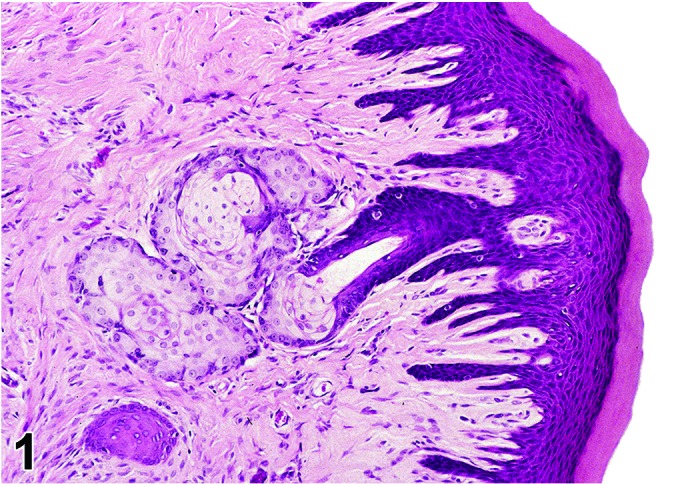
Rat gingiva. Ectopic sebaceous glands (Fordyce’s granules).
Figure 2.
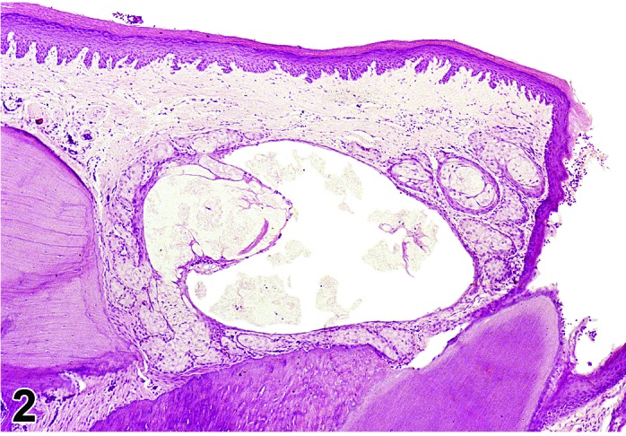
Rat gingiva. Cystic dilation of duct in ectopic sebaceous glands.
Synonym: Fordyce’s granules
Pathogenesis: This developmental change consists of aggregates of ectopic dermal sebaceous glands (Fordyce’s granules) in the oral cavity of rats (Yoshitomi et al. 1990) and is associated with gingival mucosa. This is most common in the F344 strain and seen primarily in males.
Diagnostic features
May appear grossly as white nodule or cyst in the gingival mucosa.
Most commonly observed in gingival mucosa of upper incisors.
Consists of normal sebaceous gland acini with ducts opening to mucosal surface.
Presence of cysts (dilated ducts) with or without inflammation may be components of these ectopic glands.
Differential diagnosis
Sebaceous gland adenoma: absence of common duct/lumen and presence of mitotic figures; adenoma of ectopic sebaceous gland in the gingiva of rats has not been reported.
Comment: This finding is more likely observed in routine nasal cavity sections that may include teeth and gingival tissue. With the exception of its correlation to a gross observation when a cyst/inflammation has developed, this finding is of little or no apparent pathologic significance.
Cleft palate
Synonyms: Palatoschisis; congenital malformation
Pathogenesis: Defect in the fusion of the bone and overlying mucosa of the hard palate.
Diagnostic features
Midline space defect in oral mucosa and hard palate.
Visible grossly or microscopically.
No evidence of trauma.
Differential diagnosis
Trauma: Evidence of necrosis, fracture, or inflammatory response.
Comment: Cleft palate is a longitudinal defect in bone and mucosa of the midline of the hard palate resulting from failure of fusion of the lateral palatine shelves from the maxillary processes (Jones et al. 1997).Cleft palate in mice and rats has been attributed to maternal treatment with high doses of vitamin A (Kalter and Warkany 1961) and recently been reported as a genetic mutation in the mouse (Stottmann et al. 2010). This condition has also been produced in mice (Era et al. 2009) and rats with in utero chemical exposure or as an effect from puncture of the amniotic sac (Ferguson 1981; Schuepbach and Schroeder 1984). Diagnosis is based primarily on the gross observation.
Dilatation, esophagus
Figure 3.
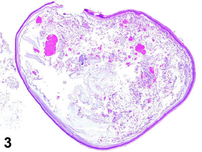
Rat esophagus. Dilation.
Figure 4.
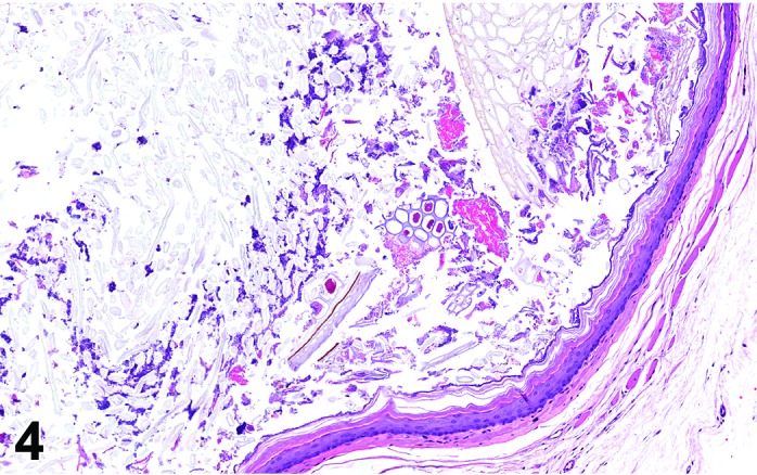
Rat esophagus. Detail of dilated esophagus in Figure 3. Dilation characterized by lumen distended with food content.
Synonyms: Distention; megaesophagus; dilatation; impaction
Pathogenesis: May be spontaneous/idiopathic, due to food impaction, or secondary to an alteration of the esophageal neuromuscular function, decreased secretion of the salivary glands or an altered swallowing reflex.
Diagnostic features
Differential diagnosis
Thinning of the muscle and mucosal layers as result of distention of the esophagus with food/bedding material.
Differential diagnosis
None.
Comment: This condition is typically related to a grossly distended esophagus containing food/bedding material and has also been diagnosed as impaction because of the gross appearance. This has been described as a spontaneous or idiopathic change (Harkness and Ferguson 1979; Ruben et al. 1983) in rats and also attributed to feeding a powdered food diet (Brown and Hardisty 1990). Esophageal dilation associated with obstruction occurred in rats administered scopolamine (National Toxicology Program 1997a) and as a background finding in several mouse strains (Haines et al. 2001; Mahler et al. 2000), in particular in mice with an abnormal myogenic plexus (Randelia and Lalitha 1988; Randelia et al. 1990). Esophageal dilation, described as megaesophagus, has also been reported as an induced finding in mice with intrauterine exposure to diethylnitrosamine (Ghaisas et al. 1989) and in rats secondary to effects of scopolamine on salivary secretion and the swallowing reflex (National Toxicology Program 1997b).
Diverticulum, esophagus
(Figure 5)
Figure 5.
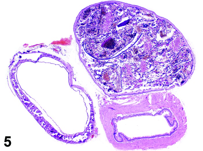
Rat esophagus. Diverticulum distended with food content.
Synonym: Pharyngoesophageal diverticulum
Pathogenesis: Usually caused by mechanical or biochemical disruption of esophageal wall structures resulting in loss of extracellular matrix integrity and internal projection of surface lining into deeper layers; may alternatively represent a congenital abnormality.
Diagnostic features
Irregular cyst-like extension of the mucosal lumen into or through the muscularis layer in the esophagus.
Lumen of diverticulum that opens to mucosal surface.
Focal.
Differential diagnosis
Cyst: Isolated from mucosal surface.
Comment: Diverticulum of the esophageal mucosa has been reported in rats (Brown and Hardisty 1990).
Malformation
Synonym: Congenital malformation
Pathogenesis: Rare developmental defects have been reported for the esophagus. Duplication of the esophagus has been reported in an adult male rat as a spontaneous occurrence (Canpolat et al. 1998) and tracheal-esophageal malformation represents one of many teratogenic effects reported in mice with intrauterine exposure to adriamycin (Dawrant, et al. 2007).
Diagnostic features
Gross observation of congenital/developmental structural alterations.
Differential diagnosis
None.
b. Cellular Degeneration, Injury and Death
Cyst
Figure 6.
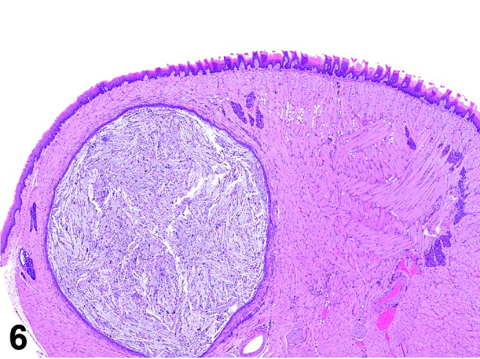
Mouse tongue. Cyst.
Figure 7.
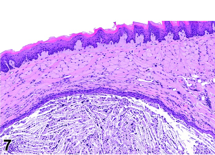
Mouse tongue. Detail of cyst in Figure 6. Lined by squamous epithelium, this cyst likely originated from an obstructed salivary gland duct in the tongue.
Pathogenesis: Epithelial cysts may occur as downgrowth of entrapped surface mucosa or a dilated salivary duct in the oral cavity/pharynx, tongue, or esophagus and have been observed in the oral cavity/pharynx of rats with dilated glands/ducts associated with ectopic sebaceous glands (Yoshitomi et al. 1990).
Diagnostic features
Dilated gland/ducts lined by simple squamous epithelium.
Lumen of cyst may contain sebaceous secretion or keratin debris.
Inflammation associated with larger or ruptured cysts.
Differential diagnosis
Esophageal diverticulum: Lumen opens into the esophagus lumen, epithelial connection to the surface mucosa of the esophagus.
Atrophy, squamous epithelium
(Figure 8)
Figure 8.
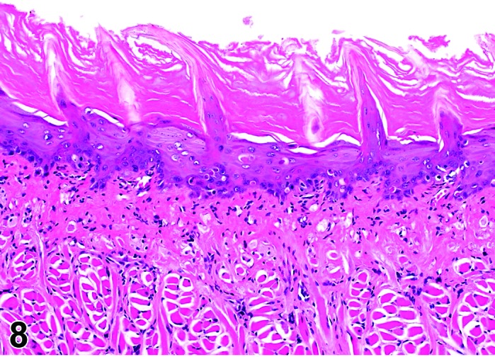
Rat tongue. Atrophy, epithelium and apoptosis.
Synonym: Atrophy
Pathogenesis: Decrease in normal thickness/cellularity of squamous mucosa of tongue, pharynx or esophagus.
Diagnostic features
Decreased thickness of mucosal epithelial layer.
Focally extensive or diffuse lesion.
Differential diagnosis
Erosion/ulcer: Focal or focally extensive absence of superficial or, in severe cases, of all epithelial layers with extension of the alteration into the muscularis. Unlike atrophy, this finding is typically associated with surface cellular debris and an inflammatory cell infiltrate in mucosa or as a surface exudate.
Erosion/ulcer
Figure 9.
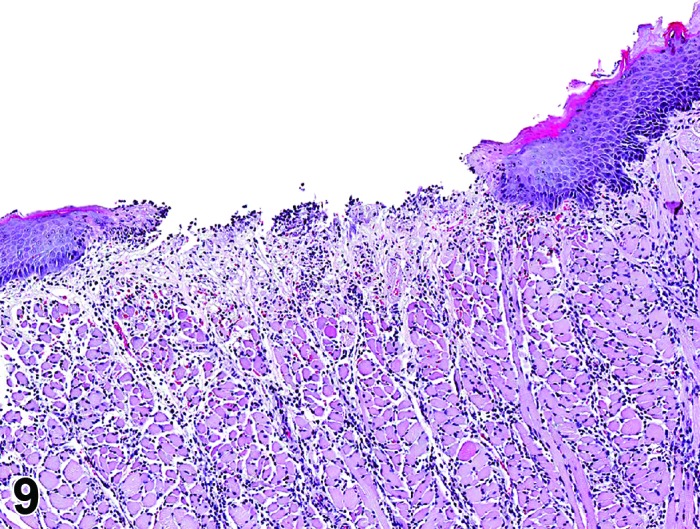
Rat tongue. Ulcer with mixed inflammatory cell infiltrate in muscle layer.
Figure 10.
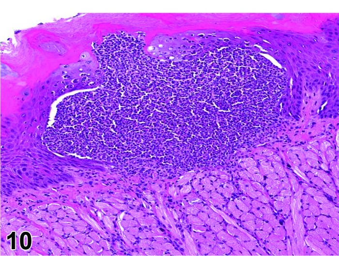
Rat tongue. Ulcer (borderline erosion) with inflammation, neutrophilic.
Synonyms: Erosion; ulcer; ulceration
Pathogenesis: Localized disruption in the uniform thickness/continuity of the squamous epithelium in the oral cavity/pharynx, tongue, or esophagus with either partial (erosion) or full penetration of the squamous epithelium (ulcer).
Diagnostic features
Focal or focally extensive loss of surface epithelium in the oral cavity/pharynx, tongue, or esophagus.
Basal lamina may remain intact (erosion) or in more severe lesions the defect in the mucosa may extend into the muscularis (ulcer).
Intraepithelial inflammatory cell infiltrate frequently associated with more severe lesions.
Differential diagnoses
Squamous cell carcinoma: may present with erosion/ulcer appearance on the mucosal surface. The presence of this neoplasm would preclude diagnosis of an associated erosion/ulcer.
Atrophy, epithelial: Reduced thickness of all layers of the squamous epithelium or, in severe cases, absence of basal germinative layers; usually no inflammatory cell infiltrate.
Comment: Although there are specific morphologic criteria to differentiate the more superficial lesion (erosion; confined to the epithelial surface) from the deeper ulcer (extension of the epithelial defect through basal cell layer/basal lamina), the plane of section through smaller, focal lesions often impacts on the appearance (depth of the epithelial loss) of this finding. In most instances it is not practical to separate these changes and they are typically diagnosed under the single, combined term of erosion/ulcer with an appropriate severity grade.
Erosion/ulcer usually is the result of epithelial necrosis. Because of its unique topography adjacent to the lumen and often involving multiple organ layers, which uniquely affects the progression and resolution of the lesion (e.g. margin hyperplasia, granulomatous tissue, inflammation), it is useful to record erosion/ulcer separately from epithelial necrosis.
A deep ulcer of the esophagus may be perforating if it destroys all layers of the esophagus wall, and may be designated as such by a free text entry. However, a perforation resulting from a physical insult, e.g. gavage-related, typically is a macroscopic lesion and generally not recorded on the light microscopic level.
Apoptosis, squamous epithelium
Figure 11.
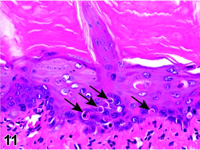
Rat tongue Apoptosis of epithelial cells (arrows). Higher magnification of Figure 8.
Synonyms: Apoptotic cell death
Pathogenesis: Gene regulated, energy dependent process leading to formation of apoptotic bodies which are phagocytosed by adjacent cells; typically associated with cytotoxic chemotherapeutics that affect the mucosal epithelium of the tongue, esophagus and/or pharynx.
Diagnostic features
Single cells or small clusters of cells.
Cell shrinkage.
Hypereosinophilic cytoplasm.
Nuclear shrinkage, pyknosis, karyorrhexis.Intact cell membrane.
Apoptotic bodies.
Cytoplasm retained in apoptotic bodies.
Phagocytosis of apoptotic bodies tissue macrophages or other adjacent cells.
No inflammation.
Differential diagnoses
Necrosis, epithelium: The morphologic features of cell death clearly fit with necrotic diagnostic criteria (cells and nuclei swollen, pale cytoplasm etc.).
Apoptosis/necrosis, epithelium: Both types of cell death are present and there is no requirement to record them separately or a combined term is preferred for statistical reasons. The term is also used when the type of cell death cannot be determined unequivocally.
Erosion/ulcer: Focal or focally extensive absence of superficial or, in severe cases, of all epithelial layers with extension of the alteration into the muscularis.
Comment: The approach for the nomenclature and diagnostic criteria of cell death applied here is based on a draft recommendation of the INHAND Cell Death Nomenclature Working Group.
Apoptosis is not synonymous with necrosis. The main morphological differences between these two types of cell death are cell shrinkage with nuclear fragmentation and tingible body macrophages in apoptosis versus cell swelling, rupture and inflammation in necrosis; however, other morphologies (e.g. nucelar pyknosis and karyorrhexis) overlap. In routine H&E sections where the morphology clearly represents apoptosis or single cell necrosis, or whereby special procedures (e.g. TEM or IHC for caspases) prove one or the other, individual diagnoses may be used. However, because of the overlapping morphologies, necrosis and apoptosis are not always readily distinguishable via routine examination, and the two processes may occur sequentially and/or simultaneously depending on intensity and duration of the noxious agent (Zeiss 2003). This often makes it difficult and impractical to differentiate between both during routine light microscopic examination. Therefore, the complex term apoptosis/necrosis may be used in routine toxicity studies.
A differentiation between apoptosis and single cell necrosis may be required in the context of a given study, in particular if it aims for mechanistic investigations. Transmission electron microscopy is considered to represent the gold standard to confirm apoptosis. Other confirmatory techniques include DNA-laddering (easy to perform but insensitive), TUNEL (false positives from necrotic cells) or immunohistochemistry for caspases, in particular caspase 3. These techniques are reviewed in detail by Elmore (Elmore 2007). Some of these confirmatory techniques detect early phases of apoptosis, in contrast to the evaluation of H&E stained sections, which detects late phases only; overall interpretation of findings should take into account these potential differences. Thus, low grades of apoptosis may remain unrecognized by evaluation of H&E stained slides only.
Certain studies may require the consideration of forms of programmed cell death other than apoptosis which requires specific confirmatory techniques (Galluzzi et al. 2012).
Necrosis, squamous epithelium
Synonyms: Oncotic cell death; oncotic necrosis; necrosis
Modifiers: Single cell
Pathogenesis: Unregulated, energy independent, passive cell death with leakage of cytoplasm into surrounding tissue and subsequent inflammatory reaction; may be induced by direct contact with a test article after oral uptake/administration.
Diagnostic features
Cells swollen with pale eosinophilic cytoplasm.
Loss of nuclear basophilia, pyknosis and/or karyorrhexis that affects aggregates of cells.
In more severe cases, there may be clefting and detachment of the epithelium from the submucosa.
Typically, the presence of degenerative cells is a component of necrosis.
Minimal or slight inflammatory cell infiltrates may be present as a feature of necrosis.
Single cell
Only single cells affected.
Differential diagnoses
Apoptosis, epithelium: The morphologic features of cell death clearly fit with apoptosis (cell and nucleus shrunken, hypereosinophilic cytoplasm, nuclear pyknosis etc.) and/or special techniques demonstrate apoptosis.
Apoptosis/necrosis: Both types of cell death are present and there is no requirement to record them separately or a combined term is preferred for statistical reasons. The term is also used when the type of cell death cannot be determined unequivocally. Atrophy, epithelium: Reduced thickness of all layers of the squamous epithelium or, in severe cases, absence of basal germinative layers; usually no cellular degeneration or necrosis.
Erosion/ulcer: Focal or focally extensive absence of superficial or, in severe cases, of all epithelial layers with distention of alteration into the muscularis.
Comments: Epithelial necrosis usually develops into erosion/ulcer and in the past has frequently been recorded as such. A separate recording of necrosis as the initial event in this process is recommended when the structure of the mucosa is still intact.
Apoptosis/necrosis, squamous epithelium
Synonym: Cell death
Pathogenesis: Unregulated, energy independent, passive cell death with leakage of cytoplasm into surrounding tissue and subsequent inflammatory reaction (single cell necrosis) and/or gene regulated, energy dependent process leading to formation of apoptotic bodies which are phagocytosed by adjacent cells (apoptosis); typically associated with cytotoxic chemotherapeutics that affect the mucosal epithelium of the tongue, esophagus and/or pharynx.
Diagnostic features
Both types of cell death are present and there is no requirement to record them separately or a combined term is preferred for statistical reasons.
The type of cell death cannot be determined unequivocally.
Differential diagnoses
Apoptosis, epithelium: The morphologic features of cell death clearly fit with apoptosis (cell and nucleus shrunken, hypereosinophilic cytoplasm, nuclear pyknosis etc.) and/or special techniques demonstrate apoptosis and there is a requirement for recording apoptosis and necrosis separately.
Necrosis, epithelium: The morphologic features of cell death clearly fit with necrotic diagnostic criteria (cells and nuclei swollen, pale cytoplasm etc.) and there is a requirement for recording apoptosis and necrosis separately.
Erosion/ulcer: Focal or focally extensive absence of superficial or, in severe cases, of all epithelial layers with distention of alteration into the muscularis.
Comments: Necrosis and apoptosis are not always readily distinguishable via routine examination, and the two processes may occur sequentially and/or simultaneously depending on intensity and duration of the noxious agent (Zeiss 2003). This often makes it difficult and impractical to differentiate between both during routine light microscopic examination. In these cases, the combined term apoptosis/necrosis may be used. It is recommended to explain in detail the use of this combined term in the narrative part of the pathology report.
Degeneration/necrosis, muscle
Figure 12.
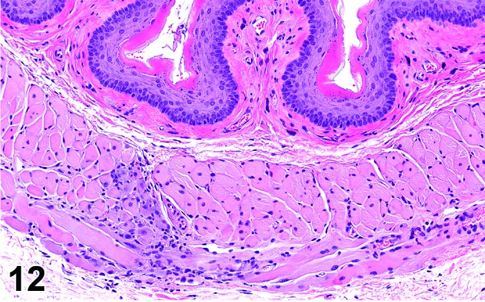
Rat esophagus. Degeneration/necrosis, muscle.
Pathogenesis: Degeneration and necrosis of muscle fibers in the tongue or esophagus.
Diagnostic features
Myocyte degeneration/necrosis of muscle fibers characterized typically by the presence of both swollen or contracted hyperchromatic or pale eosinophilic degenerative fibers and necrotic fibers with coagulative necrosis and nuclear pyknosis or lysis.
Usually a focal change of minimal severity.
Amphophilic muscle fibers consistent with a regenerative response may be present and a primary feature of this finding.
Differential diagnosis
Degeneration/necrosis or regeneration of muscular layer in conjunction with inflammation, edema and/or hemorrhage associated with gavage-related injury (discussed below under inflammation, mixed cell, gavage-related).
Comment: This change can occur as a focal lesion in the esophagus that also variably includes the presence of several degenerative and/or regenerative muscle fibers. While potentially a test article-related or incidental finding, it may be related to very minor physical effects from the gavage procedure. Morphologic features more conclusive of gavage-related injury such as perforation and inflammation are not present. In the absence of such clear, gavage-related inflammatory changes, degeneration/necrosis is the preferred diagnosis for this esophagus finding. Degeneration/necrosis of muscle fibers in the tongue may be related to test article administration (Greaves 2012).
Amyloid
Figure 13.
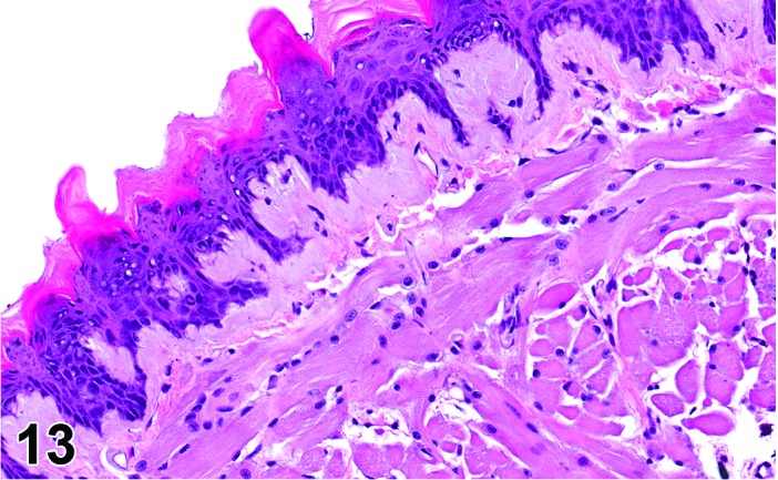
Mouse tongue. Subepithelial amyloid deposition.
Synonyms: Amyloidosis; amyloid deposition
Pathogenesis: Extracellular deposits of polypeptide fragments of a chemically diverse group of glycoproteins.
Diagnostic features
Affects medium or larger vessels (arterioles) of the tongue.
Accumulates as a thin layer of homogenous and pale eosinophilic amyloid material in basal lamina of mucosa in the tongue and esophagus.
Green birefringence using polarized light with Congo red stain.
Differential diagnosis
Fibrinoid change (necrosis) in vessel walls: Deeply eosinophilic homogenous or coagulative appearance of the vascular media.
Comment: Amyloid deposition occurs in the tongue and esophagus of mice, although much less frequently at these locations as compared to other locations (e.g. kidney, intestine).
The characteristic morphologic appearance and location of amyloid in H&E sections that is usually adequate to make this diagnosis. Confirmation of the deposits as amyloid can be accomplished with light microscopy using special stains such as Congo red. Amyloid appears apple green under polarized light with this stain.
The term “hyaline” should be used in case of uncertainties about the nature of extracellular deposits of hyaline material in H&E sections and the unavailability of a Congo red stain. Hyaline is also the appropriate term for homogenously eosinophilic extracellular deposits that react negative with Congo red.
Mineralization
Figure 14.
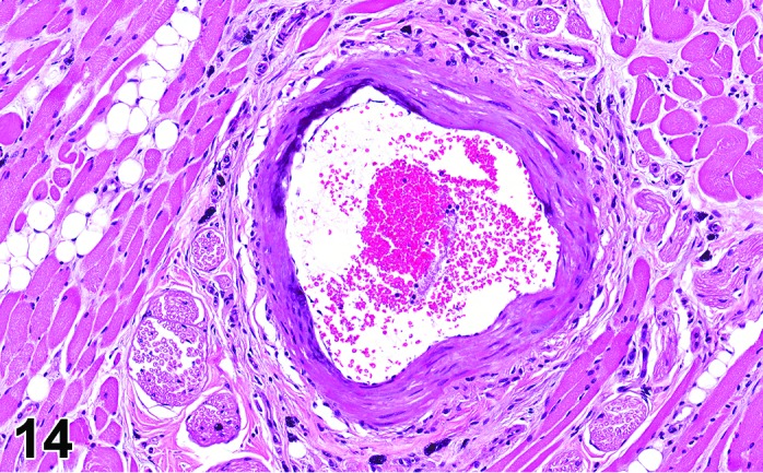
Rat tongue. Mineralization, vessel.
Synonyms: Calcification; mineral deposition
Pathogenesis: Mineralization of blood vessel walls and muscle fibers of the oral cavity/pharynx, tongue, or esophagus.
Diagnostic features
Basophilic granular deposits in the wall of arteries as well as within individual muscle fibers.
Mineral deposits usually easily identified in routine sections but can be further characterized by special stains (Alizarin red, von Kossa).
Differential diagnosis
None.
Pigment
Synonyms: Pigment deposition; pigment
Pathogenesis: Increased pigment has been described in the tongue muscle of aging rats (Bodner et al. 1991) and in the oral mucosa of rats (AG strain) given antimalarial drugs (Savage et al. 1986).
Diagnostic features
Yellow/brown pigment (lipofuscin) in muscle fibers of the tongue.
Brown/black pigment (melanin) within oral mucosa.
Differential diagnosis
None.
Comment: Pigment (increased pigmentation) is not a common finding in the oral cavity/pharynx, tongue, and esophagus. When this is present, histochemistry, immunohistochemistry, ultrastructural, or other laboratory methods may be required to characterize this material.
Hyperkeratosis
(Figures 15 and 16)
Figure 15.
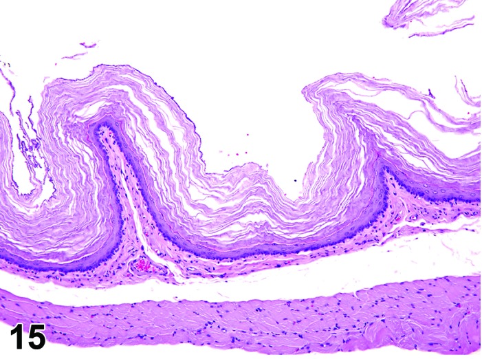
Rat esophagus. Hyperkeratosis, orthokeratotic.
Figure 16.
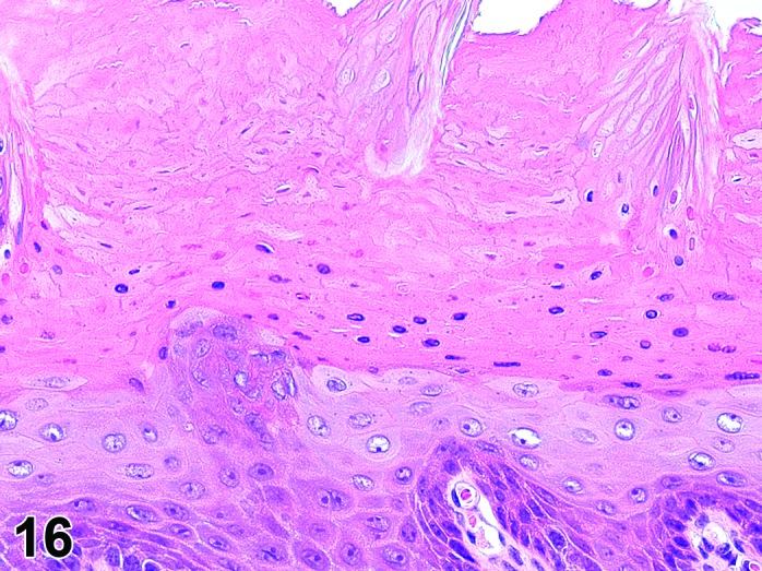
Rat, tongue. Hyperkeratosis, parakeratotic.
Synonyms: Hpyerkeratosis, orthokeratotitc may have been identified as hyperkeratosis
Hyperkeratosis, parakeratotic may have been identified as parakeratosis
Modifiers: Orthokeratotic, parakeratotic
Pathogenesis: Increased production and/or retention of keratin by squamous epithelium of the mucosa with normal (orthokeratotic) or abnormal (parakeratotic) maturation of the keratin layer.
Diagnostic features
Increase in keratin on the surface of the mucosa.
Focal or diffuse.
Often accompanied by hyperplasia of squamous epithelium.
Orthokeratotic
Thickened keratin layer with non-nucleated keratinized cells.
Parakeratotic
Thickened keratin layer with nucleated keratinized cells.
Differential diagnoses
Normal: Keratin layer that is normally less than the thickness of the mucosal squamous epithelium.
Hyperplasia, squamous cell: Proliferation and thickening of the stratum spinosum.
Comment: Parakeratosis of the esophagus has been described in zinc-deficient rats (Barney et al. 1968). It may also be induced by vitamin imbalances or hydroxymethylglutaryl-CoA reductase inhibitors (Sigler et al. 1992).
c. Inflammatory Lesions
It is recommended that when possible, descriptive terminology should be used to best characterize the type (e.g., neutrophil, mixed cell, mononuclear cell, lymphocyte, etc.) of cellular component(s)/infiltrates rather than a specific type/duration of inflammation (e.g., acute, ulcerative, suppurative, subacute, chronic) for changes in the oral cavity, pharynx, tongue and esophagus. The presence of various inflammatory cell infiltrates in the absence of other features of inflammation should be diagnosed as an ‘infiltrate’ with predominate/appropriate cell type(s) identified. However, when other prominent morphologic features (congestion, hyperemia, hemorrhage, edema, necrosis, fibrosis, foreign material, bacterial organisms, etc.) are present, it is preferable to characterize the change as an inflammatory process (inflammation) with specific descriptors. The reader is referred to the Nomenclature and Review of Basic Principles Document for discussion on the preferred use of descriptive, rather than diagnostic terminology (Mann et al. 2012). Listed below are descriptive diagnostic terms and examples of inflammatory cell infiltrates or types of inflammation. Many of the examples provided in this manuscript are more consistent with inflammation, as indicated by figure legends. Subsequently we describe common types of inflammation in an exemplary manner, while we give only a general description of inflammation for other parts of the digestive tract.
Infiltrate
(Figures 17 and 18)
Figure 17.
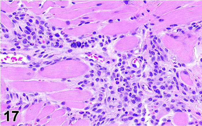
Rat tongue. Infiltrate, mononuclear cell.
Figure 18.
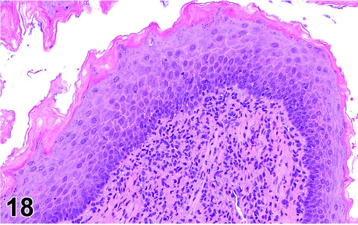
Rat esophagus. Infiltrate, mononuclear cell.
Synonyms: Infiltrate inflammatory; infiltrate inflammatory cell; infiltration (plus modifier); infiltration, inflammatory; infiltration, inflammatory cell
Modifiers: Type of inflammatory cell that represents the predominant cell type in the infiltrate
Pathogenesis: Infiltrate of lymphocytes, plasma cells, macrophages, neutrophils, eosinophils or mixtures thereof within the mucosa or muscle layers of the oral cavity/pharynx, tongue, or esophagus without other histological features of inflammation as noted above.
Diagnostic features
Focal, multifocal or diffuse cellular infiltrate in mucosa/muscle layers.
Presence of mononuclear or polymorphonuclear leucocytes but no other histological criteria of inflammation.
Lesion may be associated with overlying ulceration, distended/ruptured cyst, self-induced traumatic lesions or dysplasia/neoplasia of the teeth in the oral cavity/pharynx.
Differential diagnoses
Inflammation: In addition to the infiltrate, other morphologic features of inflammation such as edema, hemorrhage, necrosis and/or fibroplasia will be present.
Granulocytic leukemia: Infiltration of neutrophil precursors and/or abnormal neutrophils will probably be present along with mature neutrophils; infiltration of similar neoplastic cells is likely present in other organs.
Lymphoma: Infiltration of monomorphic population of lymphocytes (often with atypical, increased and/or abnormal mitoses); infiltration of similar neoplastic cells is likely present in other organs.
Comment: Other examples of infiltrates based on cell type include: Infiltrate, mononuclear cell (representing lymphocytes and macrophages); Infiltrate, lymphocyte, Infiltrate, macrophage; Infiltrate, mixed (lymphocytes, neutrophils, and/or macrophages).
Inflammation
(Figures 19, 20, 21 and 22)
Figure 19.
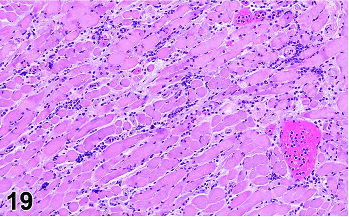
Rat tongue. Inflammation, neutrophilic, muscle.
Figure 20.
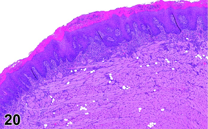
Rat tongue. Inflammation, mixed with associated reactive hyperplasia of adjacent mucosal epithelium.
Figure 21.
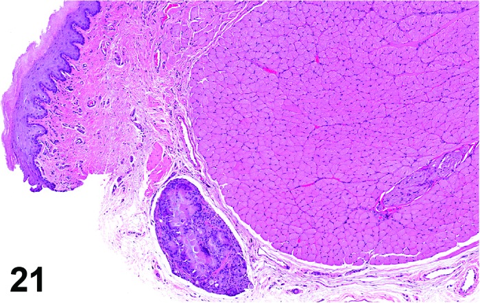
Rat tongue. Inflammation, foreign body.
Figure 22.
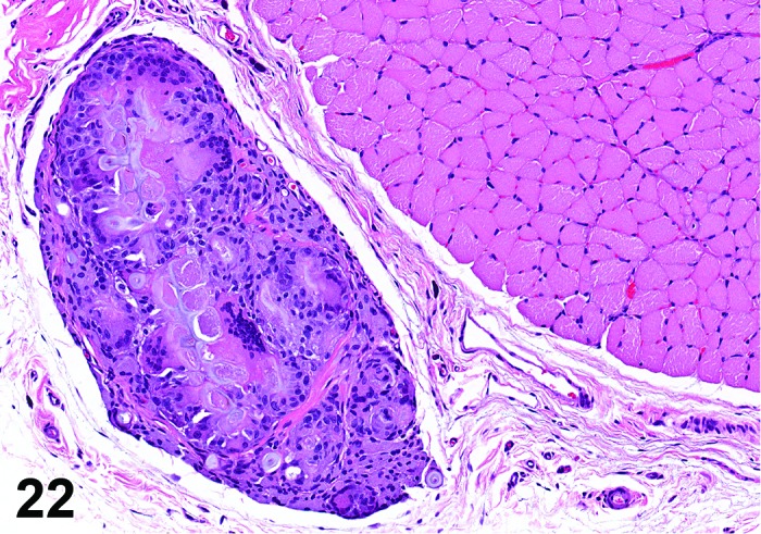
Rat tongue. Inflammation, foreign body.
Synonyms: Stomatitis; glossitis; pharyngitis; esophagitis - dependent on localization
Modifiers: Type of inflammatory cell that represents the predominant cell type in the inflammation
Inflammation, mononuclear cell
Pathogenesis: Inflammation in mucosa or muscle layers of the oral cavity/pharynx, tongue, or esophagus.
Diagnostic features
Inflammatory cell infiltrate comprised primarily of lymphocytes and macrophages.
Presence of other histological criteria of inflammation e.g. fibroplasia or fibrosis.
Fibrosis may be a prominent component in a more chronic inflammatory change.
May be associated with overlying ulcer, distended/ruptured cyst, or self-induced traumatic lesions. In the oral cavity/pharynx, the process may be associated with dysplasia/neoplasia of the teeth.
Differential diagnoses
Infiltrate, lymphocytes OR Infiltrate mononuclear cell: Other morphological features of inflammation such as edema, hemorrhage, necrosis and/or fibroplasia will be absent.
Lymphoma: Infiltration of monomorphic population of lymphocytes (often with atypical, increased and/or abnormal mitoses); infiltration of similar neoplastic cells is likely present in other organs.
Comment: When a mononuclear cell infiltrate is considered to represent an inflammatory process (inflammation, mononuclear cell) and other morphologic features (fibrosis, mineralization) associated with the infiltrate suggest an ongoing/continuing inflammatory process, the modifier ‘chronic’ may be included in the diagnosis (i.e. inflammation, mononuclear cell, chronic) to better characterize the finding.
Inflammation, mixed cell
Pathogenesis: Inflammatory cell response within the mucosa or muscle layers of the oral cavity/pharynx, tongue, or esophagus.
Diagnostic features
Mixed inflammatory cell infiltrate consisting of variable numbers of neutrophils, lymphocytes or macrophages without predominance of one cell type.
Edema, congestion/hemorrhage, and/or cell debris are components of the mixed cell infiltrates more consistent with an inflammatory process.
May be associated with overlying ulcer, distended/ruptured cyst, self-induced traumatic lesions or dysplasia/neoplasia of the teeth in the oral cavity/pharynx.
In the oral cavity/pharynx, the inflammatory process may be associated with dysplasia/neoplasia of the teeth. Reactive/regenerative hyperplasia of the overlying mucosal epithelium may also be a feature of this inflammatory response.
Differential diagnosis
Infiltrate, neutrophils: Other morphological features of inflammation such as edema, hemorrhage, necrosis and/or fibroplasia will be absent.
Comment: When a mixed cell infiltrate is considered to represent an inflammatory process (inflammation, mixed cell) with the presence of other morphologic features (fibrosis; granulomatous inflammation, granulation tissue and a prominence of neutrophils), the modifier ‘chronic’ may be included in the diagnosis (inflammation, mixed cell, chronic) to better characterize the finding.
Inflammation, mixed cell is commonly observed with gavage-related injury (Figures 21 and 22). It may have variable histopathological appearance, dependent on extent of damage and duration/onset of the lesion to include: perforation (typically seen macroscopically), bacterial organisms, or foreign material in the pharyngeal / esophageal or peripharyngeal / periesophageal tissue with the variable presence of degeneration/necrosis and/or regeneration of muscular layer in conjunction with inflammation, edema or hemorrhage. If the lesion is interpreted to be secondary to gavage-related injury, this may be addressed in the pathology narrative, or alternatively may be diagnosed as inflammation, mixed cell, gavage-related to distinguish from spontaneous and/or test article-related findings.
Inflammation, foreign body
(Figures 21 and 22)
Pathogenesis: Inflammatory cell response to presence of foreign material; observed primarily in the tongue.
Diagnostic features
Focal inflammatory lesion, most often seen in mucosa, ducts of minor salivary glands or muscle layers of the tongue.
Mixed inflammatory cell infiltrate (macrophage/multinucleated giant cell, lymphocyte, neutrophil) sometimes associated with an imbedded hairshaft fragment or other ingested foreign material (food/bedding material).
Differential diagnoses
Abscess: Circumscribed and grossly visible globular process with characteristic zonation of inner cell debris, neutrophils, and outer capsule of fibrosis/fibroplasias with numerous capillaries (granulation tissue).
Inflammation, mixed cell: The inflammatory process lacks foreign material and/or multinucleated giant cells.
d. Vascular Lesions
Inflammation, vessel
Figure 23.
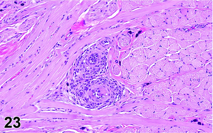
Rat tongue. Inflammation, vessel.
Synonyms: Arteritis; vasculitis; polyarteritis
Pathogenesis: Inflammation of vascular wall of the oral cavity/pharynx, tongue, or esophagus.
Diagnostic features
Typically affects muscular wall of arteries in tongue.
Variable mixed inflammatory cell infiltrate within and around vessel.
Possible presence of fibrinoid necrosis of vessel wall.
Proliferation of endothelium and thickening (fibrosis) of vessel wall.
Differential diagnoses
Infiltrate, inflammatory cell: May occur perivascular, but the vessel wall itself is unaffected.
Comment: More information and differential diagnoses are given in the INHAND publication of nomenclature and diagnostic criteria of the cardiovascular system (Berridge B et al. in preparation).
Edema
Pathogenesis: Accumulation of tissue fluid in the interstitium resulting from increased vascular permeability.
Diagnostic features
Increased eosinophilic fluid (interstitial fluid) within interstitium.
Differential diagnosis
Inflammation, neutrophils: Usually associated with tissue and vascular damage resulting in exudative edema and inflammatory cell infiltrate.
Comment: Edema accompanying an inflammatory process should be recorded separately if it is a predominant lesion.
Hemorrhage
Figure 24.
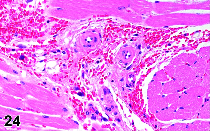
Rat tongue. Hemorrhage.
Pathogenesis: Increased vascular permeability (diapedesis) or rupture of blood vessels.
Diagnostic features
Presence of erythrocytes in tissues or lumina of the upper digestive tract.
Differential diagnoses
Angiectasis: Blood present within dilated vascular lumina.
Artifact: Erythrocytes on the surface of tissues or within lumina as a result of necropsy procedures.
Comment: Hemorrhage of the tongue is a frequent finding in studies with blood sampling from lingual veins.
e. Non-neoplastic Proliferative Lesions
Hyperplasia, squamous cell
(Figures 25, 26 and 27)
Figure 25.
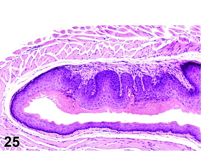
Rat esophagus. Hyperplasia, squamous cell.
Figure 26.
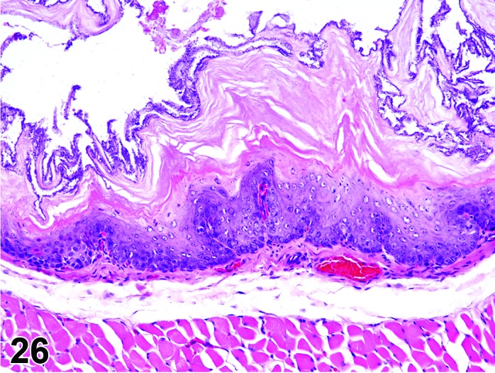
Rat esophagus. Hyperplasia, squamous cell.
Figure 27.
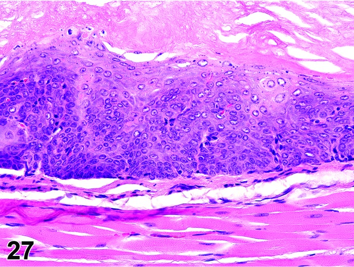
Rat esophagus. Hyperplasia squamous cell, atypical.
Synonyms: Hyperplasia, squamous cell, atypical may have been identified as: Dysplasia; hyperplasia with atypia
Modifier: Atypical
Histogenesis: Squamous epithelium (including the basal cells) of mucosa in the oral cavity/pharynx, tongue, or esophagus.
Diagnostic features
Thickening of well-differentiated squamous epithelium.
Focal, multifocal, or diffuse.
Growth pattern may be plaque-like, with blunt papillary projections or endophytic.
Papillary projections not or only minimally branched and without fibrovascular stroma.
Broad-based hyperplastic foci may have multiple straight or minimally branched projections.
Endophytic growth with cyst-like structures in the mucosa may occur. Cysts lined by well-differentiated squamous epithelium and filled with keratin; may extend into the submucosa, but basement membrane is intact.
Rete peg formation may be exaggerated, i.e. papillary body may show undulation due to enlarged germinal layers.
Often accompanied by hyperkeratosis.
Atypical
Thickening of squamous epithelium with presence of irregular shaped or nodular foci of basal or undifferentiated epithelial cells within squamous mucosa.
Increased nuclear to cytoplasmic ratio.
Abnormal cell shape/size/nuclear morphology.
Keratinization of cells within deeper layers of mucosa.
Differential diagnoses
Papilloma, squamous cell: Generally larger with a prominent stromal fibrovascular stalk.
Carcinoma, squamous cell: Invasion with loss of basement membrane integrity.
Hyperplasia, basal cell: Downgrowth of basal cells is the predominant component; no cellular or nuclear atypia.
Hyperplasia, squamous cell, atypical: Presence of nuclear or cellular atypia.
Hyperplasia, basal cell
Histogenesis: Basal layer of the stratified squamous epithelium.
Diagnostic features
Proliferation of the basal cell layer, basophilic staining increased.
Focal or diffuse.
Endophytic growth pattern.
No alteration of basement membrane integrity.
Differential diagnoses
Hyperplasia, squamous cell: Thickening of the epithelium with all normally existing layers.
Carcinoma, squamous cell: Evidence for lost basement membrane integrity, spinous cells and keratinized cells proliferate, cellular atypia.
Comment: Basal cell hyperplasia and squamous cell hyperplasia may occur concurrently.
Isolated nests of basal cells may occur in the lamina propria, dependent on the plane of section through the rete peg structures. However, their discrete border indicates an intact basement membrane.
f. Neoplasms
Neoplastic lesions in the oral cavity/pharynx, tongue, and esophagus are usually correlated to a gross observation. In addition to the proliferative lesions of squamous cell origin, neoplasms of the bone, tooth, or adjacent soft tissues (malignant schwannoma, Zymbal’s gland tumor) may extend into the oral cavity and be associated with a gross observation at this location. These tumors are described in separate INHAND publications on “Musculoskeletal system”, “Soft tissue”, and “Mammary, Zymbal’s, and Clitorial Glands”.
Papilloma, squamous cell
Figure 28.
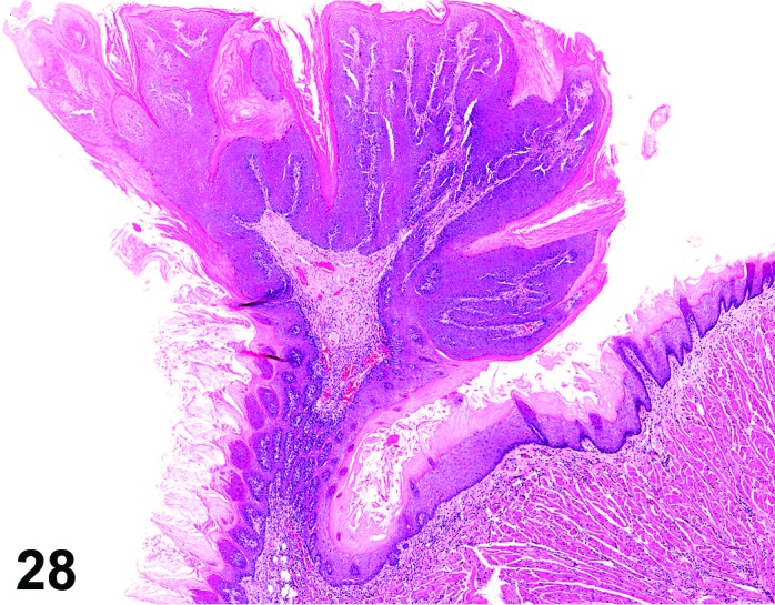
Mouse tongue. Papilloma, squamous cell.
Histogenesis: Epithelial squamous cells of the mucosa in the oral cavity/pharynx, tongue, and esophagus.
Diagnostic features
Central fibrovascular stalk with multiple finger/frond-like projections covered with variably thick squamous epithelium.
Squamous epithelium often heavily keratinized.
Cells show orderly maturation.
When occurring on the tongue, are generally dorsally located.
Differential diagnoses
Hyperplasia, squamous cell: Generally smaller, well-differentiated cells, absence of a prominent stromal component, particularly stalk.
Carcinoma, squamous cell: Invasion with lost basement membrane integrity.
Comment: Clusters of epithelial cells may occur in the stalks as a result of tangential sectioning but are not indicative of malignant change.
Carcinoma, squamous cell
(Figures 29, 30, 31 and 32)
Figure 29.
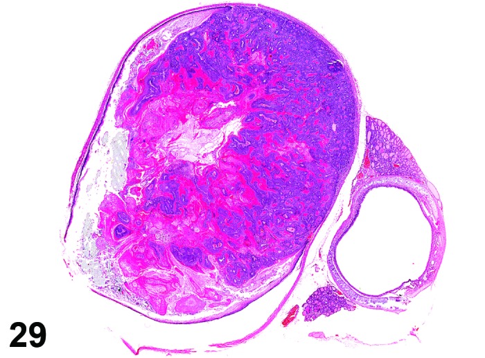
Rat esophagus. Carcinoma, squamous cell.
Figure 30.
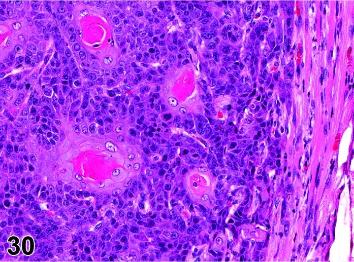
Rat esophagus. Detail of carcinoma, squamous cell in Figure 29.
Figure 31.
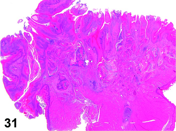
Rat tongue. Carcinoma, squamous cell.
Figure 32.
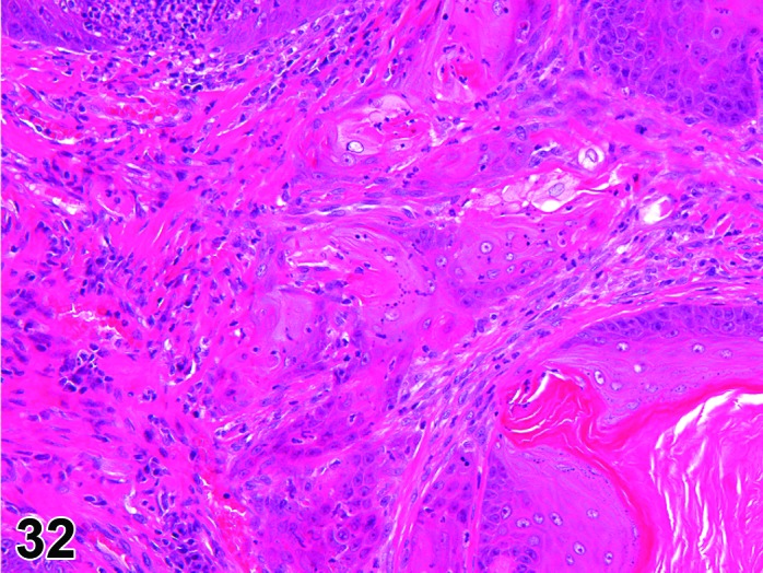
Rat tongue. Carcinoma, squamous cell. Detail of carcinoma, squamous cell in Figure 31.
Histogenesis: Epithelial squamous cells of the mucosal surface in the oral cavity/pharynx, tongue, and esophagus.
Diagnostic features
Invasion with destruction of basement membrane, often in adjacent tissue.
Keratin masses on the surface and in crypts between foldings of malignant squamous epithelium.
Strands of malignant cells may form keratin pearls.
Many mitotic figures.
Cellular and nuclear pleomorphism.
Spindle shaped or ovoid cells and multinucleated cells occur in poorly differentiated tumors.
Individual cells often loose polarity.
Differential diagnoses
Papilloma, squamous cell: Generally smaller, well-differentiated cells, presence of a prominent stromal component, particularly as a component of a stalk, no destruction and/or invasion of basement membrane.
Hyperplasia, (atypical), squamous cell: Generally smaller, well-differentiated cells, no destruction and/or invasion of basement membrane.
Tumor, granular cell, benign
(Figures 33 and 34)
Figure 33.
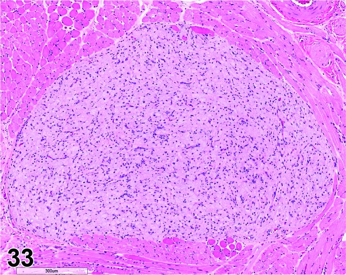
Rat tongue. Tumor, granular cell, benign.
Figure 34.
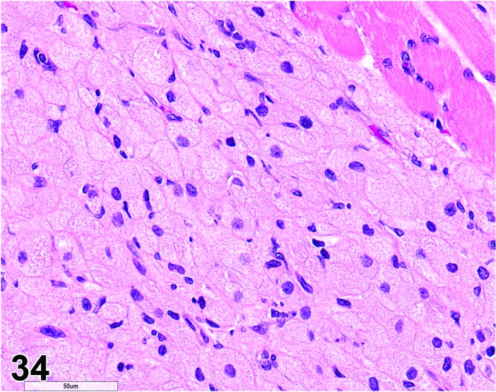
Rat tongue. Tumor, granular cell, benign. Higher magnification of Figure 33.
Histogenesis: Uncertain origin for this benign neoplasm; initially considered as being derived from mesenchymal/striated muscle (myoblastoma) but recent reports have suggested most likely a neural/schwann cell origin.
Diagnostic features
Consists of a solid, irregularly rounded, micronodular proliferation in the muscle layer of the tongue.
Minimal compression of surrounding musculature but striated muscle fibers may be present in the periphery of the tumor.
Relatively homogeneous population of polygonal cells with central to eccentric, round to oval-shaped nuclei with finely dispersed chromatin pattern. A prominent nucleolus may be present. Tumor cells are usually closely associated with minimal separation by a delicate fibrovascular stroma. The cytoplasm is typically finely stippled with abundant eosinophilic granules based on H&E staining.
Some tumor cells may be elongated with irregular nuclei and/or small and rounded with sparsely granular cytoplasm and chromatin-dense nuclei.
Mitotic figures are generally absent in this benign neoplasm.
Differential diagnoses
Aggregates, granular cell (although there are no reports this has been observed in the tongue, this likely could occur early in the process, as described in the INHAND CNS/PNS and Female Reproductive systems): Few scattered cells to small aggregates with minimal disturbance of normal tissue architecture and no compression of adjacent tissue.
Tumor, granular cell malignant (although there are no reports this has been observed in the tongue, there is the potential for this lesion to progress to malignancy, as described in the INHAND CNS/PNS and Female Reproductive systems): Prominent pleomorphism and/or invasion of surrounding tissue.
Comment: Benign granular cell tumor (GCT) of the tongue is rare and has not been previously reported in rodents. We are currently aware of several cases of granular cell tumors in the tongue of Wistar rats (4 females and one male) and one case in a Sprague Dawley rat (male) but are unaware of this neoplasm occurring in the tongue of mice. GCTs have been reported in the tongue as well as other sites in the dog, cat, horse, and a bird (Patnaik 1993). Although uncommon, GCTs most frequently occur in the head and neck region (often in the tongue) of humans with a slight predominance in females (Becelli et al. 2001; van de Loo et al., 2015). GCTs in the meninges of rats (Kaufmann et al., 2012) and the reproductive tract (uterus/cervix/vagina) of rats and mice have also been described (Dixon et al, 2014) but their relationship to the GCT observed in the tongue of rats is unknown.
III. Stomach
The rodent stomach is morphologically different from the stomach of other laboratory species and from that of humans. In the rat and mouse the proximal portion, the nonglandular stomach (synonym forestomach), is lined by stratified squamous epithelium and comprises approximately half of the total area of the stomach. It is separated from the distal glandular stomach by the limiting ridge (squamocolumnar junction), where the squamous epithelium is thicker than elsewhere in the stomach.
As humans lack a nonglandular stomach, the relevance of test article-related changes in this tissue for humans can be challenged. However, it is likely that the squamous mucosa of the esophagus (in species without the nonglandular stomach), will react to test articles in a similar way as the nonglandular stomach epithelium, if equivalent exposure levels are attained. Therefore, interpretations of changes in the nonglandular region need to take account of retention time of a test article in the stomach and physiological functioning of the stomach (Greaves 2012). Exposure to the rodent nonglandular stomach by gavage can mimic aspects of skin painting studies, i.e. direct contact of test chemical to the keratinized squamous epithelium.
A. Morphology (Anatomy)
The glandular stomach of rodents has histological and functional similarities to that of other mammals with both corpus and antral regions. It anatomically differs from other non-rodent laboratory animal species and humans by the lack of a notable cardiac region in the glandular mucosa and the absence of the dorsal/anterior pouch in the glandular region known as the fundus. In rodents, the analogous dorsal/anterior pouch is nonglandular although the term glandular fundus is frequently used for the corpus. The term “fundic gland” has been generically applied to the human stomach to denote glands containing parietal (oxyntic) and/or chief (zymogenic) cells, regardless of anatomic site. Rodents do not have glands in their fundus; however, the ubiquitous use of the term in medical pathology has led to its frequent use in rodent studies. Similarly, pylorus is sometimes used to denote the antrum; however, its use should be limited to designating the portion of the antrum adjoining the gastroduodenal junction.
B. Morphology (Histology)
The surface epithelium of the glandular mucosa is a simple columnar mucous epithelium that extends into the gastric pits; the depth of the gastric pit varies based on location in the corpus or antrum, being most shallow close to the limiting ridge and becoming deeper approaching the antrum. Accordingly, the proportion of fundic glands (Synonym: oxyntic glands) contributing to the thickness of the corpus mucosa is greatest near the limiting ridge on the greater curvature and progressively diminishes toward the corpus/antral junction.
The fundic glands are continuous with the gastric pit and have a simple tubular structure with isthmus, neck and base regions. There are 4 cell types in the corpus: mucous neck cells which produce mucus; parietal or oxyntic cells which produce hydrochloric acid and intrinsic factor; chief or zymogenic cells which produce pepsinogen; and argentaffin or neuroendocrine cells that produce a variety of endocrine and paracrine hormones. The isthmus region serves as the site of proliferation with mucous neck cells acting as progenitor cells that differentiate luminally to become mucous epithelium and basally to become the specialized cells of the fundic glands (Karam, 1999). This is reflected in differences of the turnover rate, which is at around 3 days in mucous cells compared to 194 day in oxyntic and 54 day in parietal cells (Karam, 1999). In the antrum, the glands are lined by mucous cells and neuroendocrine cells including those that produce gastrin. The turnover rate of mucus cells is much faster at around 3 days than oxyntic or parietal cells (about 194 and 54, respectively) (Karam, 1999).
The mucus phenotype of gastric epithelium and glandular cells can be identified by histochemical methods, e.g. the combination Alcian blue pH 2.5 followed by PAS stain (Table 1).
Table 1. Mucin Profiles of Gastric Epithelial and Glandular Cells.
| Cell type | Mucin produced | Histochemical staining properties | |
|---|---|---|---|
| PAS | Alcian blue pH 2.5 | ||
| Cardiac glands | Neutral and small amount of sialomucin | Positive | Occasional positive cells |
| Corpus: Surface epithelium | Neutral | Positive | Negative |
| Corpus: Mucus neck cells | Mostly Neutral | Scant positivity | Negative |
| Antrum/pylorus | Mixed, becoming more acidic towards duodenum | Positive | Positive |
| Corpus: mucus metaplasia, parietal cell layer (foamy cells on H&E) | Mixed with increased neutral and variable acidic | Positive increased | Positive |
| Corpus: antral (“pseudoduopyloric”) metaplasia* (cuboidal to columnar non-foamy cells on H&E) |
Mixed neutral and acidic | Positive increased | Positive |
*Intestinal metaplasia with Cdx2+ surface epithelial and/or goblet cells also will present with a mixed mucin profile, but is rare in rodents.
The mucosa is supported by the connective tissue of the lamina propria and the continuous muscle layer of the muscularis mucosa. Beneath the muscularis mucosa is the relatively loose connective tissue of the submucosa, which contains larger blood vessels, lymphatics and nerves, and the tunica muscularis. The external surface is covered by the serosal membrane.
C. Physiology
The esophagus enters into the nonglandular region of the rodent stomach. Functionally, the nonglandular stomach serves as a temporary storage organ and, due to prolonged exposure, is potentially a major site for interaction with test articles entering the body via oral ingestion. As a result of this storage function, the presence of irritants in the diet may quickly lead to damage and inflammation in this area. The lower pH in the nonglandular stomach when compared to the esophagus may be of major importance, as it may influence the diffusion of test articles into the lipophilic squamous epithelium.
In the stomach, food undergoes mechanical and chemical breakdown to form chyme which then passes into the duodenum for further digestion. Mechanical breakdown is achieved through the churning action of the tunica muscularis and chemical breakdown results from the action of digestive juices secreted from the glandular mucosa. The glandular mucosa produces an acidic watery secretion containing pepsinogen. Pepsinogen is converted into active pepsin in the acid environment which then hydrolyses proteins into polypeptide fragments. Self digestion of the stomach mucosa is prevented by a layer of surface mucous which has a higher pH than the gastric juice due to secretion of bicarbonate ions by the surface mucous cells. Changes in, or loss of, the mucous layer by test articles may quickly lead to erosion and ulceration of the glandular epithelium. On the other hand complete inhibition of acid secretion by test articles will lead to hypergastrinemia which, if maintained, leads to proliferation of the enterochromaffin-like cells.
D. Necropsy & Trimming
Attention should be paid to the amount and nature of the stomach contents at necropsy. A stomach abnormally distended with food, particularly in a rodent fasted overnight, may indicate that the test article and/or vehicle is inhibiting gastric function, and this may have lead to reflux of stomach contents and entry of gastric contents into the respiratory tract (Damsch et al. 2011). Conversely, a stomach grossly distended with gas is often an indication in a rodent that there has been a blockage in the upper respiratory tract resulting in dyspnoea, mouth breathing and aerophagia.
In its natural configuration, the non-distended stomach has an irregularly folded mucosa and submucosa. Consistent opening and positioning during fixation and trimming are necessary to obtain standardized sections of both glandular corpus and antrum and nonglandular stomach with correct orientation of the muscularis, submucosa and mucosa.
At necropsy, the stomach should be opened along its greater curvature to the proximal part of the duodenum, any ingesta washed off with isotonic saline and then pinned to a rigid surface (e.g. cork). The stomach should be gently but sufficiently stretched to produce a flattened surface of both nonglandular and glandular regions but excessive stretching should be avoided. Alternatively, the pylorus may be ligated and the stomach inflated with fixative for a short period, before opening and flattening between filter paper prior to further fixation (Mahler et al. 2000) However, an excessive volume must not be used to avoid excessive stretching.
Trimming of stomach should be undertaken as specified in the goRENI trimming guide (website.reni.item.fraunhofer.de/reni/trimming) so that standardized sections of nonglandular epithelium, corpus, antrum and pyloric-duodenal junction are obtained. The ratios of parietal and chief cells in the fundic glands and the height of the mucosa vary between lesser and greater curvature so inter-animal variation in trimming needs to be avoided and a sensitivity to topography maintained. Furthermore it is important to recognize that the glandular corpus mucosa may be minimal or absent along the lesser curvature with the antral mucosa extending to the limiting ridge.
In the following sections, lesions that may affect the nonglandular and glandular regions in a similar manner are described only once.
E. Nomenclature, Diagnostic Criteria and Differential Diagnosis
a. Congenital Lesions
Congenital/developmental lesions occur occasionally in rodents. Most occur as isolated cases and the pathologist must distinguish such background changes from test article-induced lesions.
Cyst, squamous
(Figures 35 and 36)
Figure 35.
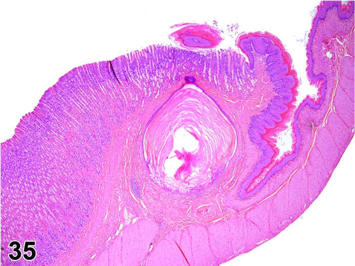
Rat glandular stomach. Cyst squamous.
Figure 36.
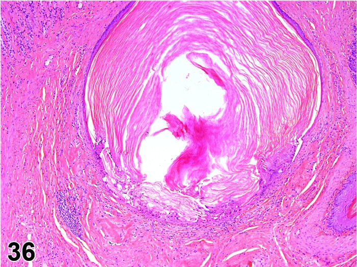
Rat glandular stomach. Cyst squamous. Higher magnification of Figure 35.
Synonym: Cyst, epithelial
Pathogenesis: Invagination of stratified squamous epithelium into the wall of the nonglandular stomach.
Diagnostic features
Most commonly seen in nonglandular stomach or near the limiting ridge in glandular mucosa.
Keratinized stratified squamous epithelium.
Mucous cells occasionally present.
May extend into submucosa/tunica muscularis.
Lumen frequently contains keratinized material which may be mineralized.
May be surrounded by chronic inflammation.
Differential diagnosis
Carcinoma, squamous cell: Epithelial atypia and invasive growth patterns with penetration of basement membrane by single cells or nests of neoplastic cells.
Comment: While squamous cysts are most likely to be of congenital origin they may potentially form during life secondary to derangement of the epithelium e.g. in an inflammatory process. If post-inflammatory extension is the suspected pathogenesis, the pathologist may choose to interpret the focal change as a diverticulum, rather than a congenital cyst.
Ectopic tissue
(Figures 37, 38, 39 and 40)
Figure 37.
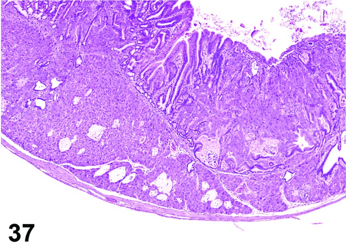
Mouse glandular stomach. Hepatocytes, ectopic; note presence of hepatocytes in the submucosa and mucosa.
Figure 38.
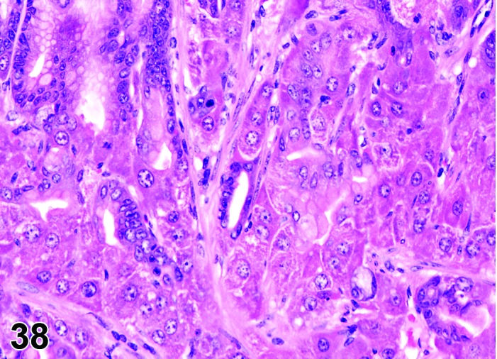
Mouse glandular stomach. Hepatocytes, ectopic. Higher magnification of Figure 37.
Figure 39.
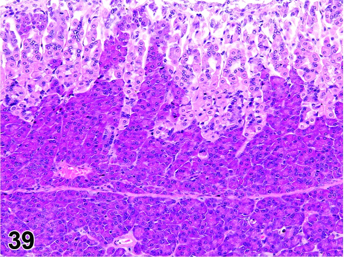
Mouse glandular stomach. Pancreas ectopic.
Figure 40.
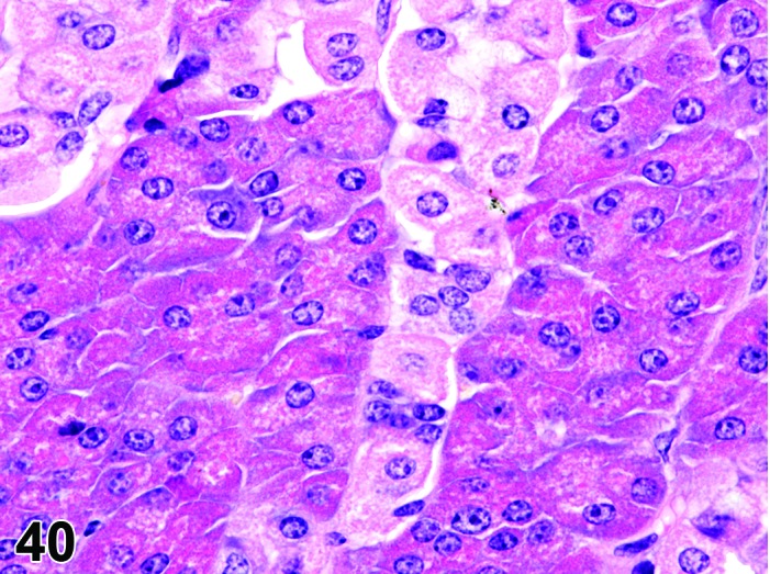
Mouse glandular stomach. Pancreas ectopic. Higher magnification of Figure 39.
Modifiers: Hepatocytes, pancreas
Pathogenesis: Presence of pancreatic or hepatic tissue in the mucosa or submucosa.
Diagnostic features
Normally formed pancreatic acini in mucosa or submucosa. Islets of Langerhans may also be present.
Normally formed hepatocytes present in cords or nests in mucosa or submucosa; binucleate cells may be present.
Differential diagnosis
Adenocarcinoma: Formation of hepatocyte-like cells has been reported in human gastric adenocarcinoma.
Comment: Ectopic pancreatic acini or hepatocytes are rare incidental findings in rats and mice (Maekawa et al. 1996; Brown and Hardisty 1990), but ectopic differentiation may be a feature of some engineered mice (Fukuda et al. 2006). In one publication, hepatocytes accompanied fundic gland abnormalities in the mouse and it was not clear whether they represented metaplasia or congenital heterotopia (Leininger et al. 1990); the same conundrum may apply to ectopic pancreatic foci. Metaplasia to a pancreatic cell type was reported as part of a hypertrophic gastropathy in a strain of rat (WTC-dfk) which has a deletion of the potassium channel Kcnq1 gene (Kuwamura et al. 2008). Ectopic pancreatic tissue also may occur rarely in the pyloric submucosa of mice (Maekawa et al. 1996).
b. Cellular Degeneration, Injury and Death of the Nonglandular Stomach
Atrophy, squamous epithelium
(Figures 41 and 42)
Figure 41.
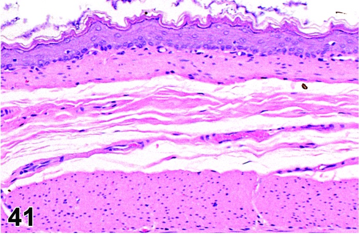
Mouse nonglandular stomach. Control.
Figure 42.
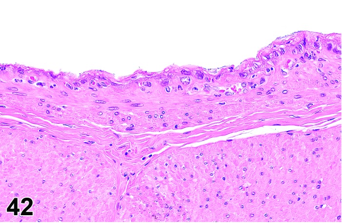
Mouse nonglandular stomach. Atrophy, squamous epithelium; compare to Figure 41.
Pathogenesis: Decrease in normal thickness/cellularity of squamous mucosa.
Diagnostic features
Focally extensive or diffuse lesion.
Decreased thickness of mucosal epithelial layer.
Differential diagnoses
Erosion/ulcer: Focal or focally extensive absence of superficial or, in severe cases (ulcer) of all epithelial layers.
Artifact: Excessive stretching due to food contents or stretching stomach at necropsy leads to decreased thickness of all layers of the gastric wall.
Vacuolation, squamous epithelium
Figure 43.
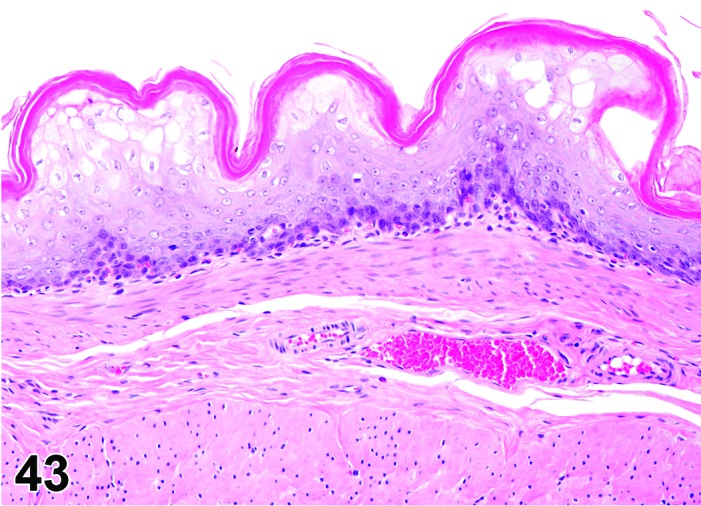
Rat nonglandular stomach. Vacuolation, squamous epithelium, in addition to hyperplasia, squamous cell.
Pathogenesis: Degenerative change in the squamous epithelial cells which may precede necrosis, erosion or ulceration of the nonglandular epithelium.
Diagnostic features
Cells of squamous epithelium have a pale staining, vacuolated appearance.
May be accompanied by edema in the submucosa and vacuolation of the muscularis.
Differential diagnosis
Artifacts.
Comment: Epithelial vacuolation and vesiculation along with submucosal edema was described in rats after administration of ethyl acrylate as a forerunner to necrosis and erosion/ulcer (Ghanayem et al. 1985).
Certain compounds may specifically cause vacuolation of the squamous epithelium adjacent to the limiting ridge.
Apoptosis, squamous epithelium
Synonyms: Apoptotic cell death
Pathogenesis: Gene regulated, energy dependent process leading to formation of apoptotic bodies which are phagocytosed by adjacent cells; typically associated with cytotoxic chemotherapeutics that affect the mucosal epithelium of the nonglandular stomach.
Diagnostic features
Single cells or small clusters of cells.
Cell shrinkage.
Hypereosinophilic cytoplasm.
Nuclear shrinkage, pyknosis, karyorrhexis.Intact cell membrane.
Apoptotic bodies.
Cytoplasm retained in apoptotic bodies.
Phagocytosis of apoptotic bodies tissue macrophages or other adjacent cells.
No inflammation.
Differential diagnoses
Necrosis, epithelium: The morphologic features of cell death clearly fit with necrotic diagnostic criteria (cells and nuclei swollen, pale cytoplasm etc.).
Apoptosis/necrosis, epithelium: Both types of cell death are present and there is no requirement to record them separately or a combined term is preferred for statistical reasons. The term is also used when the type of cell death cannot be determined unequivocally.
Erosion/ulcer: Focal or focally extensive absence of superficial or, in severe cases, of all epithelial layers with extension of the alteration into the muscularis.
Comment: The approach for the nomenclature and diagnostic criteria of cell death applied here is based on a draft recommendation of the INHAND Cell Death Nomenclature Working Group.
Apoptosis is not synonymous with necrosis. The main morphological differences between these two types of cell death are cell shrinkage with nuclear fragmentation and tingible body macrophages in apoptosis versus cell swelling, rupture and inflammation in necrosis; however, other morphologies (e.g. nucelar pyknosis and karyorrhexis) overlap. In routine H&E sections where the morphology clearly represents apoptosis or single cell necrosis, or whereby special procedures (e.g. TEM or IHC for caspases) prove one or the other, individual diagnoses may be used. However, because of the overlapping morphologies, necrosis and apoptosis are not always readily distinguishable via routine examination, and the two processes may occur sequentially and/or simultaneously depending on intensity and duration of the noxious agent (Zeiss 2003). This often makes it difficult and impractical to differentiate between both during routine light microscopic examination. Therefore, the complex term apoptosis/necrosis may be used in routine toxicity studies.
A differentiation between apoptosis and single cell necrosis may be required in the context of a given study, in particular if it aims for mechanistic investigations. Transmission electron microscopy is considered to represent the gold standard to confirm apoptosis. Other confirmatory techniques include DNA-laddering (easy to perform but insensitive), TUNEL (false positives from necrotic cells) or immunohistochemistry for caspases, in particular caspase 3. These techniques are reviewed in detail by Elmore (Elmore 2007). Some of these confirmatory techniques detect early phases of apoptosis, in contrast to the evaluation of H&E stained sections, which detects late phases only; overall interpretation of findings should take into account these potential differences. Thus, low grades of apoptosis may remain unrecognized by evaluation of H&E stained slides only.
Certain studies may require the consideration of forms of programmed cell death other than apoptosis which requires specific confirmatory techniques (Galluzzi et al. 2012).
Necrosis, squamous epithelium
Synonyms: Oncotic cell death; oncotic necrosis; necrosis
Modifiers: Single cell
Pathogenesis: Unregulated, energy independent, passive cell death with leakage of cytoplasm into surrounding tissue and subsequent inflammatory reaction; may be induced by direct contact with a test article after oral uptake/administration.
Diagnostic features
Cells swollen with pale eosinophilic cytoplasm.
Loss of nuclear basophilia, pyknosis and/or karyorrhexis that affects aggregates of cells.
In more severe cases, there may be detachment of the epithelium from the submucosa.
Typically, the presence of degenerative cells is a component of necrosis.
Minimal or slight inflammatory cell infiltrates may be present as a feature of necrosis.
Single cell
Only single cells affected.
Differential diagnoses
Apoptosis, squamous epithelium: The morphologic features of cell death clearly fit with apoptosis (cell and nucleus shrunken, hypereosinophilic cytoplasm, nuclear pyknosis etc.) and/or special techniques demonstrate apoptosis.
Apoptosis/necrosis, squamous epithelium: Both types of cell death are present and there is no requirement to record them separately or a combined term is preferred for statistical reasons. The term is also used when the type of cell death cannot be determined unequivocally.
Atrophy, squamous epithelium: Reduced thickness of all layers of the squamous epithelium or, in severe cases, absence of basal germinative layers; usually no cellular degeneration or necrosis.
Erosion/ulcer: Focal or focally extensive absence of superficial or, in severe cases, of all epithelial layers with distention of alteration into the muscularis.
Comments: Epithelial necrosis usually develops into erosion/ulcer and in the past has frequently been recorded as such. A separate recording of necrosis as the initial event in this process is recommended when the structure of the mucosa is still intact.
Apoptosis/necrosis, squamous epithelium
Synonym: Cell death
Pathogenesis: Unregulated, energy independent, passive cell death with leakage of cytoplasm into surrounding tissue and subsequent inflammatory reaction (single cell necrosis) AND/OR gene regulated, energy dependent process leading to formation of apoptotic bodies which are phagocytosed by adjacent cells (apoptosis); typically associated with cytotoxic chemotherapeutics that affect the mucosal epithelium of the nonglandular stomach.
Diagnostic features
Both types of cell death are present and there is no requirement to record them separately or a combined term is preferred for statistical reasons.
The type of cell death cannot be determined unequivocally.
Differential diagnoses
Apoptosis, squamous epithelium: The morphologic features of cell death clearly fit with apoptosis (cell and nucleus shrunken, hypereosinophilic cytoplasm, nuclear pyknosis etc.) and/or special techniques demonstrate apoptosis and there is a requirement for recording apoptosis and necrosis separately.
Necrosis, squamous epithelium: The morphologic features of cell death clearly fit with necrotic diagnostic criteria (cells and nuclei swollen, pale cytoplasm etc.) and there is a requirement for recording apoptosis and necrosis separately.
Erosion/ulcer: Focal or focally extensive absence of superficial or, in severe cases, of all epithelial layers with distention of alteration into the muscularis.
Comments: Necrosis and apoptosis are not always readily distinguishable via routine examination, and the two processes may occur sequentially and/or simultaneously depending on intensity and duration of the noxious agent (Zeiss 2003). This often makes it difficult and impractical to differentiate between both during routine light microscopic examination. In these cases, the combined term apoptosis/necrosis may be used. It is recommended to explain in detail the use of this combined term in the narrative part of the pathology report.
Erosion/ulcer
Figure 44.
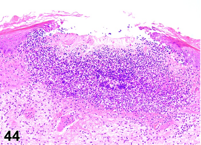
Rat nonglandular stomach. Ulcer.
Pathogenesis: Localized loss of mucosa with either partial mucosal penetration (erosion) or with full penetration of the mucosa to the muscularis mucosa (ulcer).
Diagnostic features
Focal or multifocal.
Ulcers will be associated with acute/chronic inflammatory cell infiltration in submucosa.
Basal lamina may remain intact (erosion) or in the more severe cases defect in the mucosa and inflammation will extend to the tunica muscularis and serosa (ulcer).
Hyperplasia is often present in the surrounding epithelium.
Hemorrhage may be seen with larger ulcers.
Differential diagnoses
Carcinoma, squamous cell: May show secondary ulceration, but always penetration of basement membrane by single cells or nests of neoplastic cells.
Necrosis, squamous epithelial: No superficial or deep loss of epithelial cells.
Artifacts: Mucosal loss due to manual or processing artifact – inflammation will be absent.
Comment: Ulcers in the nonglandular epithelium are often seen macroscopically as multiple small dark depressed areas surrounded by raised areas of whitened epithelium(Maekawa 1994). Whilst ulcers have been induced by a wide range of irritant agents, the cause of ulceration in control animals is often unclear, although advanced age, parasitism, infection, diet, feeding regimen, generalized debility and stress can play a role (Maekawa 1994; Maekawa et al. 1996; Greaves 2012). Protein restriction and starvation have also been shown to produce ulcers (Boyd et al. 1970).
Although there are specific morphologic criteria to differentiate the more superficial lesion (erosion; confined to the epithelial surface) from the deeper ulcer (full penetration of the mucosa to the muscularis mucosa), the plane of section through smaller, focal lesions often impacts on the appearance (depth of the epithelial loss) of this finding. In most instances it is not practical to separate these changes and they are typically diagnosed under the single, combined term of erosion/ulcer with an appropriate severity grade.
Erosion/ulcer usually is the result of epithelial necrosis. Because of its unique topography adjacent to the lumen and often involving multiple organ layers, which uniquely affects the progression and resolution of the lesion (e.g. margin hyperplasia, granulomatous tissue, inflammation), it is useful to record erosion/ulcer separately from epithelial necrosis.
A deep ulcer may be perforating if it destroys all layers of the gastric wall, and may be designated as such by a free text entry. However, a perforation resulting from a physical insult, e.g. gavage-related, typically is a macroscopic lesion and generally not recorded on the light microscopic level.
Hyperkeratosis
Figure 45.
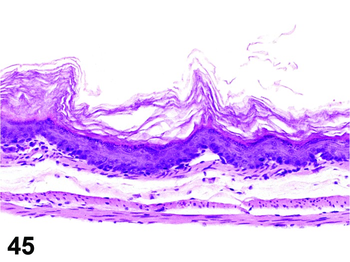
Mouse nonglandular stomach. Hyperkeratosis, orthokeratotic; compare to Figure 41.
Figure 46.
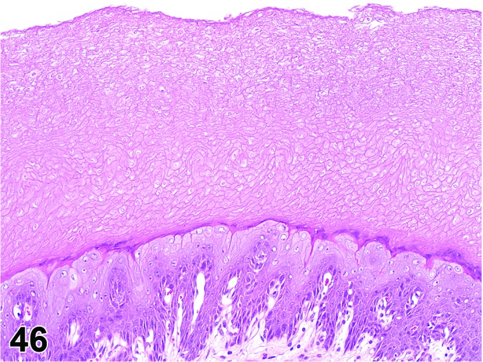
Rat nonglandular stomach. Hyperkeratosis, parakeratotic.
Figure 47.
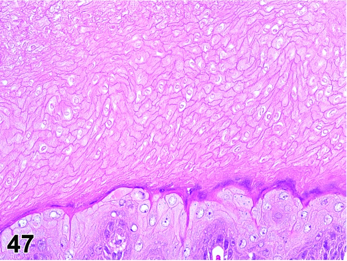
Rat nonglandular stomach. Hyperkeratosis, parakeratotic. Higher magnification of Figure 46.
Synonyms: Hpyerkeratosis, orthokeratotic may have been identified as hyperkeratosis
Hyperkeratosis, parakeratotic may have been identified as parakeratosis
Modifiers: Orthokeratotic, parakeratotic
Pathogenesis: Keratinization of squamous epithelium faster than desquamation of keratinized layers with normal (hyperkeratotic) or abnormal (parakeratotic) maturation of the keratin layers
Diagnostic features
Increase in the thickness of the keratin layer on the luminal epithelial surface.
Orthokeratotic
Thickened keratin layer with non-nucleated keratinized cells.
Parakeratotic
Thickened keratin layer with nucleated keratinized cells.
Differential diagnoses
Hyperplasia, squamous cell: Proliferation and thickening of the stratum spinosum.
Comments: Hyperkeratosis occurs often in association with hyperplasia of underlying epithelium. Alternatively, it may be observed in the absence of hyperplasia under circumstances suggesting anorexia and a presumptive lack of mechanical abrasion.
Hyperkeratosis can be a frequent observation attributable to local irritation of orally administered test articles in toxicity studies (Til et al. 1988). The local irritant effect is presumably potentiated by increased duration of exposure associated with intragastric storage and increased contact, if test article precipitates in the gastric compartment.
c. Cellular Degeneration, Injury and Death of the Glandular Stomach
Dilatation, glands
(Figures 48 and 49)
Figure 48.
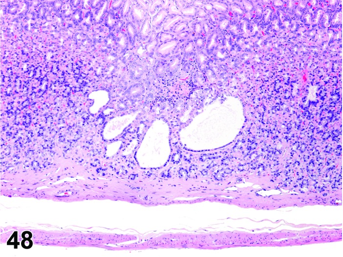
Rat glandular stomach. Dilatation glands.
Figure 49.
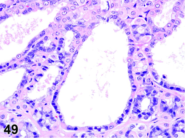
Rat glandular stomach. Dilatation glands. Higher magnification of Figure 48; note well differentiated cuboidal epithelium.
Synonyms: Glandular dilatation, glandular dilation
Pathogenesis: Dilatation within glands.
Diagnostic features
Lined by well-differentiated cuboidal to flattened epithelial cells; no atypia.
Lumen may contain mucus.
Usually multifocal.
Differential diagnoses
Cysts, glandular: Marked dilatation of glands lined by flattened epithelial cells.
Diverticulum, cystic OR atypical, cystic: extension of dilated glands through muscularis mucosae, into submucosa and further in some cases.
Atrophy, mucosa: Dilated glands may be present as part of the atrophic changes.
Comment: Dilatation, glands is a common observation in the rat stomach and usually not reported by toxicological pathologists unless it shows a treatment-related trend
Cyst, glandular
Figure 50.
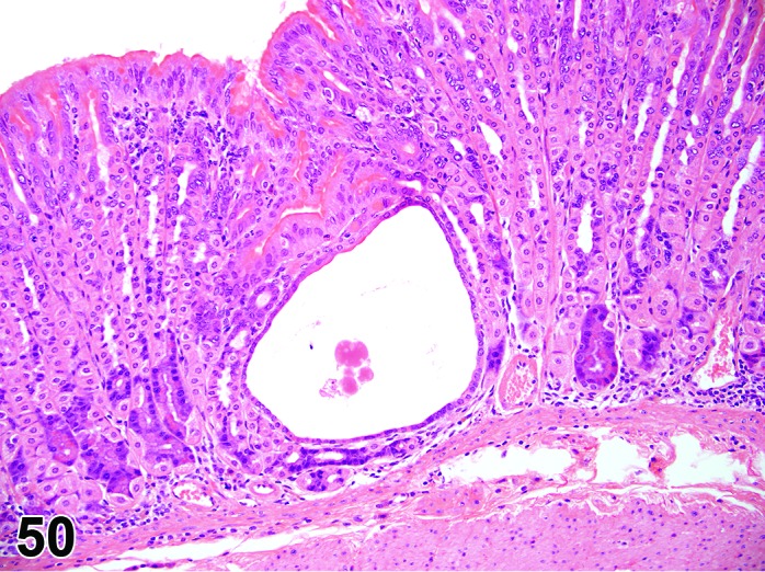
Mouse glandular stomach. Cyst.
Pathogenesis: Dilatation within glands.
Diagnostic features
Marked dilatation of glands.
Lined by well-differentiated flattened epithelial cells. No atypia.
Lumen may contain mucus.
Luminal contents may be mineralized.
May be surrounded by chronic inflammation.
Very occasionally squamous metaplasia is present in cysts.
Differential diagnoses
Diverticulum, cystic OR atypical, cystic: Extension of glands through muscularis mucosae, into submucosa and further in some cases.
Hyperplasia, atypical: Focal hyperchromatic lesion with cellular atypia and pleomorphism.
Adenocarcinoma: Multiple layers of lining epithelium with varying degrees of dysplasia. True infiltrative growth with loss of basement membrane integrity and often with a scirrhous stromal reaction.
Atrophy, mucosa: Cysts may be present as part of the atrophic changes.
Comment: Glandular cysts may be congenital but most are acquired as their incidence increases with age in both rats and mice (Brown and Hardisty 1990; Maekawa et al. 1996). In the Fischer rat they are more common in the antrum than in the fundus (Brown and Hardisty 1990). When cysts extend through the muscularis mucosae, the diagnostic term “diverticulum, cystic” or “diverticulum, atypical, cystic” should be used.
Atrophy
(Figures 51 and 52)
Figure 51.
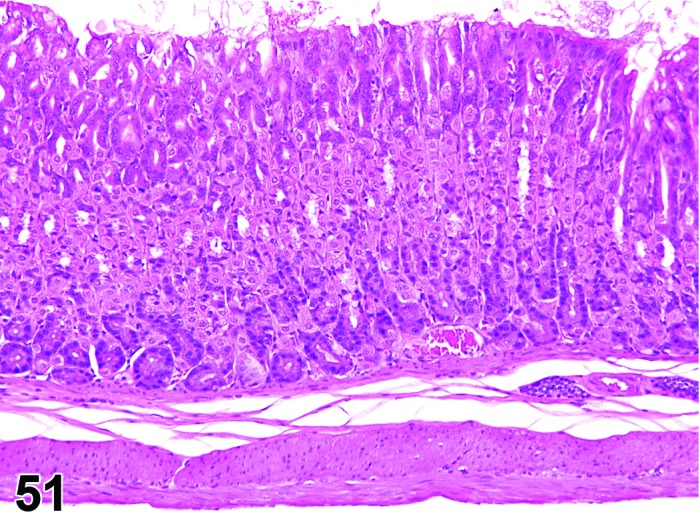
Mouse fundic mucosa. Normal – compare to Figure 52.
Figure 52.
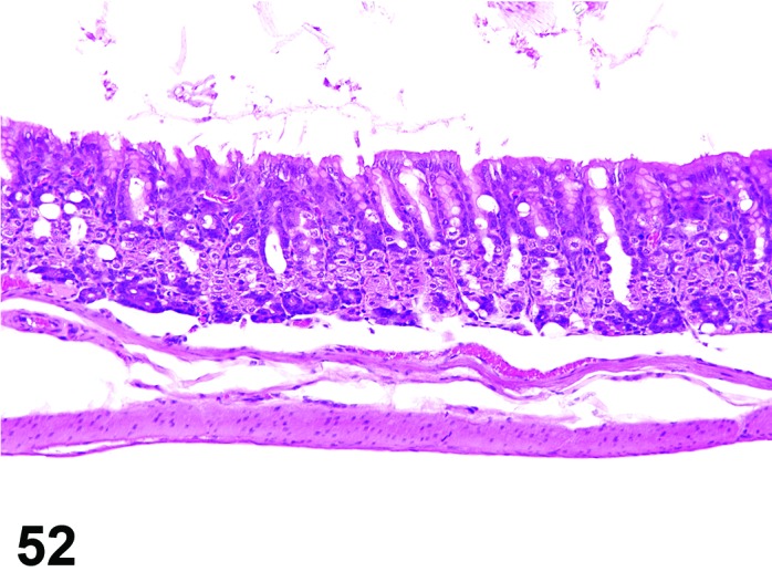
Mouse fundic mucosa. Atrophy mucosal diffuse. Same magnification as Figure 51.
Modifiers: Mucosa or specific cell type (e.g., chief cell, parietal or mucous cell)
Pathogenesis: Decreased number and/or size of epithelial cells reducing the mucosal thickness and function of the glandular mucosa. In the fundic glands, it may affect one or all cell types. In most instances chief cells are lost before other cell types.
Diagnostic features
Decreased numbers of a one or more cell types. Remaining cells may be small and/or poorly differentiated.
Reduced mucosal thickness in advanced cases, although compensatory hyperplasia of less differentiated epithelial cells can mask this.
Dilatation of atrophic glands.
Focal areas of atrophy may be seen with aging.
Differential diagnoses
Artifact: Over stretched stomach during ‘pinning’ at necropsy leading to a reduction in thickness of all layers of the gastric wall.
Secretory depletion, mucus: Reduction of mucous cell size due to reduced amounts of mucus but no loss of cells.
Comment: At least two different types of atrophy can be distinguished. In one type, atrophy broadly affects all mucosal cell types resulting in an overall reduction in mucosal height with maintenance of relatively normal cellular composition. Under other circumstances a specific cell type may be atrophic with the loss of specific functionality.
Atrophy affecting all cell types of the gastric mucosa can be produced by starvation, antrectomy or deletion of the gastrin gene which removes the trophic stimulation of gastrin (Greaves 2012). Analogous changes may be produced by test articles that block the secretion or activity of gastrin (Dethloff et al. 1997). In contrast, chronic inflammation and some xenobiotics can cause loss of specific cell types (Greaves 2012). In addition, in the lesions associated with helicobacter infections in mice it has been noted that chief cells are lost before parietal cells (Rogers and Houghton 2009; Rogers 2012). Focal or diffuse atrophy of the mucosa can occur with age in rodents where there is partial replacement of the glands by fibrous connective tissue (Brown and Hardisty 1990).
Vacuolation, epithelium
Modifiers: Mucosa or specific cell type (e.g., chief cell, parietal or mucous cell)
Pathogenesis: Vacuolation with/without degeneration of epithelial cells. May affect all cell types or be primarily chief, parietal or mucous cell.
Diagnostic features
Cytoplasmic vacuolation, swelling.
Occasional Apoptosis/single cell necrosis may also be present.
Differential diagnoses
Autolysis: Destruction and loss of cell types but often occurs preferentially at the luminal surface.
Depletion, mucus: Only mucous cells affected, no other cytological changes.
Comment: Vacuolation with no or minimal evidence of cell loss can be induced by agents that inhibit gastric acid secretion (Dethloff et al. 1997; Karam and Alexander 2001). In addition, degeneration is observed with anticancer cytotoxic agents, ulcerogenic agents and agents that decrease mucosal blood supply, or test articles that sequester in parietal cells (Ito et al. 2000; Bertram et al. 2013; Greaves 2012). Depending on the nature and severity of the insult, degeneration will be followed by necrosis, erosion/ulcer of the epithelium, inflammation and/or hemorrhage.
Secretory depletion
(Figures 53 and 54)
Figure 53.
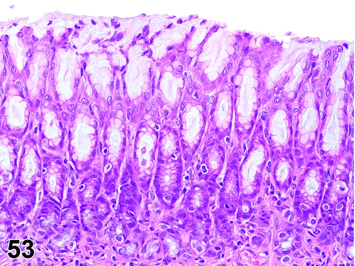
Rat antral mucosa. Normal – compare to Figure 54.
Figure 54.
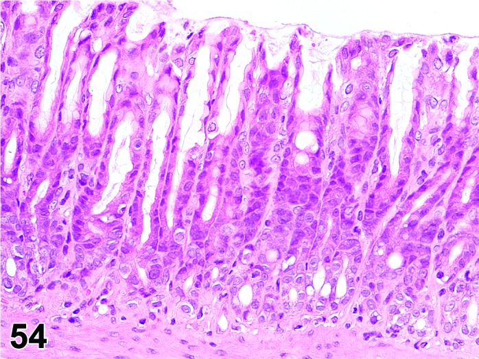
Rat antral mucosa. Secretory depletion. Same magnification as Figure 53.
Pathogenesis: Decreased mucus content of mucous cells.
Diagnostic features
Intact epithelial layer.
Replacement of normal clear cytoplasm with more basophilic cytoplasm containing little or no mucus.
Differential diagnoses
Degeneration, epithelial: Additional cytological features apart from reduction in mucus.
Atrophy, mucous cell: Reduced number of mucus cells in addition to mucus depletion.
Comment: Mucus depletion may develop secondary to spontaneous inflammatory lesions and drug-induced lesions (Greaves 2012). Qualitative changes in mucus composition may accompany depletion as described following administration of aspirin, anti-inflammatory agents and adrenocortical steroids (Ishihara et al. 1984; Bertram et al. 2013; Greaves 2012). These substances may also alter the phospholipids present in the mucus layer thereby reducing the protective hydrophobic barrier properties and a loss of functional integrity. Administration of histamine H2 receptor antagonists and proton pump inhibitors that reduce gastric acid secretion have also been shown to reduce total and sulphated glycoproteins along with reduction in mucus (Yoshimura et al. 1996).
Eosinophilic globules
(Figures 55, 56 and 57)
Figure 55.
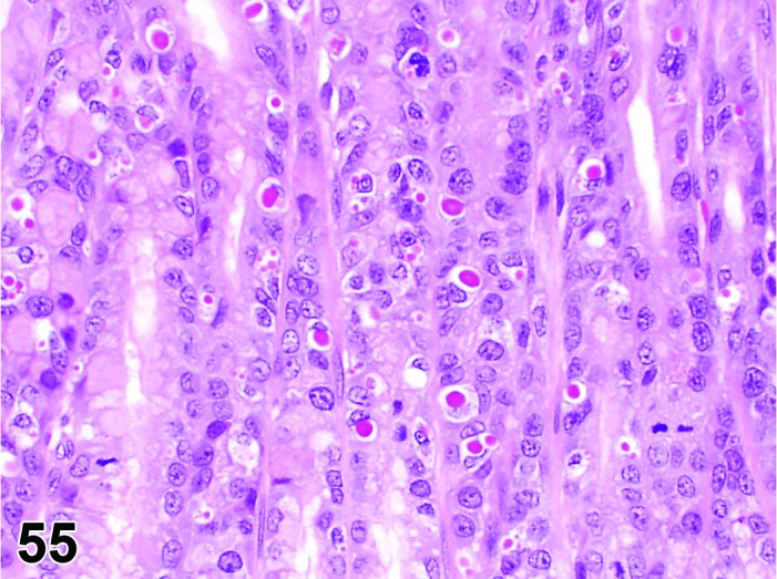
Rat, glandular stomach, eosinophilic globules.
Figure 56.
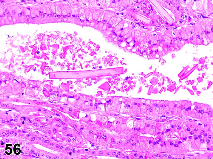
Mouse glandular stomach. Eosinophilic globules. Note intracellular eosinophilic globules and extracellular eosinphilic crystals.
Figure 57.
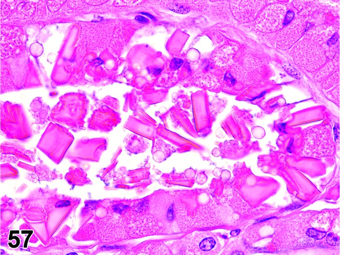
Mouse glandular stomach. Eosinophilic globules. Note eosinophilic crystals also in epithelial cells that undergo cell death.
Synonyms: Hyalinosis; eosinophilic chief cells; eosinophilic change; eosinophilic droplets
Pathogenesis: Transformation of any glandular epithelial cell to produce hyaline cytoplasm.
Diagnostic features
Intensely pink-red cytoplasmic droplets and/or crystals in surface/mucous cells, most often adjacent to the limiting ridge.
May extend into and/or replace fundic glands in advanced cases.
Cells are usually hypertrophic.
Can be single or multiple (focal or multifocal).
Crystals if present may be intracellular or extracellular.
Differential diagnosis
None.
Comment: Mucous epithelial cells distended with bright, hyaline eosinophilic cytoplasm occur infrequently in rodents either as a spontaneous change or associated with another lesion e.g. inflammation (especially eosinophilic) or lymphoma infiltration (Bertram et al. 1996). They are usually observed in mucus neck cells of the glandular mucosa near the limiting ridge and occasionally may be accompanied by hyaline eosinophilic crystals. Eosinophilic globules may be considered a precursor of these crystals. Formation of these cells is also associated with the administration of gastric antisecretory agents although the incidence is not proportional to the neuroendocrine cell hyperplasia which also occurs with these agents (Betton et al. 1988). The eosinophilic granules are similar to those seen in other epithelia e.g. upper and lower respiratory tract or pancreas (Leininger et al. 1999; Renne et al. 2009). Once thought to be accumulation of mucosubstances or pepsinogen, they have been shown to be composed of a Ym1/Ym2 a chitinase-like protein that may be produced in response to mucosal irritation (Ward et al. 2001; Rogers and Houghton 2009). The use of the term hyalinosis for this lesion will potentially cause confusion in safety assessment as the same term may also be used for a distinctly different clinical disorder in humans and can be used to describe vascular and renal glomerular changes.
Apoptosis
Synonyms: Apoptotic cell death
Pathogenesis: Gene regulated, energy dependent process leading to formation of apoptotic bodies which are phagocytosed by adjacent cells.
Diagnostic features
Single cells or small clusters of cells.
Cell shrinkage.
Hypereosinophilic cytoplasm.
Nuclear shrinkage, pyknosis, karyorrhexis. Intact cell membrane.
Apoptotic bodies.
Cytoplasm retained in apoptotic bodies.
Phagocytosis of apoptotic bodies tissue macrophages or other adjacent cells.
No inflammation.
Differential diagnoses
Necrosis, mucosa: The morphologic features of cell death clearly fit with necrotic diagnostic criteria (cells and nuclei swollen, pale cytoplasm etc.).
Apoptosis/necrosis, mucosa: Both types of cell death are present and there is no requirement to record them separately or a combined term is preferred for statistical reasons. The term is also used when the type of cell death cannot be determined unequivocally.
Erosion/ulcer: Focal or focally extensive penetration of the mucosa, either partial (erosion) or full to the muscularis mucosa (ulcer).
Comment: The approach for the nomenclature and diagnostic criteria of cell death applied here is based on a draft recommendation of the INHAND Cell Death Nomenclature Working Group.
Apoptosis is not synonymous with necrosis. The main morphological differences between these two types of cell death are cell shrinkage with nuclear fragmentation and tingible body macrophages in apoptosis versus cell swelling, rupture and inflammation in necrosis; however, other morphologies (e.g. nucelar pyknosis and karyorrhexis) overlap. In routine H&E sections where the morphology clearly represents apoptosis or single cell necrosis, or whereby special procedures (e.g. TEM or IHC for caspases) prove one or the other, individual diagnoses may be used. However, because of the overlapping morphologies, necrosis and apoptosis are not always readily distinguishable via routine examination, and the two processes may occur sequentially and/or simultaneously depending on intensity and duration of the noxious agent (Zeiss 2003). This often makes it difficult and impractical to differentiate between both during routine light microscopic examination. Therefore, the complex term apoptosis/necrosis may be used in routine toxicity studies.
A differentiation between apoptosis and single cell necrosis may be required in the context of a given study, in particular if it aims for mechanistic investigations. Transmission electron microscopy is considered to represent the gold standard to confirm apoptosis. Other confirmatory techniques include DNA-laddering (easy to perform but insensitive), TUNEL (false positives from necrotic cells) or immunohistochemistry for caspases, in particular caspase 3. These techniques are reviewed in detail by Elmore (Elmore 2007). Some of these confirmatory techniques detect early phases of apoptosis, in contrast to the evaluation of H&E stained sections, which detects late phases only; overall interpretation of findings should take into account these potential differences. Thus, low grades of apoptosis may remain unrecognized by evaluation of H&E stained slides only.
Certain studies may require the consideration of forms of programmed cell death other than apoptosis which requires specific confirmatory techniques (Galluzzi et al. 2012).
Necrosis, mucosa
Figure 58.
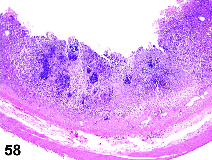
Rat glandular stomach. Necrosis, mucosa.
Synonyms: Oncotic cell death; oncotic necrosis; necrosis
Modifiers: Single cell
Pathogenesis: Unregulated, energy independent, passive cell death with leakage of cytoplasm into surrounding tissue and subsequent inflammatory reaction; may be induced by direct contact with a test article after oral uptake/administration.
Diagnostic features
Cells swollen with pale eosinophilic cytoplasm.
Loss of nuclear basophilia, pyknosis and/or karyorrhexis that affects aggregates of cells.
In more severe cases, there may be detachment of the epithelium from the submucosa.
Typically, the presence of degenerative cells is a component of necrosis.
Minimal or slight inflammatory cell infiltrates may be present as a feature of necrosis.
Single cell
Only single cells affected.
Differential diagnoses
Apoptosis, mucosa: The morphologic features of cell death clearly fit with apoptosis (cell and nucleus shrunken, hypereosinophilic cytoplasm, nuclear pyknosis etc.) and/or special techniques demonstrate apoptosis.
Apoptosis/necrosis, mucosa: Both types of cell death are present and there is no requirement to record them separately or a combined term is preferred for statistical reasons. The term is also used when the type of cell death cannot be determined unequivocally.
Atrophy: Reduced thickness of all layers of the squamous epithelium or, in severe cases, absence of basal germinative layers; usually no cellular degeneration or necrosis.
Erosion/ulcer: Focal or focally extensive penetration of the mucosa, either partial (erosion) or full to the muscularis mucosa (ulcer).
Comments: Epithelial necrosis usually develops into erosion/ulcer and in the past has frequently been recorded as such. A separate recording of necrosis as the initial event in this process is recommended when the structure of the mucosa is still intact.
Apoptosis/necrosis, mucosa
Synonym: Cell death
Pathogenesis: Unregulated, energy independent, passive cell death with leakage of cytoplasm into surrounding tissue and subsequent inflammatory reaction (single cell necrosis) and/or gene regulated, energy dependent process leading to formation of apoptotic bodies which are phagocytosed by adjacent cells (apoptosis).
Diagnostic features
Both types of cell death are present and there is no requirement to record them separately or a combined term is preferred for statistical reasons.
The type of cell death cannot be determined unequivocally.
Differential diagnoses
Apoptosis: The morphologic features of cell death clearly fit with apoptosis (cell and nucleus shrunken, hypereosinophilic cytoplasm, nuclear pyknosis etc.) and/or special techniques demonstrate apoptosis and there is a requirement for recording apoptosis and necrosis separately.
Necrosis, mucosa: The morphologic features of cell death clearly fit with necrotic diagnostic criteria (cells and nuclei swollen, pale cytoplasm etc.) and there is a requirement for recording apoptosis and necrosis separately.
Erosion/ulcer: Focal or focally extensive penetration of the mucosa, either partial (erosion) or full to the muscularis mucosa (ulcer).
Comments: Necrosis and apoptosis are not always readily distinguishable via routine examination, and the two processes may occur sequentially and/or simultaneously depending on intensity and duration of the noxious agent (Zeiss 2003). This often makes it difficult and impractical to differentiate between both during routine light microscopic examination. In these cases, the combined term apoptosis/necrosis may be used. It is recommended to explain in detail the use of this combined term in the narrative part of the pathology report.
Erosion/ulcer
Figure 59.
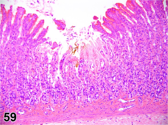
Rat glandular stomach. Erosion, focal.
Pathogenesis: Loss of superficial mucosa with preservation of the muscularis mucosa (erosion) or with penetration of muscularis mucosa and exposure of submucosa (ulcer).
Diagnostic features
Focal or multifocal.
Epithelial cells are absent but muscolaris mucosa is intact (erosion).
Epithelial cells are absent and muscularis mucosa has been destroyed (ulcer).
Ulceration will be associated with acute/chronic inflammatory cell infiltration in submucosa.
In the more severe cases ulceration and/or inflammation will extend into the tunica muscularis and serosa.
Hyperplasia is often present in the surrounding epithelium.
May be associated with foreign bodies (plant fibers) in submucosa.
Hemorrhage may be seen with larger ulcers.
Differential diagnoses
Adenocarcinoma: May show secondary ulceration, but penetration of basement membrane by single cells or nests of neoplastic cells always present.
Artifacts: Mechanical loss of mucosa during necropsy; plane of section due to orientation of tissue in block.
Autolysis: All tissues in stomach affected but there is often preferential destruction and loss of cells at the luminal surface.
Comment: Erosions and ulcers develop in rodents following stress, bile reflux, changes in acid secretion and hypoxia (Bertram et al. 2013; Greaves 2012; Haschek et al. 2010). Certain types of xenobiotics e.g. non-steroidal anti-inflammatory drugs and ethyl alcohol are well known ulcerogenic agents but ulcers have also been induced by innocuous substances e.g. saline glucose when given as hyperosmolar solutions (Puurunen et al. 1980). Ulcers can also reflect systemic disease e.g. associated with uremia. Small focal erosions can occur in the glandular corpus of both rats and mice following overnight fasting.
Although there are specific morphologic criteria to differentiate the more superficial lesion (erosion; confined to the epithelial surface) from the deeper ulcer (full penetration of the mucosa to the muscularis mucosa), the plane of section through smaller, focal lesions often impacts on the appearance (depth of the epithelial loss) of this finding. In most instances it is not practical to separate these changes and they are typically diagnosed under the single, combined term of erosion/ulcer with an appropriate severity grade.
Erosion/ulcer usually is the result of epithelial necrosis. Because of its unique topography adjacent to the lumen and often involving multiple organ layers, which uniquely affects the progression and resolution of the lesion (e.g. margin hyperplasia, granulomatous tissue, inflammation), it is useful to record erosion/ulcer separately from epithelial necrosis.
A deep ulcer may be perforating if it destroys all layers of the gastric wall, and may be designated as such by a free text entry. However, a perforation resulting from a physical insult, e.g. gavage-related, typically is a macroscopic lesion and generally not recorded on the light microscopic level.
Hypertrophy, mucous cell
Figure 60.
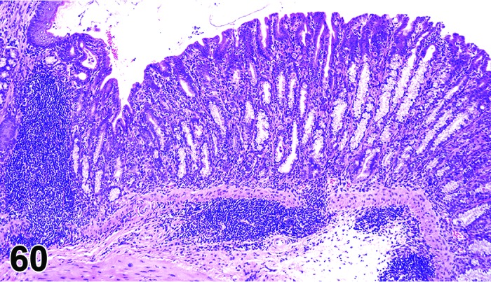
Rat glandular stomach. Hypertrophy, mucus cell.
Pathogenesis: Enlargement of mucous cells that line gastric glands, accompanied by loss of parietal and/or chief cells. The mucous cells may manifest foamy cytoplasm.
Diagnostic features
Parietal and/or chief cells in corpus mucosa replaced by round, foamy, lightly basophilic cells resembling duodenal Brunner’s gland cells. Concurrent loss of oxyntic glandular cells implicates an effect on the differentiation of the glandular epithelium.
Increased numbers of such cells may be present (hyperplasia).
Histochemical staining with Alcian blue/PAS pH 2.5 shows that cells express increased gastric-type neutral mucins (red) as well as antral gland and intestinal-type acid mucins (blue).
Differential diagnosis
None.
Comment: Evaluation in H&E sections limits the characterization of this mucosal change to hypertrophy, hyperplasia and/or vacuolation. This may occur spontaneously or in response to chronic injury. Histochemical stains and immunochemical markers facilitate further characterization of the change as a metaplastic shift in the mucous secretory phenotype of the cells. Several classes of metaplasia have been identified based on these additional techniques, which may have relevance for modeling human gastric pathogenesis. True intestinal metaplasia (metaplasia with development of a genuine intestinal phenotype) is very rare in rodents but has been induced by PCBs in mice (Morgan et al. 1981). “Pseudopyloric” metaplasia where the cells resemble antral cells morphologically and histochemically is much more common. Authentic intestinal metaplasia is best confirmed by immunohistochemistry for the intestinal-specific nuclear protein Cdx2 (Fox et al. 2007). Pseudopyloric/intestinal metaplasia occurs in the rodent stomach as a change associated with some experimentally induced cancers (e.g. following administration of polychlorinated biphenyls), localized ionizing radiation, injection of xenogenic stomach antigens and the non carcinogen, iodoacetamide (Greaves 2012), as well as infectious models in mice such as H. felis and H. pylori (Leininger et al. 1999; Rogers and Fox 2004).
Amyloid
(Figures 61 and 62)
Figure 61.
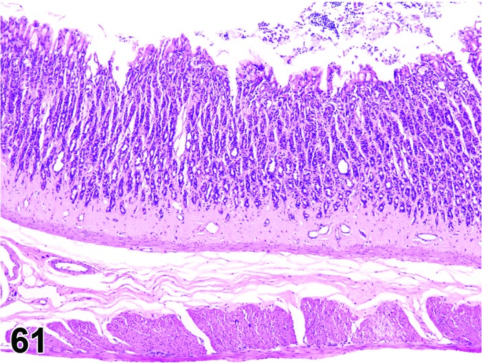
Rat glandular stomach. Amyolid.
Figure 62.
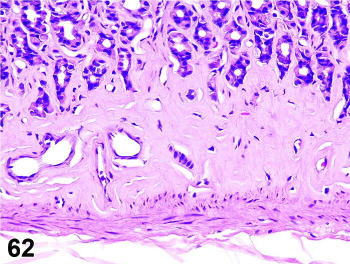
Rat glandular stomach. Amyloid. Higher magnification of Figure 61.
Synonyms: Amyloidosis; amyloid deposition
Pathogenesis: Extracellular deposits of polypeptide fragments of a chemically diverse group of glycoproteins, in various tissues including stomach mucosa.
Diagnostic features
Deposition of pale amorphous, homogenous eosinophilic material.
Location is extracellular in connective tissue and/or blood vessel walls.
Green birefringence with Congo red stain when viewed under polarized light.
Differential diagnoses
Hyaline connective tissue: Congo red negative.
Fibrinoid necrosis of blood vessels: Other evidence of tissue damage in section.
Comment: Amyloid deposition may occur in the stomach mucosa as part of systemic amyloidosis but it is not one of the predilection sites. Amyloidosis is extremely rare in rats but common in some strains of mice. In susceptible strains a number of factors including gender, diet, housing conditions, stress, endocrine status and microbiological status can influence the occurrence of amyloidosis (Lipman et al. 1993). For as yet unidentified reasons the previously susceptible CD-1 strain in Europe now rarely develops amyloidosis.
The characteristic morphologic appearance and location of amyloid in H&E sections that is usually adequate to make this diagnosis. Confirmation of the deposits as amyloid can be accomplished with light microscopy using special stains such as Congo red. Amyloid appears apple green under polarized light with this stain.
The term “hyaline” should be used in case of uncertainties about the nature of extracellular deposits of hyaline material in H&E sections and the unavailability of a Congo red stain. Hyaline is also the appropriate term for homogenously eosinophilic extracellular deposits that react negative with Congo red.
Mineralization
Figure 63.
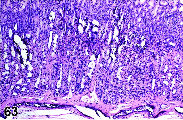
Rat glandular stomach. Mineralization.
Synonym: Calcification
Pathogenesis: Deposition of mineral in muscle, fibrous or elastic tissues secondary to necrosis (dystrophic mineralization) or hypercalcemia (metastatic mineralization).
Diagnostic features
Mineral may be found in the muscle layers, blood vessels, basement membranes and/or in the interstitium of mucosa and submucosa.
Interstitial mineral occurs predominantly in a band paralleling the parietal cell-rich region of the corpus mucosa.
Acellular basophilic material.
Mucosal mineralization can be limited to small focal deposits.
Diffuse mineralization of the corpus mucosa may be accompanied by degenerative changes or mucous cell hyperplasia in neighboring glands.
Differential diagnoses
Artifact: Hematoxylin stain precipitate.
Bacteria: Colonies of bacteria may be confused with mineral due to their basophilic staining. Ante mortem bacterial invasion will be accompanied by necrosis and usually inflammation; postmortem bacterial invasion will be accompanied by autolysis.
Osseous metaplasia: Osteoid present.
Comment: Presence of mineral can be confirmed with von Kossa or Alizarin red stains but these are rarely needed to make a diagnosis. Multifocal or diffuse mineralization occurs secondary to chronic renal disease in rodents (Leininger et al. 1999; Brown and Hardisty 1990) and in situations where serum calcium and/or phosphate balances are perturbed e.g. following intravenous administration of salts of rare earth metals such as gadolinium chloride (Rees et al. 1997). Mineralization can sometimes be observed in combination with inflammation and neoplasia.
d. Inflammatory Lesions
Infiltrate
Figure 64.
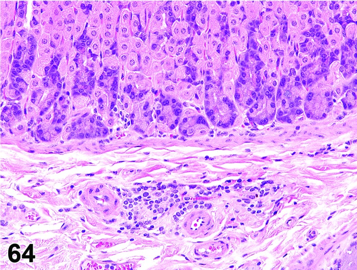
Mouse glandular stomach. Infiltrate mononuclear focal.
Synonyms: Infiltrate inflammatory; infiltrate inflammatory cell; infiltration (plus modifier); infiltration, inflammatory; infiltration, inflammatory cell
Modifiers: Type of inflammatory cell that represents the predominant cell type in the infiltrate
Pathogenesis: Infiltration of lamina propria and/or submucosa with neutrophils (Infiltrate, neutrophil), eosinophils (Infiltrate, eosinophil), mononuclear cells (Infiltrate, mononuclear cell) or a combination of more than one type (Infiltrate, mixed) without other histological features of inflammation e.g. hemorrhage, edema, fibroplasia.
Diagnostic features
Focal, multifocal or diffuse infiltration of cells in the lamina propria and/or submucosa.
Presence of mononuclear or polymorphonuclear leucocytes but no other histological criteria of inflammation.
Differential diagnoses
Inflammation: Other histological features of inflammation such as edema, hemorrhage, necrosis and/or fibroplasia will be present.
Granulocytic leukemia: Infiltration of neutrophil precursors and/or abnormal neutrophils will probably be present along with mature neutrophils; infiltration of similar neoplastic cells is likely present in other organs.
Lymphoma: Infiltration of monomorphic population of lymphocytes (often with atypical, increased and/or abnormal mitoses); infiltration of similar neoplastic cells is likely present in other organs.
Comment: A minimal/mild infiltration of eosinophilic leucocytes is common in the submucosa of the corpus of rats and the infiltrate may also extend into the non-glandular region near the limiting ridge (McInnes 2012). The stimulus for this infiltration is unknown.
Inflammation
Figure 65.
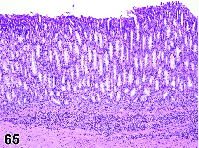
Mouse glandular stomach. Inflammation neutrophil with hypertrophy, mucus cell.
Modifiers: Type of inflammatory cell that represents the predominant cell type in the infiltrate
Pathogenesis: Infiltration of lamina propria and/or submucosa with neutrophils (Inflammation, neutrophil) or mononuclear cell (Inflammation, mononuclear cell) or a combination (Inflammation, mixed) with additional histological features of inflammation e.g. hemorrhage, edema, fibroplasia.
Diagnostic features
Focal, multifocal or diffuse infiltration of cells in lamina propria and/or submucosa.
Presence of other histological criteria of inflammation e.g. hemorrhage, edema, fibroplasia.
Differential diagnoses
Infiltrate, inflammatory cell: Other morphologic features of inflammation such as edema, hemorrhage, necrosis and/or fibroplasia will be absent.
Granulocytic leukemia: Infiltration of neutrophil precursors and/or abnormal neutrophils will probably be present along with mature neutrophils; infiltration of similar neoplastic cells is likely present in other organs.
Lymphoma: Infiltration of monomorphic population of lymphocytes (often with atypical, increased and/or abnormal mitoses); infiltration of similar neoplastic cells is likely present in other organs.
Comment: Inflammation in the both glandular and nonglandular stomach is most often seen in combination with erosion or ulceration.
e. Infectious Diseases
Helicobacter
Pathogenesis: Presence of Helicobacter spp. in mice.
Diagnostic features
Spiral shaped bacteria in the glands primarily usually near border of antrum and corpus.
Infection by H. felis and H. pylori in some strains of mice results in inflammatory, dysplastic and eventually neoplastic changes.
Proliferation of gut associated lymphoid tissue (GALT), resembling human MALT lymphoma, may occur in certain strains of mice (e.g. BALB/c).
Differential diagnosis
Inflammation: Non-helicobacter opportunists may colonize the achlorhydric rodent stomach (e.g. gastrin-deficient mice) and invoke a similar chronic inflammatory disease.
Comment: The progression from infection by H. pylori and H. felis to the formation of gastric adenocarcinoma in various mouse strains has been well described (Rogers and Fox, 2004; Rogers and Houghton 2009). Host immunity plays a crucial role in determining the outcome of gastric helicobacter infection. Mouse strains such as C57BL/6 that mount a strong Th1 responses to helicobacter infection demonstrate lower colonization but increased inflammation and hyperplasia/dysplasia compared to strains such as BALB/C with Th2 predominant responses (Rogers and Houghton 2009). Silver stains e.g. Wartin-Starry can be used to confirm the presence of helicobacter in tissues section.
Yeast
Figure 66.
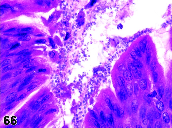
Mouse glandular stomach. Yeast.
Pathogenesis: Infection of the glandular stomach in compromised host, e.g. immunodeficiency.
Diagnostic Features
Presence of a layer of round yeast-like structures along the glandular stomach epithelial surface lining and in the gastric contents.
Maybe be large numbers of yeasts or few.
Budding maybe seen.
Associated inflammation is usually not observed but may be seen.
Stains with Giemsa stain.
Differential diagnosis:
Bacteria: Do not show budding.
Cryptosporidia: Are more within or intergrated with the surface epithelium.
Comments: This infection appears to be rare histologically but may be more commonly found by culture (Mackinnon 1959, Savage and Dubos 1967). A variety of similar organisms have been reported in many species (Kurtzman et al. 2005). These organisms usually are not pathogenic but may be under specific conditions such as immunodeficiency.
f. Vascular Lesions
Edema and hemorrhage of the stomach are similar to those described for the upper digestive tract. Other vascular lesions in the stomach and mesentery are dealt with in the INHAND monograph on the cardiovascular system.
g. Non-neoplastic Proliferative Lesions of the Nonglandular Stomach
Hyperplasia, squamous cell
(Figures 67 and 68)
Figure 67.
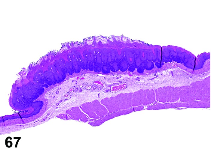
Rat nonglandular stomach. Hyperplasia squamous cell focal.
Figure 68.
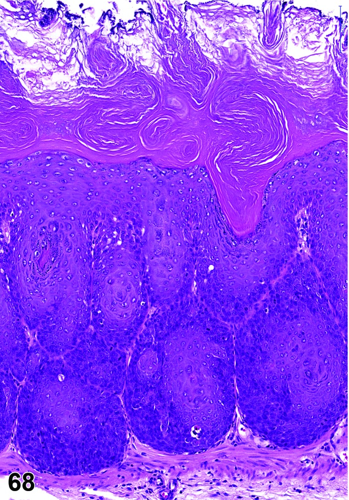
Rat nonglandular stomach. Hyperplasia squamous cell focal. Higher magnification of Figure 67 indicating maintained polarity of epithelium and complete differentiation to keratinocytes.
Synonyms: Squamous hyperplasia; squamous cell hyperplasia
Histogenesis: Squamous epithelium of the nonglandular stomach.
Modifier: Reactive
Diagnostic features
Proliferation and thickening of the stratum spinosum, often in association with a thickened keratin layer (parakeratotic or hyperkeratotic).
Focal, multifocal or diffuse.
Exophytic (papillary hyperplasia) or endophytic growth pattern.
Papillary projections not or only minimally branched and without fibrovascular stroma.
Broad-based hyperplastic foci might have multiple straight or minimally branched projections.
Polarity of differentiation to keratinocytes is orderly and complete.
Rete peg formation may be exaggerated, i.e. papillary body may show undulation due to enlarged germinal layers.
Endophytic growth with cyst-like structures may occur; cysts lined by well-differentiated squamous epithelium and filled with keratin; may extend into the submucosa, but basement membrane is intact.
Mitotic activity may be increased.
Dysplasia of spinous and basal cells may occur.
Reactive as a descriptive term: Evidence for a primary irritating process, e.g. ulcer, irritating chemical, foreign material.
Differential diagnoses
Papilloma, squamous cell: Papillary projection with a single and complex branching fibrovascular connective tissue core.
Hyperplasia, basal cell: Endophytic proliferation of basal cells; cytoplasm of proliferative cells is basophilic.
Carcinoma, squamous cell: True invasion, i.e. lost basement membrane integrity; in the poorly differentiated type: Lost polarity of differentiation to keratinocytes.
Comments: There may be a morphologic continuum from hyperplasia to papilloma and the distinction between severe hyperplasia and papilloma may in these cases be subjective.
The squamous epithelium at the border of nonglandular and glandular stomach (limiting ridge) is generally thickened relative to the adjacent squamous mucosa. This area may be folded and, thus, cut tangentially or obliquely, if the stomach was not properly stretched prior to fixation.
Hyperkeratosis or parakeratosis alone should be distinguished from squamous hyperplasia. Acanthosis is a synonym for hyperplasia of the squamous cell epithelium of the skin and should not be used in this organ. Diffuse squamous cell hyperplasia is relatively common in oral gavage studies when the test article has an irritating potential.
The modifier “reactive” should be used if proliferation of stratified squamous epithelium is associated with or secondary to local erosion, ulcer or inflammation. The absence of an ulcer or erosion in the plane of section does not preclude the presence of such in adjacent unexamined tissue.
Florid hyperplasia of the nonglandular stomach epithelium without evidence of cellular atypia can be completely reversible following the withdrawal of an inciting stimulus. Nonglandular stomach carcinogens may induce dysplasia of spinous and basal cells, especially if the hyperplasia is a precursor to a papilloma or squamous cell carcinoma. Squamous cell hyperplasia and basal cell hyperplasia may occur concurrently. This change has been observed in association with test article exposure (e.g. butylated hydroxyanisole) or chronic vitamin A deficiency (Klein-Szanto et al. 1982).
Hyperplasia, basal cell
(Figures 69, 70 and 71)
Figure 69.
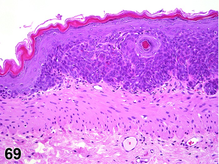
Rat nonglandular stomach. Hhyperplasia basal cell, focal. Note unaltered suprabasal epithelial layers.
Figure 70.
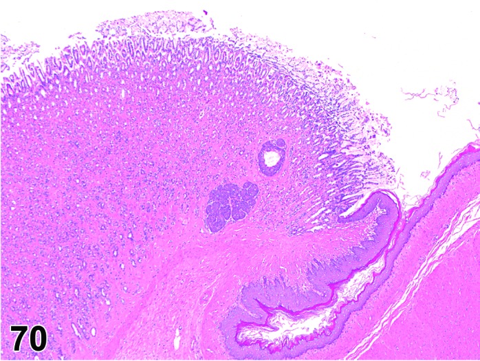
Rat nonglandular and glandular stomach. Foci of hyperplastic basal cells in the glandular mucosa close to the limiting ridge. As this lesion is considered to originate from nonglandular stomach it should be collected under the term “nonglandular stomach, hyperplasia, basal cell”.
Figure 71.
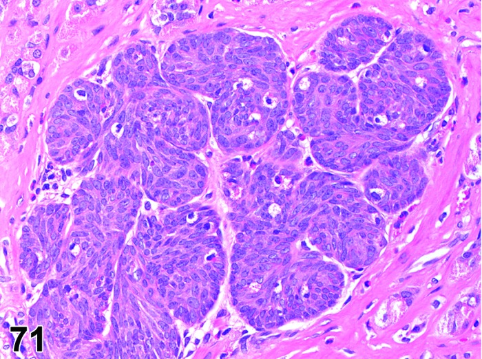
Higher magnification of Figure 70, showing the basal cell character of the lesion.
Histogenesis: Basal layer of the stratified squamous epithelium.
Diagnostic features
Proliferation of the basal cell layer, basophilic staining increased.
Focal or diffuse.
Endophytic growth pattern.
No alteration of basement membrane integrity.
Papillary body shows marked undulation but rete peg structure still present.
Differential diagnoses
Hyperplasia, squamous cell: Thickening of the epithelium with all normally existing layers.
Carcinoma, squamous cell: Evidence for lost basement membrane integrity, spinous cells and keratinized cells proliferate, cellular atypia.
Tumor, basal cell, benign: Circumscribed proliferation of basal cells with loss of rete peg structure and leading to compression of surrounding tissue or prominent elevation of overlying epithelial layers.
Tumor, basal cell, malignant: Basal cells predominate, keratinization is absent, evidence for lost basement membrane integrity.
Comment: Basal cell hyperplasia and squamous cell hyperplasia may occur concurrently.
Isolated nests of basal cells may occur in the lamina propria, dependent on the plane of section through the rete peg structures. However, their discrete border indicates an intact basement membrane.
Basal cell hyperplasia and dysplasia are more frequently associated with chemicals (e.g., butylated hydroxyanisole) that result in neoplasms of the nonglandular mucosa.
Dysplasia of basal cells, characterized by keratinization within the basal layer, may be observed.
Foci of basal cell hyperplasia may be found in the mucosa of the glandular stomach in the vicinity to the limiting ridge (Figure 49b and c). Due to their similarity to basal cell hyperplasia of the nonglandular stomach and their specific location they are considered to originate from the nonglandular stomach. Such lesions should be recorded under “nonglandular stomach, hyperplasia, basal cell”.
h. Neoplasms of the Nonglandular Stomach
Papilloma, squamous cell
Figure 72.
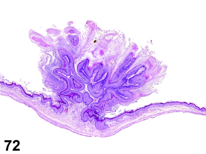
Mouse nonglandular stomach. Papilloma, squamous cell.
Synonym: Papilloma
Histogenesis: Stratified squamous epithelium.
Diagnostic features
Solitary or multiple.
Exophytic or endophytic (usually exophytic).
Formation of a single complex branching fibrovascular core by exophytic tumors; the stroma typically forms secondary branches or finger-like projections which blend with the underlying lamina propria.
Well-differentiated squamous cell layer (orderly maturation of the epithelium), often with prominent hyperkeratosis or parakeratosis.
Penetration of muscularis mucosae is possible (papillomas extend into the submucosa), but no evidence for lost basement membrane integrity.
Lack of cellular atypia.
Mitotic figures are rare.
Local inflammatory reactions are common (lymphocytic infiltrations in the stromal stalk).
Differential diagnoses
Hyperplasia, squamous cell: Papillary projections are minimally branched at the most and lack a fibrovascular stromal stalk.
Carcinoma, squamous cell: Evidence for lost basement membrane integrity is obligatory; invading tumor cells generally with varying degrees of structural and cellular atypia.
Comment: There is a morphologic continuum from hyperplasia to papilloma and the distinction between severe hyperplasia and papilloma is sometimes subjective.
Also comparative diagnosis and distinction of well differentiated squamous cell carcinomas from papillomas may be difficult: The tumor tissue of papillomas may extend into the submucosa; a growth pattern suggestive of intact basement membranes and the lack of cellular and structural atypia distinguishes it from squamous cell carcinomas.
Invasive squamous cell carcinomas may have papillary lesions on their luminal surface. In carcinomas arising within papillomas, invasion of the fibrovascular connective tissue stalk is present.
The spontaneous papilloma in the nonglandular stomach is a rare event in rats, but more common in mice and in some lines of genetically-engineered mice (Sundberg et al. 1992). Papillomas occur more often in gavage studies than with other routes of exposure.
Carcinoma, squamous cell
(Figures 73 and 74)
Figure 73.
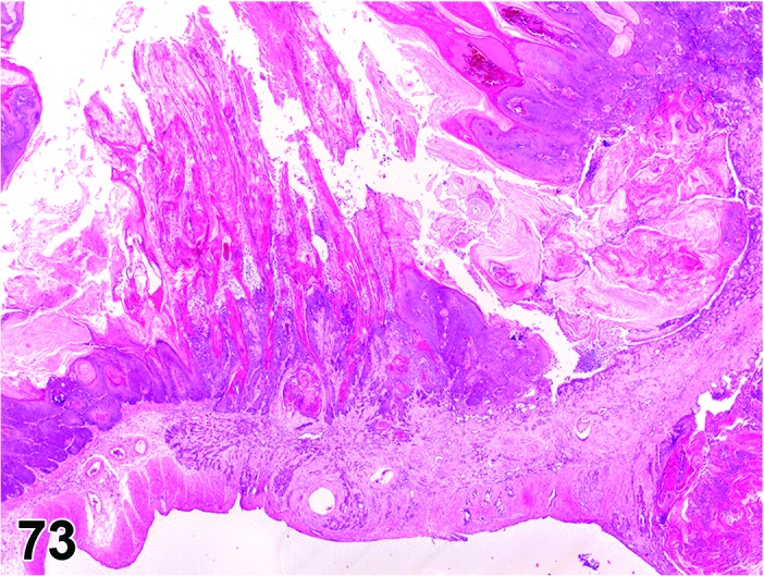
Rat nonglandular stomach. Carcinoma squamous cell, arising from diffuse severe hyperplasia, squamous cell.
Figure 74.
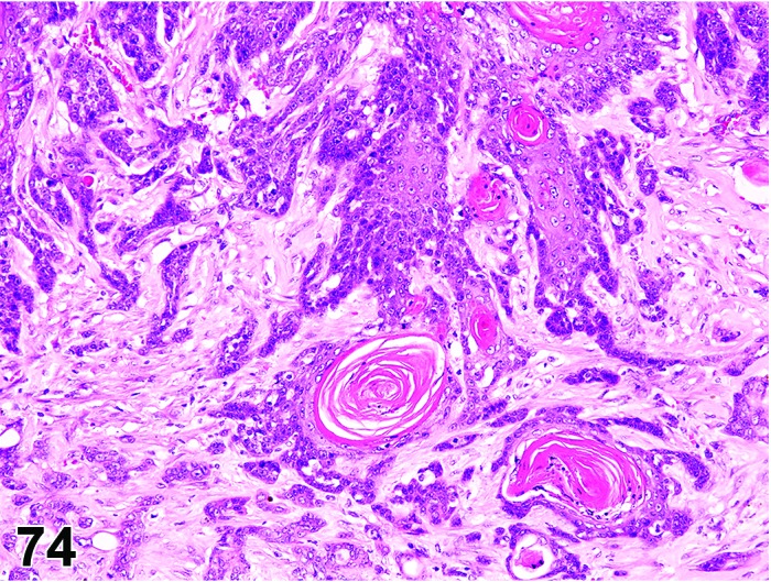
Rat nonglandular stomach. Carcinoma squamous cell; infiltrative growth with fibroblastic stromal response.
Synonyms: Carcinoma epidermoid; carcinoma squamous
Histogenesis: Stratified squamous epithelium.
Diagnostic features
Exophytic and/or endophytic growth pattern.
Penetration of basal membrane by single cells or nests of neoplastic cells.
Loss of cellular differentiation, anaplasia.
Submucosa, muscle layer and serosa can be infiltrated.
Well-differentiated type: nearly normal squamous epithelium with irregular structure of the papillary body, frequently showing foci with central hyperkeratinization (keratin pearls); in areas of invasion, predominance of polygonal and pleomorphic cells with varying degrees of keratinization. Poorly differentiated type (anaplastic): solid sheets or strands of spindle cells leading to varying degrees of desmoplastic appearance; keratinization may be difficult to detect.
Cells show varying size (frequently larger than normal) and shape; nuclei are hyperchromatic, enlarged and have prominent nucleoli.
Mitotic figures may be numerous.
Superficial ulceration and inflammation or fibroplasia of the lamina propria or submucosa are common features.
May metastasize to the abdominal cavity, regional lymph nodes, or the lung.
Differential diagnoses
Papilloma, squamous cell: No evidence of lost basement membrane integrity or invasion and no cellular atypia.
Tumor, basal cell, benign OR tumor, basal cell, malignant: Exclusively endophytic growth pattern and a typical arrangement of the small basophilic basal cells in garlands or rows; poorly differentiated squamous cell carcinomas can have a prominent basal cell component but this is often associated with extensive invasion and a scirrhous response.
Hyperplasia, squamous cell: No evidence of lost basement membrane integrity or of invasive growth although muscularis mucosae may be displaced; no evidence of cellular atypia.
Hyperplasia, basal cell: Can form round or oval clusters which extend into the underlying lamina propria; these clusters usually have discrete borders (suggestive of maintained basement membrane integrity) and no cellular atypia.
Comment: Depending on the plane of section, it can be difficult to distinguish a well-differentiated carcinomas from a papilloma. Papillomas may also extend into the submucosa and thereby displace the muscularis mucosa. The presence of carcinomatous foci in an otherwise benign appearing papilloma is indicative of malignancy (Fukushima and Ito, 1985; Tsukamoto et al. 2007). In general, the lack of cellular atypia allows a distinction to be made. Many squamous cell carcinomas in the nonglandular stomach are exophytic which can result in partial or total occlusion of the lumen. The exophytic portion can be papillary resembling papilloma.
Chemically-induced (e.g. butylated hydroxyanisole, 1,2-dibromo-ethane) squamous cell carcinomas may contain mostly basal or spindle cells with little squamous differentiation. These tumors are considered a morphological variant of the more typical squamous cell carcinoma. Ultrastructurally, squamous cell carcinomas contain typical desmosomes and bundles of electron-dense tonofilaments. Some chemicals especially when given by gavage cause highly malignant squamous cells carcinomas in rats which metastasize to the peritoneal cavity and lungs (Olson et al. 1973).
Tumor, basal cell, benign
Histogenesis: Basal layer of the stratified squamous epithelium.
Diagnostic features
Endophytic growth pattern.
Closely packed small or oval cells from the stratum germinativum forming garlands or nests with peripherally palisade-like cells.
Loss of rete peg structure.
Growth pattern suggests that basement membrane is intact.
Compression of surrounding tissue or elevation of overlying epithelium which itself does not show neoplastic alteration.
No keratinization.
Differential diagnoses
Hyperplasia, basal cell: Proliferation of basal cells with preservation of rete peg structure; no or only slight compression of surrounding tissue or elevation of overlying epithelial layers.
Carcinoma, basal cell: Growth pattern suggests that basement membrane integrity is lost.
Carcinoma, squamous cell: Growth pattern suggests that basement membrane integrity is lost, spinous cells and keratinized cells proliferate.
Comment: The absence of differentiation and therefore the predominant proliferation of the basal cell is a criterion for the basal cell tumor diagnosis. This tumor type is very rare, and no images were available at point of publication.
Tumor, basal cell, malignant
Synonym: Carcinoma, basal cell
Histogenesis: Basal layer of the stratified squamous epithelium.
Diagnostic features
Endophytic growth pattern.
Superficial layers almost normal, but sometimes ulcerated, while neoplastic basal cells infiltrate into the lamina propria of the mucosa and submucosa.
Round to oval closely packed nonspinous cells with dark nuclei and little cytoplasm, often forming palisade-like garlands or chains.
Keratinization generally absent.
Growth pattern suggests that basement membrane integrity is lost.
Frequent mitotic figures.
Differential diagnoses
Carcinoma, squamous cell: Well differentiated squamous cell carcinomas with prominent keratinization; poorly differentiated variants can have a prominent basal cell component but this is generally associated with extensive invasion and a scirrhous response.
Hyperplasia, basal cell: Proliferation of basal cells with preservation of rete peg structure; no or only slight compression of surrounding tissue or elevation of overlying epithelial layers; growth pattern indicates that basement membrane integrity is maintained.
Tumor, basal cell, benign: Growth pattern indicates that basement membrane integrity is maintained.
Comment: The absence of differentiation and therefore the predominant proliferation of the basal cell is a criterion for the basal cell tumor diagnosis. This tumor type is very rare, and no images were available at point of publication.
i. Non-neoplastic Proliferative Lesions of the Glandular Stomach
Diverticulum
(Figures 75, 76, 77, 78, 79 and 88)
Figure 75.
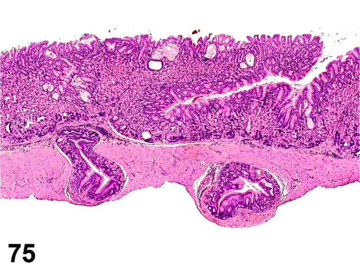
Mouse glandular stomach. Diverticulum. Well-differentiated mucosa present in submucosal to subserosal location.
Figure 76.
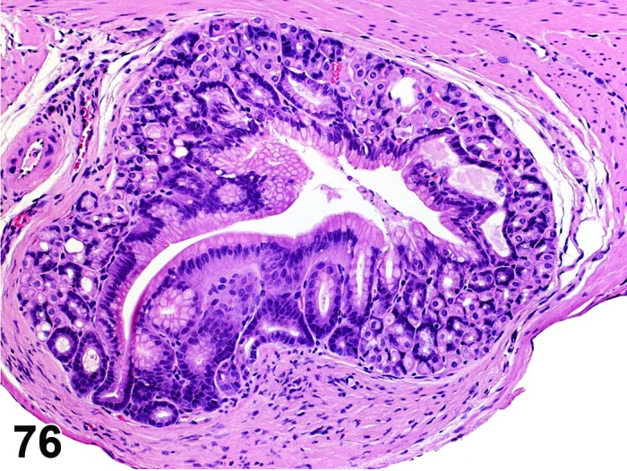
Mouse glandular stomach. Diverticulum. Higher magnification of Figure 75. Note growth pattern indicating maintained basement membrane integrity.
Figure 77.
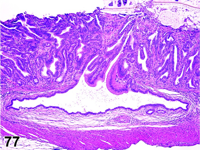
Mouse glandular stomach. Diverticulum. Penetration of muscularis mucosae by well differentiated single-layered epithelium with maintained basement membrane integrity.
Figure 78.
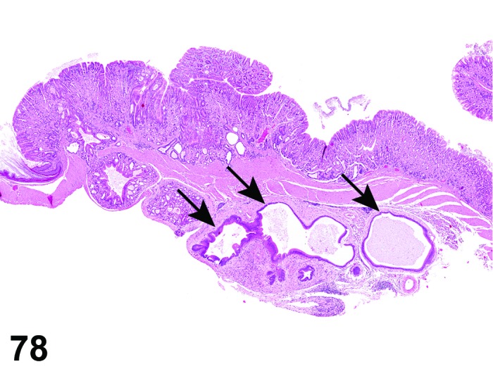
Mouse glandular stomach. Diverticula, some of them atypical (arrows).
Figure 79.
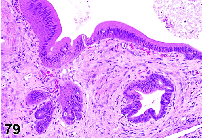
Mouse glandular stomach. Diverticula atypical. Higher magnification of Figure 78.
Figure 88.
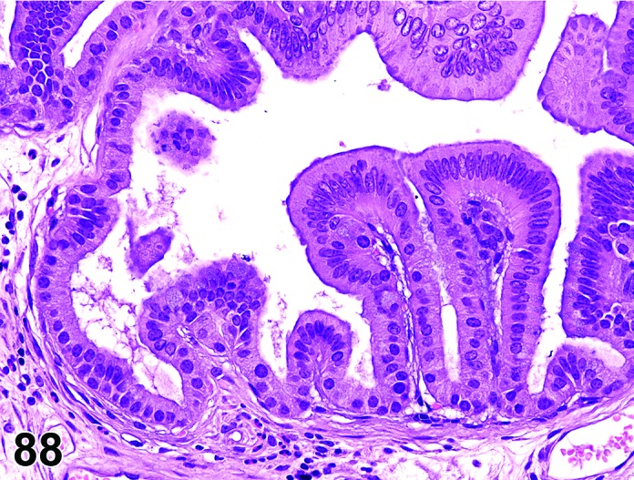
Mouse glandular stomach. Diverticulum atypical. Higher magnification of Figure 87. Atypia evidenced by increased cellularity and absence of intracellular mucus. Note intact basement membrane.
Synonyms: Cystic adenomatous hyperplasia; diverticulosis;Diverticulum, atypical, may have been identified as: Atypical cystic hyperplasia; cystic hyperplasia with growth into the gastric wall; cystic adenomatous hyperplasia; herniation atypical; “pseudoinvasion”, atypical; hamartoma, atypical
Modifiers: Cystic, atypical
Pathogenesis: Usually caused by mechanical or biochemical disruption of gastric wall structures resulting in loss of extracellular matrix integrity and internal projection of surface lining into deeper layers; may alternatively represent a congenital abnormality.
Diagnostic features
Extension of glands through muscularis mucosae, into submucosa and further in some cases.
Morphology of epithelial lining is variable, ranging from single layer cuboidal or columnar cells to complete mucosa.
Epithelium may show features of regeneration like increased basophilia, increased nucleus-to-cytoplasm ratio and gradual loss of polarity, but atypia is minimal at the most.
Basement membrane integrity is always maintained.
More often seen in the antrum of mice.
Often accompanied by inflammation and regenerative / reparative processes.
May contain ingesta, hair, or other foreign material.
Cystic:
Rounded cystic structures in lamina propria or deeper layers.
Compression of surrounding tissue, some degree of compression atrophy of the epithelium lining the cyst.
May contain mucus.
Atypical:
Extension of atypical/dysplastic mucosal glands into or through muscularis mucosa, submucosa, and deeper in some cases.
Hyperplastic atypical (dysplastic) epithelial cells forming cystic glands that may appear to be “invading” into deeper layers of the stomach.
Atypical change often focal in an otherwise normal appearing epithelium.
Differential diagnoses
Adenoma: Polypoid or papillary growth into gastric lumen obligatory, but pseudo-invasion into stalk may be seen.
Adenocarcinoma: Multiple layers of lining epithelium with varying degrees of dysplasia. True infiltrative growth with loss of basement membrane integrity and often with a scirrhous stromal reaction.
Comment: Glandular diverticula of the stomach, frequently observed in mice (especially in genetically-engineered mice and in mice with chronic experimental gastric infections), may be associated with local mucosal defects (e.g. post-ulcerative repair), suggesting opportunistic expansion, but these have also been observed within submucosal veins in the absence of mucosal defects, suggesting there may be a vascular route of penetration from the mucosa.
These structures are typically lined by a simple cuboidal mucus epithelium, which may have an antral or intestinal mucus phenotype that can be characterized by histochemical stains (e.g. Alcian blue). These focal gastric lesions do not resemble diverticulosis in human colon which may be focal, multifocal or diffuse. They do not appear to be neoplastic, although they do create an increased risk of rupture and septic peritonitis.
It is not recommended to record “diverticulum” (plus modifier) as a separate finding, if it occurs in association with another proliferative process (e.g. hyperplasia, adenoma).
Small glandular diverticula lined by a single layer of cuboidal or columnar epithelium resemble “crypt herniation” of human gastrointestinal pathology (e.g. Rex et al. 2012). Such herniation of crypts through the muscularis mucosae is interpreted as “pseudoinvasive” or “inverted” growth pattern. Although the process-related term “crypt herniation” is common in literature (e.g. Betton et al. 2001), the term “diverticulum” is considered to better fit to the INHAND principle to use descriptive terms and to avoid process-related ones (Mann et al. 2012). Also the misnomer “adenomatous diverticulum” has been used to describe these structures and the prefix “adenomatous” should be avoided.
Atypical diverticula must be distinguished from genuine neoplastic invasion (e.g. loss of basement membrane and other signs of malignancy in the latter). They are associated with preexisting mucosal atypical hyperplasia/dysplasia as seen under specific experimental conditions, tumor-associated gastric toxins, genetically-engineered mice and chronic Helicobacter pylori infection (Andersson et al. 2005; Boivin et al. 1996; Boivin et al. 2003; Fernandez-Salguero et al. 1997; Miyoshi et al. 2002; Rogers and Houghton 2009). Whereas they often do not progress to unequivocal adenocarcinoma, atypical glandular and cellular features are consistent with neoplastic potential.
Atypical diverticula may be associated with atypical hyperplasia and adenoma, but usually not with adenocarcinoma. As noted above for the simple diverticula, they should not be recorded as a separate diagnostic entity when associated with another proliferative process.
Hyperplasia, focal, mucosa
(Figures 80 and 81)
Figure 80.
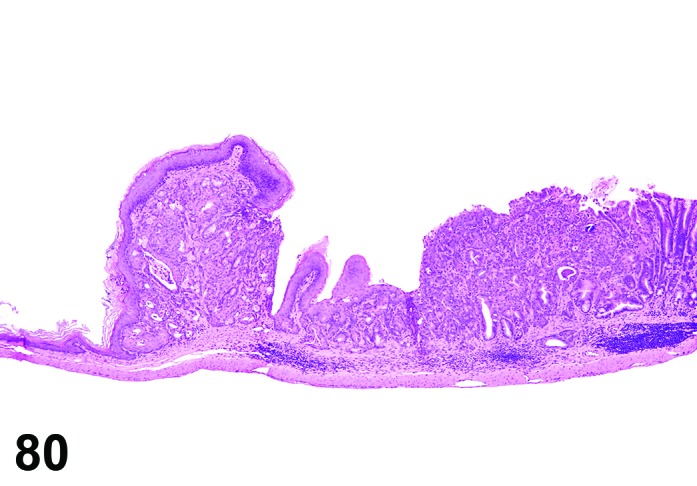
Mouse glandular stomach. Hyperplasia, focal, mucosa, atypical.
Figure 81.
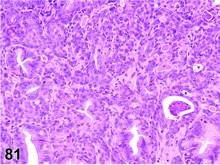
Mouse glandular stomach. Hyperplasia, focal, mucosa, atypical. Higher magnification of Figure 80.
Synonyms: Regenerative hyperplasia, focal Hyperplasia, focal (mucosa), atypical may have been identified as: Adenomatous hyperplasia, dysplasia, or gastric intraepithelial neoplasia (GIN)
Histogenesis: Glandular and/or surface epithelial cells.
Modifier: Atypical
Diagnostic features
Focal or multifocal due to underlying injury (e.g. ulcer); mucosal defects/injury and inflammation may be observed in tandem with proliferative changes.
Confined to the gastric mucosa.
No significant compression of adjacent tissue.
Proliferation may be manifested as an increase in mucosal thickness or penetration of glands through the muscularis mucosae to the submucosa, tunica muscularis, or serosa forming cystic or glandular diverticula. These structures are typically lined by a simple cuboidal mucus epithelium.
With the exception of diverticula, no distortion of regular mucosal structure by the proliferative process itself.
May be associated with a reduction of parietal cells.
Eosinophilic globules may be present in the epithelial cell cytoplasm.
Mitoses may be increased.
Atypical:
Abnormal structure of gastric glands.
Focal penetration by glandular diverticula into lamina propria or deeper layers but basement membrane is always intact.
Hyperchromatic foci with cellular atypia and pleomorphism.
Loss of epithelial polarity.
Differential diagnoses
Adenoma: Typically arise as singular foci in the distal antral/pyloric region, often with polypoid architecture. Nodular (polypoid, papillary) lesions growing into the lumen of the stomach; distortion of glandular architecture and in case of non-papillary adenomas also compression of surrounding tissue.
Adenocarcinoma: Invasion of nests and cords of neoplastic cells into lamina propria or deeper layers, indicating loss of basement membrane integrity; cellular atypia.
Comment: Focal hyperplasia may be accompanied by metaplasia of the mucus epithelium, characterized by a shift from a neutral mucin phenotype of the surface epithelium to an acidic antral or intestinal mucin phenotype. This may be associated with a reduction in parietal cells (atrophy) and a presumptive shift in local pH. Distinct patterns of hyperplastic changes may arise in the glandular stomach because of the cellular complexity, regional differences in architecture, and selectivity of stimuli.
Focal atypical hyperplasia represents an altered proliferation of mucosal epithelium. It may be associated with chronic inflammation. An example is the Helicobacter pylori mouse model. The lesion may progress into an adenoma or adenocarcinoma. Although frequently considered as preneoplastic by nature, this needs to be proven in a given case in order to be classified as such. Focal intra-epithelial atypical proliferations resembling morphologically an invasive adenocarcinoma have been found in specific genetically modified mouse lines and after chemical induction. They have been termed Gastric Intraepithelial Neoplasia (GIN). Other terms occasionally used but not preferred as synonyms are microadenoma, carcinoma in situ, and intra-epithelial carcinoma.
Hyperplasia, diffuse, mucosa
Figure 82.
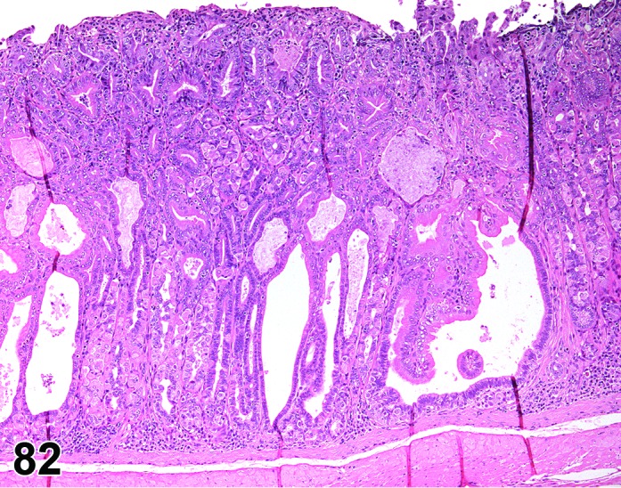
Mouse glandular stomach. Hyperplasia, diffuse, mucosa. Age-related diffuse hyperplasia of fundic mucosa.
Synonyms: Regenerative hyperplasia diffuse; hypertrophic gastritis; mucosal hypertrophy
Histogenesis: Glandular and/or surface epithelial cells.
Diagnostic features
May be diffuse within a specific regional zone (corpus, antrum, pylorus).
Mitotic activity may be increased.
Within a region of mucosa, thickening of the mucosa may affect the full thickness or may be compartmentalized within discrete glandular zones or cell types.
With increasing severity, basophilia, decreased epithelial differentiation, and structural disorganization may be observed.
Metaplasia of the mucus epithelium may arise, characterized by a shift from a neutral mucin phenotype to an intestinal mucin phenotype. This may be associated with a reduction in parietal cells and a presumptive shift in local pH.
A cause may be evident (irritating substance, gavage material).
Differential diagnosis
Adenoma: Nodular lesions growing exophytic (polypoid, papillary).
Comment: Diffuse hyperplasia may be induced by chemicals (e.g. Misprostal) or by larval (Taenia taeniaeformis) infestation in the liver. The latter results in hypergastrinemia and diffuse glandular mucosal hyperplasia.
The incidence of mitotic figures at the base of the gastric pit and the ratio of the lengths of the foveolus (gastric pit) to the deeper oxyntic region can be indices of proliferative activity; however, the latter will normally increase moving distally from the squamocolumnar junction.
Diffuse fundic hyperplasia is a common finding in aged mice, where it is often accompanied by degenerative changes like glandular cysts or diverticula.
Hyperplasia, neuroendocrine cell
(Figures 83 and 84)
Figure 83.
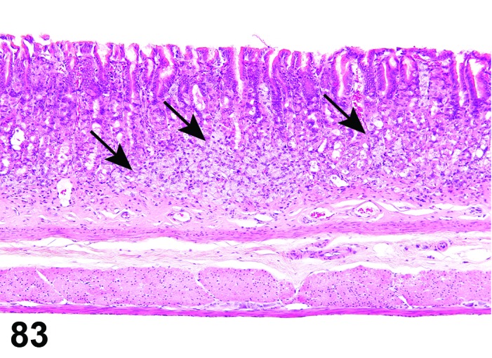
Rat glandular stomach. Hyperplasia neuroendocrine cell (arrows).
Figure 84.
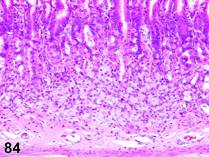
Rat glandular stomach. Hyperplasia neuroendocrine cell. Higher magnification of Figure 83. Note characteristics of neuroendocrine cells: clear cytoplasm and small, monomorphic nuclei.
Synonym: Enterochromaffin-like (ECL) cell hyperplasia
Histogenesis: Neuroendocrine cells of the gastric mucosa. The enterochromaffin-like (ECL) cell is the predominant neuroendocrine cell of the oxyntic mucosa.
Diagnostic features
Focal: aggregates of neuroendocrine cells no greater than 3 glands in diameter.
Diffuse: increased number of individual or small clusters of neuroendocrine cells scattered in the mucosa.
Cytoplasm is pale and abundant, nuclei are small and monomorphic.
Neuroendocrine cells are argyrophilic (e.g. Grimelius, Servier-Munger stains) and immunohistochemically positive for neuron-specific enolase, chromogranin A, histidine decarboxylase, and histamine. Secretory granules can be observed by electron microscopy.
Differential diagnosis
Tumor, neuroendocrine cell, benign/malignant: Discrete neoplastic foci are greater than 3 gastric glands in diameter. Larger neoplasms may form sheets or demonstrate atypical architecture (e.g. acini). Greater cellular pleomorphism, eosinophilic cytoplasm, increased nuclear size and mitotic figures. Diminished differentiation may influence the degree of immunohistochemical staining.
Comment: Proton pump-inhibitors may induce neuroendocrine hyperplasia. Small numbers of normal neuroendocrine cells can occasionally be observed in the submucosa and should not be categorized as neoplasia.
j. Neoplasms of the Glandular Stomach
Adenoma
(Figures 85, 86, 87 and 88)
Figure 85.
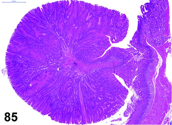
Mouse glandular stomach. Adenoma, polypoid.
Figure 86.
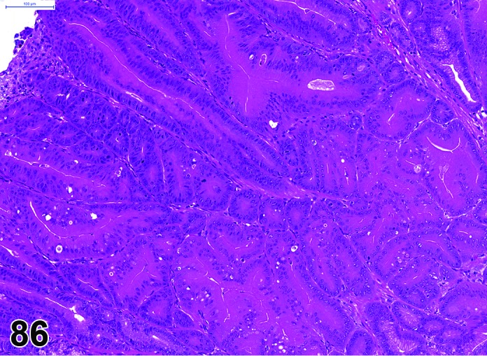
Mouse glandular stomach. Adenoma, polypoid. Higher magnification of Figure 85 demonstrating distorted mucosal architecture.
Figure 87.
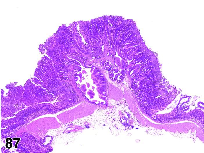
Mouse glandular stomach. Adenoma, sessile and multiple diverticula.
Synonyms: Polyp; adenomatous polyp; polypoid adenoma; exocrine adenoma
Histogenesis: Glandular and/or surface epithelial cells.
Modifiers: Polypoid, papillary, sessile
Diagnostic features
Typically arise in the antral or pyloric mucosa.
Mucosal architecture distorted.
Growth pattern often polypoid, sessile, or papillary with or without a fibrovascular stalk; in polypoid adenomas of the antral mucosa, gland architecture may be retained.
Focal penetration by glandular diverticula into lamina propria or deeper layers may be present but basement membrane is always intact.
Cells are a basophilic, less differentiated glandular epithelium, but with little atypia; polarity is maintained.
Epithelial nuclei are unilayered or organized with varying degree of stratification.
Differential diagnoses
Hyperplasia, focal (mucosal): Linear mucosal orientation without polypoid or papillary architecture.
Adenocarcinoma: Infiltration as evidenced by penetration into lamina propria or deeper layers with a growth pattern indicative of lost basement membrane integrity; scirrhous stromal reaction.
Tumor, neuroendocrine cell, benign: Discrete intramucosal foci, which typically arise in hyperplastic areas in the oxyntic mucosa (i.e. corpus region). Closely organized clusters of neuroendocrine cells with eosinophilic cytoplasm, separated by thin fibrovascular stroma. Argyrophilic and characteristic immunohistochemical phenotype.
Comment: Well-differentiated, hyperplastic glands may be entrapped in the adjoining lamina propria or submucosa, Such structures originate from herniation of glands, are well demarcated, lack a scirrhotic stromal response and should not be interpreted as neoplastic infiltration.
Adenomas, as well as atypical hyperplasia, may exhibit some degree of dysplasia and distortion of pre-existing architecture. Macroscopic visibility has been suggested as a differential diagnostic criterion by some authors. This criterion is not used in the context of this description of histologic criteria. Any lesion exhibiting a papillary or polypoid growth pattern is an adenoma, irrespective whether it was grossly visible or not. An intra-epithelial small focus with cytological characteristics seen in adenomas should NOT be diagnosed as an adenoma but as atypical hyperplasia. An adenoma must possess a polypoid, sessile or papillary growth pattern, whether grossly visible or not.
Gastric adenomas are an uncommon observation in untreated rodents, but may be frequent in some genetically modified mouse strains (Takaku et al. 1999; Miyoshi et al. 2002). They are associated with specific mutations, which may be similar to those of humans with gastric polyps.
Adenocarcinoma
(Figures 89 and 90)
Figure 89.
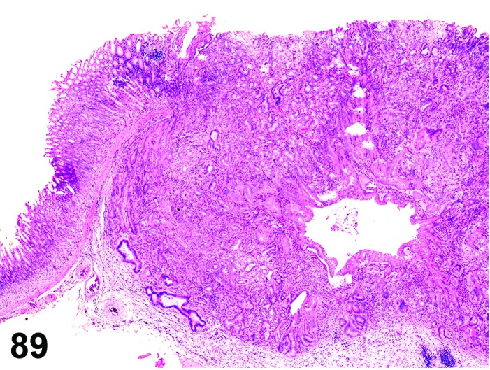
Rat glandular stomach. Adenocarcinoma.
Figure 90.
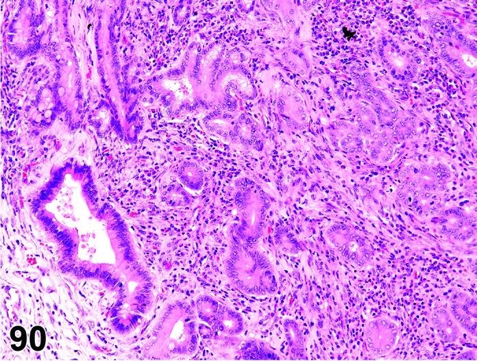
Rat glandular stomach. Adenocarcinoma. Higher magnification of Figure 89, showing true infiltrative growth with lost basement membrane integrity. Stroma immature and cellular.
Synonym: Carcinoma
Histogenesis: Glandular and/or surface epithelial cells.
Modifiers: Tubular, papillary, cystic, solid, scirrhous, mucinous, signet ring, undifferentiated. mixed
Diagnostic features
Predominantly found in the antrum.
Regular mucosal structure is lost.
Growth exophytic or endophytic.
Major morphologic types include tubular, papillary, cystic, solid, scirrhous, mucinous, and signet ring.
Invasion of nests and cords of neoplastic cells into lamina propria or deeper layers, loss of basement membrane integrity.
Varying degrees of cytologic and nuclear atypia, anaplasia, hyperchromatism, basophilic or clear cytoplasm, and increased mitotic figures.
Mucin secretion may be present even if poorly differentiated and, if strong, may result in signet ring morphology; such tumors usually grow locally highly infiltrative but tumor cells may be deceivingly inconspicuous, in which case cytokeratin stains are most helpful for both diagnosis and assessment of tumor extent.
Invasive growth may elicit fibrous/scirrhous reaction and inflammation, i.e. areas of infiltration are surrounded by loose stroma.
Differential diagnoses
Hyperplasia focal (mucosal): Proliferation with maintained mucosal structure, except of glandular diverticula; no atypia in simple hyperplasia; in case of glandular diverticula, localized and discrete penetration into lamina propria or deeper layers with maintained basement membrane integrity and surrounded by pre-existing mature tissues lacking a scirrhotic stromal response.
Hyperplasia focal (mucosal) atypical: Limited to the mucosal lining, no infiltrative growth; focal penetration by glandular diverticula into lamina propria or deeper layers may be present but basement membrane always intact.
Diverticulum: Localized and discrete penetration into lamina propria or deeper layers with maintained basement membrane integrity and surrounded by pre-existing mature tissues lacking a scirrhotic stromal response.
Adenoma: No infiltrative growth; focal penetration by glandular diverticula into lamina propria or deeper layers may be present but basement membrane always intact.
Tumor, neuroendocrine cell, malignant: Typically arise in hyperplastic areas in the oxyntic mucosa (i.e., corpus region); closely organized clusters of neuroendocrine cells with eosinophilic cytoplasm, separated by thin fibrovascular stroma; argyrophilic and characteristic immunohistochemical phenotype.
Comment: Gastric adenocarcinomas are uncommon spontaneous tumors in rodents, except of some genetically modified mouse strains. Chemically-induced tumors in routine mouse/rat strains are not common except for induction by specific chemicals, e.g. N-methyl-N’-nitrosoguanidine, NMU, MNNG in rats, while mice are more resistant (Tsukamoto et al. 2007). The morphologic type may depend on the etiology / carcinogen.
Tumor, neuroendocrine cell, benign
(Figures 91 and 92)
Figure 91.
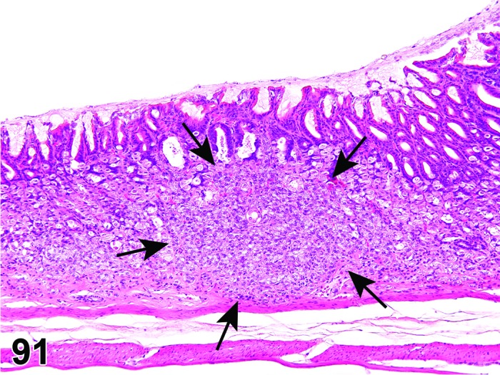
Rat glandular stomach. Tumor, neuroendocrine cell, benign; on a background of disseminated neuroendocrine hyperplasia. Tumor outlined by arrows.
Figure 92.
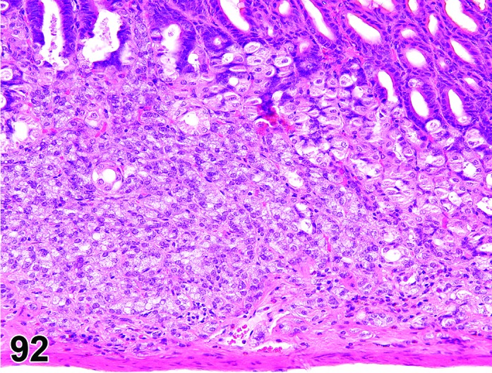
Rat glandular stomach. Tumor, neuroendocrine cell, benign. Higher magnification of Figure 91. Note increased cytoplasmic eosinophilia and mild nuclear and cellular pleomorphism.
Synonyms: Enterochromaffin-like cell tumor; APUDoma [APUD = Amine Precursor Uptake and Decarboxylase]; Intramucosal neuroendocrine cell tumor
Histogenesis: Neuroendocrine cells of the gastric mucosa; the enterochromaffin-like (ECL) cell is the predominant neuroendocrine cell of the oxyntic mucosa.
Diagnostic features
Discrete, well-demarcated aggregates of neuroendocrine cells, which may arise from hyperplastic areas in the oxyntic mucosa (i.e. corpus region).
Size of the aggregates exceeds 3 gastric glands in diameter.
Restricted to intramucosal involvement.
Compression or necrosis of adjoining mucosa glands may be observed.
Differential diagnoses
Hyperplasia, neuroendocrine cell: Scattered distribution of individual or small clusters of well-differentiated cells; discrete foci no more than 3 gastric glands in diameter.
Tumor, neuroendocrine cell, malignant: Infiltration through muscularis mucosae by neoplastic, often less differentiated cells arranged in cords or nodules OR serosal deposits OR distant metastases.
Ectopic hepatocytes: Absence of neoplastic behavior or cytologic alteration. Absence of characteristic markers for neuroendocrine cells (e.g. argyrophilia, immunohistochemical profile).
Comment: Neuroendocrine cell hyperplasia and neoplasia may be seen in response to hypergastrinemia following anti-secretory drug treatment (Spencer et al. 1989). Fundic mucosal hyperplasia is usually also evident. Small numbers of normal neuroendocrine cells can be observed in the submucosa and should not be categorized as neoplasia.
Tumor, neuroendocrine cell, malignant
(Figures 93 and 94)
Figure 93.
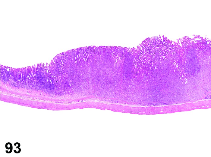
Rat glandular stomach. Tumor, neuroendocrine cell, malignant.
Figure 94.
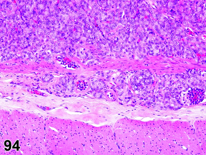
Rat glandular stomach. Tumor, neuroendocrine cell, malignant. Higher magnification of Figure 93. Note presence of neoplastic neuroendocrine cells in submucosal location indicative of infiltrative growth.
Synonyms: Neuroendocrine carcinoma; carcinoid; enterochromaffin-like cell tumor; APUDoma [APUD = amine precursor uptake, decarboxylase]; intramucosal neuroendocrine cell tumor (benign)
Histogenesis: Neoplastic transformation of neuroendocrine cells.
Diagnostic features
Aggregates of neuroendocrine cells, which may arise from hyperplastic areas in the oxyntic mucosa (i.e., corpus region).
Growth is intramucosal as nodules of pale staining or eosinophilic cells, sometimes with larger submucosal nodules.
Infiltration through muscularis mucosae by neoplastic, often less-differentiated, cells arranged in cords or nodules.
Metastasis to regional lymph nodes or liver may be present.
Differential diagnoses
Hyperplasia, neuroendocrine cell: Scattered distribution of individual or small clusters of well-differentiated cells. Discrete foci are less than 3 gastric glands in diameter.
Ectopic hepatocytes: Absence of neoplastic behavior or cytologic alteration. Absence of characteristic markers for neuroendocrine cells (e.g. argyrophilia, immunohistochemical profile).
Comment: Neuroendocrine cell hyperplasia and neoplasia may be seen in response to hypergastrinemia following anti-secretory drug treatment (Spencer et al. 1989). Fundic mucosal hyperplasia is usually also evident. Small numbers of normal neuroendocrine cells can be observed in the submucosa and should not be categorized as neoplasia.
Leiomyoma
(Figures 95 and 96)
Figure 95.
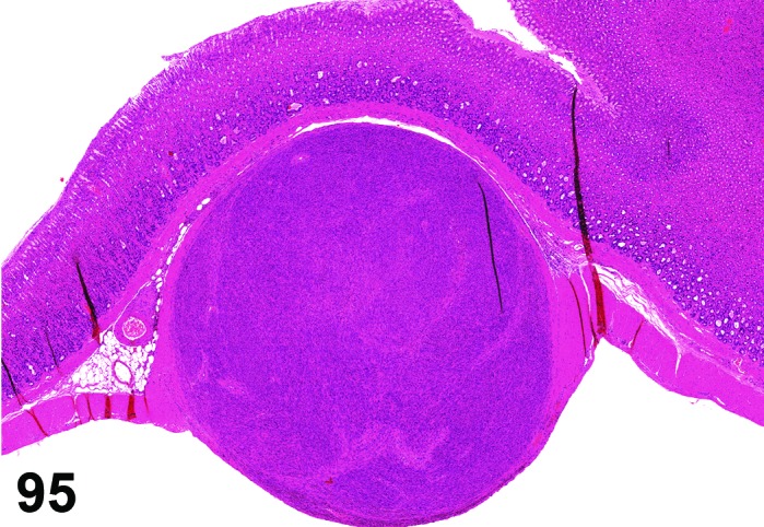
Rat glandular stomach. Leiomyoma.
Figure 96.
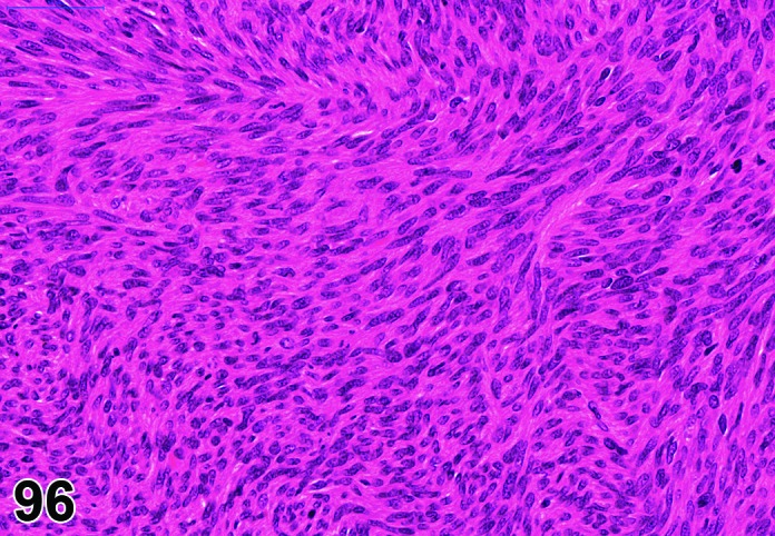
Rat glandular stomach. Leiomyoma. Higher magnification of Figure 95, demonstrating the monomorphic character of the spindle shaped tumor cells.
Histogenesis: Smooth muscle cells from tunica muscularis.
Diagnostic features
Intramural involvement.
Interlacing bundles and whorls of uniform spindle cells arranged in criss-cross patterns of bundles.
Nuclei typically blunt-ended or cigar shaped.
Eosinophilic cytoplasm contains longitudinal myofilaments and perinuclear clear space and show immunoreactivity for desmin.
Differential diagnoses
Leiomyosarcoma: Shows mitotic activity and cellular pleomorphism.
Gastrointestinal Stromal Tumor (GIST): Differentiation from other soft tissue tumors need only be made in cases of intergroup difference in the incidence of soft tissue tumors in a given study; unequivocal differentiation from other soft tissue tumors possible only by immunohistochemistry: typically CD117 positive (cytokine receptor encoded by c-kit). Spindle, epithelioid, or pleomorphic cell morphology. Indistinct cell borders and fibrillary cytoplasm; cells may be arranged in bundles with storiform architecture; spindle- or irregular-shaped nuclei.
Schwannoma: CD117 negative, S-100 positive; indistinct cell borders, elongate cells with eosinophilic cytoplasm; nuclei sometime arranged in palisades (i.e., Antoni A pattern).
Fibroma/sarcoma: CD 117 and S-100 negative; interlacing bundles of spindle cells; Masson’s trichrome-blue collagen.
Histiocytic sarcoma: Positive for histiocytic markers (ED-1 for rats, Factor 4/80 in mice), CD 117 and S-100 negative; infiltrative pattern, pleomorphic morphology.
Comment: Similar to those observed in other sites (see also INHAND-publication of soft tissue tumors, Greaves et al. 2013).
Leiomyosarcoma
(Figures 97 and 98)
Figure 97.
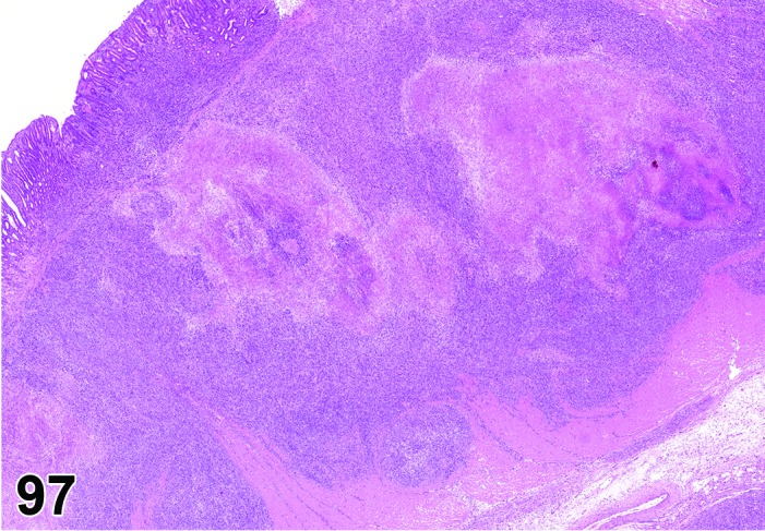
Rat nonglandular stomach. Leiomyosarcoma.
Figure 98.
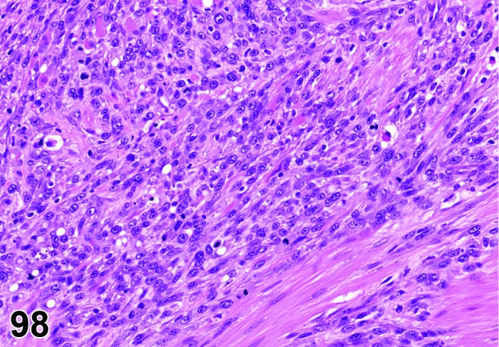
Rat nonglandular stomach. Leiomyosarcoma. Higher magnification of Figure 97. Cellular pleomorphism characterizes this tumor as malignant.
Histogenesis: Smooth muscle cells from tunica muscularis.
Diagnostic features
Intramural involvement.
Similar to leiomyoma, but more pleomorphic and with mitotic activity.
Necrosis, cystic change and mineralization are common features.
Distant metastases.
Differential diagnoses
Leiomyoma: Shows little or no cellular pleomorphism or mitotic activity.
Gastrointestinal Stromal Tumor (GIST): Differentiation from other soft tissue tumors need only be made in cases of intergroup difference in the incidence of soft tissue tumors in a given study; unequivocal differentiation from other soft tissue tumors is possible only by immunohistochemistry: typically CD117 positive (cytokine receptor encoded by c-kit). Spindle, epithelioid, or pleomorphic cell morphology. Indistinct cell borders and fibrillary cytoplasm; cells may be arranged in bundles with storiform architecture; spindle- or irregular-shaped nuclei.
Schwannoma: CD117 negative, S-100 positive; indistinct cell borders, elongate cells with eosinophilic cytoplasm; nuclei sometime arranged in palisades (i.e., Antoni A pattern).
Fibroma/sarcoma: CD 117 and S-100 negative; interlacing bundles of spindle cells; Masson’s trichrome-blue collagen.
Histiocytic sarcoma: Positive for histiocytic markers (ED-1 for rats, Factor 4/80 in mice), CD 117 and S-100 negative; infiltrative pattern, pleomorphic morphology.
Comment: Similar to those observed in other sites (see also INHAND-publication of soft tissue tumors, Greaves et al. 2013).
Gastrointestinal stromal tumor (GIST), benign
Synonyms: Interstitial Cajal cell tumor, benign; gastrointestinal pacemaker cell tumor, benign
Histogenesis: Specialized smooth muscle cells (i.e. interstitial cells of Cajal) in the tunica muscularis or myenteric plexus.
Diagnostic features
Well demarcated tumor, expansive growth.
Cells may be arranged in bundles with storiform architecture, myxoid areas may be present.
Spindle, epithelioid, or pleomorphic cell morphology. Indistinct cell borders and fibrillary cytoplasm.
Spindle- or irregular-shaped nuclei.
Typically CD117 positive (cytokine receptor encoded by c-kit).
Differential diagnoses
Gastrointestinal stromal tumor, malignant: Infiltrative growth; distant metastases may be present.
Leiomyoma: CD117 negative, desmin positive; interlacing bundles and whorls of uniform spindle cells arranged in criss-cross pattern; blunt-ended, fusiform nuclei, minimal nuclear pleomorphism; phosphotungstic acid-hematoxylin positive myofibrils.
Leiomyoscarcoma: CD117 negative, desmin positive; interlacing bundles and whorls of uniform spindle cells arranged in criss-cross pattern; blunt-ended, fusiform nuclei with hyperchromasia, pleomorphism, and high mitotic index; phosphotungstic acid-hematoxylin positive myofibrils.
Schwannoma: CD117 negative, S-100 positive; indistinct cell borders, elongate cells with eosinophilic cytoplasm; nuclei sometime arranged in palisades (i.e., Antoni A pattern).
Comment: GIST will generally not be easily differentiated from leiomyomas / leiomyosarcomas or other soft tissue tumors. However, immunohistochemical differentiation of the different tumor types, including GIST, needs to be performed in case of imbalances in the incidence of smooth muscle tumors in a given study.
Experimentally-induced GIST in the glandular stomach of rats and a spontaneous GIST in rat nonglandular stomach have been reported (Fujimoto et al. 2006; Mukaisho et al. 2006). Cecal GIST have been identified in GEM mice over-expressing a mutant KIT (Nakai et al. 2008). The question arises whether some conventionally diagnosed rodent stromal tumors (e.g., leiomyoma) were actually GIST.
Gastrointestinal stromal tumor (GIST), malignant
Synonyms: Interstitial Cajal cell tumor, malignant; gastrointestinal pacemaker cell tumor, malignant
Histogenesis: Specialized smooth muscle cells (i.e. interstitial cells of Cajal) in the tunica muscularis or myenteric plexus.
Diagnostic features
Locally infiltrative growth pattern.
Cells may be arranged in bundles with storiform architecture, myxoid areas may be present.
Spindle, epithelioid, or pleomorphic cell morphology. Indistinct cell borders and fibrillary cytoplasm.
Spindle- or irregular-shaped nuclei.
Typically CD117 positive (cytokine receptor encoded by c-kit).
Distant metastases may be present.
Differential diagnoses
Gastrointestinal stromal tumor, benign: Expansive growth pattern, no distant metastases.
Leiomyoma: CD117 negative, desmin positive; interlacing bundles and whorls of uniform spindle cells arranged in criss-cross pattern; blunt-ended, fusiform nuclei, minimal nuclear pleomorphism; phosphotungstic acid-hematoxylin positive myofibrils.
Leiomyoscarcoma: CD117 negative, desmin positive; interlacing bundles and whorls of uniform spindle cells arranged in criss-cross pattern; blunt-ended, fusiform nuclei with hyperchromasia, pleomorphism, and high mitotic index; phosphotungstic acid-hematoxylin positive myofibrils.
Schwannoma: CD117 negative, S-100 positive; indistinct cell borders, elongate cells with eosinophilic cytoplasm; nuclei sometime arranged in palisades (i.e. Antoni A pattern).
Comment: GIST will generally not be easily differentiated from leiomyomas / leiomyosarcomas or other soft tissue tumors. However, immunohistochemical differentiation of the different tumor types, including GIST, needs to be performed in case of imbalances in the incidence of smooth muscle tumors in a given study.
Experimentally-induced GIST in the glandular stomach of rats and a spontaneous GIST in rat nonglandular stomach have been reported (Fujimoto et al. 2006; Mukaisho et al. 2006). Cecal GIST have been identified in GEM mice over-expressing a mutant KIT (Nakai et al. 2008). The question arises whether some conventionally diagnosed rodent stromal tumors (e.g. leiomyoma) were actually GIST.
IV. Intestine, Small / Large
A. Morphology (Anatomy)
The small intestine in rodents, as in other mammals, is subdivided into duodenum, jejunum and ileum and is designed for the digestion and absorption of ingested products (Treuting and Dintzis, 2012). The large intestine is composed of cecum, colon and rectum. The cecum is a large blind end sac with considerable fermentation potential. The colon has an ascending section, a short transverse section and a descending section leading to the rectum which terminates at the anus. All parts, except the terminal rectum, are connected to the body wall by the mesentery which functions as a conduit for vascular, lymphatic and nervous supply. The terminal rectum lies outside the pelvic cavity and consequently lacks a surrounding serosal membrane.
Very rarely, congenital developmental abnormalities can occur, e.g. intestinal duplication (Elangbam et al. 1998). Such pathologic conditions that can be reliably diagnosed grossly are not covered by this monograph.
B. Morphology (Histology)
Histologically, the mucosa of the small intestine is composed of basal crypts (also known as crypts of Lieberkuhn) and villi which extend into the lumen. The base of the crypt is lined by stem cells; the remainder of the crypt is composed of proliferating epithelial cells which either migrate up the villi as they differentiate into absorptive cells with microvilli or goblet cells, with a turnover time of about 3 days, or remain in the crypt and differentiate into Paneth cells. The villous epithelium is composed of tall columnar cells with surface microvilli (the main absorptive cells), goblet cells containing mucus and scattered neuroendocrine cells covering a lamina propria composed of connective tissue with a rich vascular and lymphatic network. The lamina propria contains variable numbers of lymphocytes, macrophages, plasma cells, eosinophils and/or mast cells whilst occasional intraepithelial lymphocytes can also be observed. Paneth cells, which have an exocrine serous morphology and produce antimicrobial peptides, are present in higher numbers in the crypts of jejunum and ileum compared to the duodenum. Brunner’s glands, which produce a bicarbonate rich secretion, are present in the submucosa immediately following the pyloric-duodenal junction. Villi in the duodenum are taller than those in jejunum and ileum. Jejunum and ileum can be hard to differentiate in cross section except that the terminal ileum has two serosal attachments (mesentery and ligamentum ileocecale) compared to the single mesenteric attachment of the jejunum. With age, the duodenal villi tend to become shorter and thicker due to increased amounts of fibrous connective tissue in the core, whilst those in the ileum become longer probably related to reduced absorption in the proximal intestine resulting in greater amounts of nutrients passing into the distal intestine (Shackelford and Elwell 1999).
In the jejunum and ileum there are well defined aggregations of lymphoid tissue (Peyer’s patches) located opposite the mesenteric attachment; these may show formation of germinal centers. Similar aggregations are present in other parts of the gastrointestinal tract and are collectively referred to as gastrointestinal-associated lymphoid tissue (GALT).
Villi are absent from the large intestine and instead the mucosa is thrown into transverse or longitudinal folds. In mice and rats the folds of the proximal colon have a distinctive spiral orientation. The mucosa is lined by a single layer of three cell types: absorptive cells, goblet (mucous) cells and neuroendocrine cells. There is a zone of proliferation in each gland and cells either migrate outwards and differentiate into absorptive or goblet cells or remain in the basal part of the gland as secretory cells. The turnover time of the absorptive and outer goblet cells is 4.6 days, that of the deep crypt secretory cells 14 to 21 days (Karam 1999). Aggregates of lymphoid cells may be found in the mucosa and submucosa of all parts of the large intestine, but these are not limited to the antimesenteric aspect as in the small intestine.
The mucosa in both small and large intestines is supported by the connective tissue of the lamina propria and the continuous muscle layer of the muscularis mucosa. Beneath the muscularis mucosa is the relatively loose connective tissue of the submucosa which contains larger blood vessels, lymphatics and nerves and the tunica muscularis (also known as muscularis propria or muscularis externa). The external surface, except for the terminal rectum, is covered by the serosal membrane. The rectum also is enclosed by a notably thicker tunica muscularis. The enteric nervous system in the intestine is in the form of the submucosal and myenteric plexi, the latter being located between the longitudinal and circular layers of the tunica muscularis.
C. Physiology
The primary function of the small intestine is to complete the chemical digestion of food by pancreatic enzymes and bile salts and allow for efficient absorption of the products of digestion. The primary functions of the large intestine are to allow for further digestion by fermentation in the cecum, absorption of water, and temporary storage of waste material in the rectum before defecation. Disease processes or test articles that interfere with these processes will often produce diarrhea. Less commonly muscle function is compromised leading to impaction of rectum and colon and clinical constipation. The mucosa of the small intestine is a site of high enzyme activity and conjugation reactions and consequently orally administered test articles may be metabolized, activated or deactivated in the intestine. The GALT plays a primary role in the defense of the body against invasion by micro-organisms in the food and those normally resident in the lumen.
D. Necropsy & Trimming
The small intestine is the primary site for absorption of test articles given orally so it must be carefully evaluated in safety evaluation studies. Due to variations in morphology along the length of both small and large intestine it is important there is consistent sampling at necropsy and consistent orientation on the slide. The GoRENI trimming guide (website.reni.item.fraunhofer.de/reni/trimming) specifies the following sites for routine examination: 1) Duodenum: 1 cm distal to the pyloric sphincter; 2) Jejunum: central section; 3) Ileum: 1 cm proximal to cecum; 4) Cecum; 5) Colon: central section; and 6) Rectum: 2 cm proximal to the anus. For investigative studies involving induction of intestinal tumors, the entire intestine can be opened, lesion location and size recorded and individual tumors may be fixed for sectioning in additional to the 6 standard sections. Alternatively the whole length of the intestine can be fixed using the Swiss roll method. However, opening the intestine and washing away contents with normal saline brings the risk of damaging the delicate mucosa which degenerates quickly due to post mortem autolysis; tissue orientation may be less reliable during subsequent tissue trimming and blocking using this method. In addition, it is important to note the amount and quality of ingesta/digesta present in the segments of intestine sampled for histological examination as this may affect the height of the mucosa.
Attempts should be made at necropsy to sample Peyer’s patches (which are visible macroscopically) from the jejunum or ileum so that microscopic evaluation of GALT can be undertaken on the routine section of small intestines. Toxicological evaluation of the intestine should also include examination of the tunica muscularis and the submucosal and myenteric nerve plexi which occasionally show changes related to test article administration.
E. Nomenclature, Diagnostic Criteria and Differential Diagnosis
a. Congenital Lesions
Ectopic tissue
Figure 99.
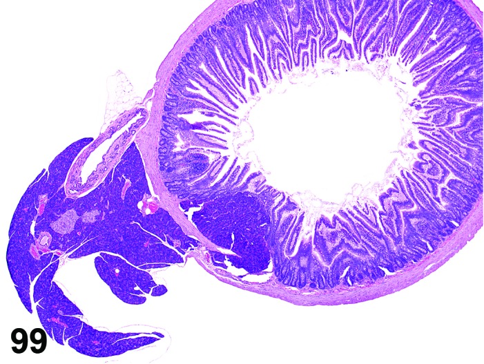
Rat duodenum. Ectopic tissue, pancreas.
Modifier: Pancreas
Pathogenesis: Presence of pancreatic cells in the mucosa or submucosa.
Diagnostic features
Normally formed pancreatic acini in mucosa or submucosa. Islets of Langerhans may also be present.
Present near the site of mesenteric attachment.
Differential diagnosis
None.
Comment: Presence of ductular (biliary or pancreatic) tissue in the duodenal wall is a normal finding in the anterior duodenum (Shackelford and Elwell 1999).
Cyst, squamous
Figure 100.
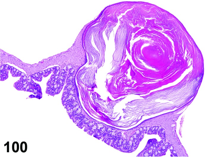
Mouse cecum. Cyst, squamous.
Pathogenesis: Presence of a cyst(s) lined by squamous cells in the mucosa, muscularis or serosa.
Diagnostic features
Cyst lined by keratinized stratified squamous epithelium.
Lumen frequently contains keratinized material.
Differential diagnosis
Diverticulum, simple, cystic OR diverticulum, atypical, cystic: Lined by glandular epithelium; no keratin formation.
Comment: While squamous cysts are most likely to be of congenital origin they may potentially form secondary to derangement of the epithelium e.g. in an inflammatory process. Squamous cysts been observed very occasionally in the muscularis of the colon and rectum of B6C3F1 mice (Shackelford and Elwell 1999). They are thought to be associated with focal weakness or discontinuity of the muscularis resulting in herniation through the submucosa and mucosa to the serosa. If post-inflammatory extension is the suspected pathogenesis, the pathologist may choose to interpret the focal change as a diverticulum, rather than a congential cyst.
b. Cellular Degeneration, Injury and Death
Atrophy, Brunner’s gland
Figure 101.
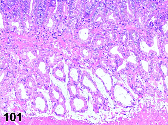
Rat duodenum. Atrophy, Brunner’s glands.
Pathogenesis: Reduction in size and number of Brunner’s glands.
Diagnostic features
Reduction in size and number of the acini that comprise the Brunner’s glands.
May be accompanied by an inflammatory reaction.
May be accompanied by fibrosis.
Differential diagnosis
None.
Atrophy
Figure 102.
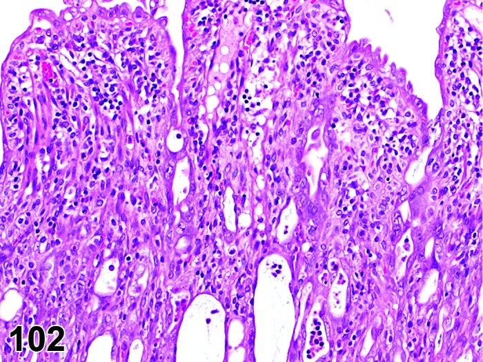
Rat jejunum. Atrophy, mucosal. Blunted villi and markedly flattened crypt epithelium.
Modifiers: Villous, mucosal
Pathogenesis: Atrophy of the mucosa related to reduced mitotic capabilities. In the small intestine, there is a reduction in villus height and crypt length; in the large intestine there is a decrease in size and number of mucous cells leading to a reduction in mucosal thickness.
Diagnostic features
Villi show reduced height but increased width.
Reduced depth of crypts in small intestine and mucous glands in large intestine.
Reduced mitotic rate and/or increased cell loss through apoptosis or necrosis.
Epithelium loses normal regular arrangement and may be flattened, cuboidal or vacuolated in appearance.
Differential diagnoses
Dilatation: Dilatation of the intestine by fluid or gas will reduce villous height and thickness of the large intestinal mucosa by mechanical means.
Autolysis: Preferential destruction and loss of cells at the tips of villi; no inflammatory reaction.
Normal topography: Villi overlying Brunner’s glands may be smaller/fewer than in neighboring areas of the duodenum.
Comment: Villous atrophy can be induced by withdrawal of food, administration of antimitotic cytotoxic agents, secondary to chronic inflammation or following rotavirus infection (Maekawa 1994; Shackelford and Elwell 1999; Percy and Barthold 2007; Kramer et al. 2010; Haschek et al. 2010). In addition, villous atrophy can be induced experimentally by hypophysectomy, thyroidectomy and in segments of surgically bypassed intestine (Greaves 2012). Villous atrophy may be accompanied by other degenerative changes e.g. lipidosis (Murgatroyd 1980). Villous and mucosal atrophy without damage to the underlying lamina propria or to the proliferative cell population is reversible. However, if there is a sustained effect on proliferation e.g. with cytotoxic agents, the atrophy will be followed by erosion/ulceration, hemorrhage and secondary infection. Mucosal atrophy induced by agents that interfere with the spindle apparatus may also show nuclear abnormalities including karyomegaly.
Hypertrophy
(Figures 103 and 104)
Figure 103.
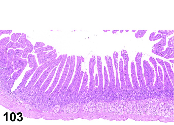
Rat duodenum. Normal mucosa (for comparison to Figure 104).
Figure 104.
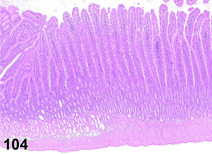
Rat duodenum. Hypertrophy, mucosal, diffuse. Same magnification as Figure 103.
Synonyms: Hyperplasia mucosa diffuse
Modifiers: Villous, mucosal
Pathogenesis: Increased in height of villi and depth of crypts.
Diagnostic features
Increased length of villi in the small intestine.
Increased mucosal thickness in the large intestine.
Increased mitotic activity may be observed.
Differential diagnosis
Artifacts: Villi are longer in the duodenum compared to jejunum and ileum, so inconsistency of sampling at necropsy could lead to an erroneous diagnosis.
Comment: Increased mucosal thickness or villous length reflects hyperplasia of all cellular elements. Villous hypertrophy has been induced by test articles (e.g. alloxan, polychlorinated aromatics, thyroid mimetic test articles), and by hormonal changes e.g. lactation and hyperthyroid states (Bertram et al. 1996; Greaves 2012). Villous length can also be influenced by nutritional factors e.g. amount of dietary fiber (Burkhardt et al. 1998).
Reduction, Paneth cell
Pathogenesis: Reduction in Paneth cell granules and/or loss of Paneth cells in small intestine.
Diagnostic features
Reduced numbers of granules in Paneth cells and/or loss of Paneth cells.
Differential diagnosis
None; however, it is necessary to verify the location of the intestinal specimen because Paneth cell density varies based on topography with considerably fewer cells present in the duodenum.
Comment: Recent literature identifies Paneth cells as particularly sensitive targets of endoplasmic reticulum stress responses and disruption of their function is a risk factor in inflammatory bowel disease (Ouellette 2010). Reduction in Paneth cells is observed in norovirus infection in certain transgenic mice strains (Cadwell et al. 2010).
Hypertrophy, Paneth cell
Figure 105.
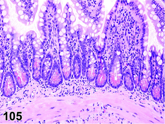
Mouse jejunum. Hypertrophy, Paneth cell.
Pathogenesis: Enlargement of Paneth cells in small intestine.
Diagnostic features
Hypertrophic Paneth cells.
May accompany villous atrophy.
Paneth cell hyperplasia may also be present.
Differential diagnosis
None.
Comment: Hypertrophy and hyperplasia of the Paneth cells, accompanying villous atrophy, were induced experimentally by surgical isolation of ileal loops (Keren et al. 1975).
Metaplasia, Paneth cell
Figure 106.
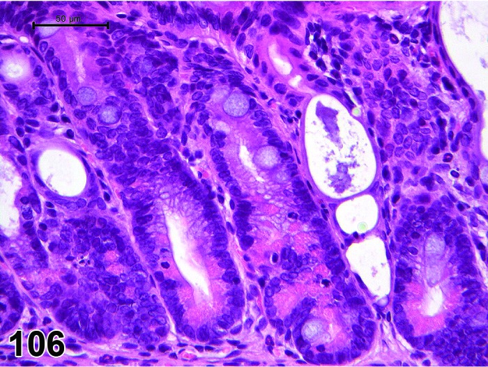
Rat colon. Metaplasia, Paneth cell. Note also epithelial atrophy in some crypts.
Pathogenesis: Presence of Paneth cells in the large intestinal mucosa.
Diagnostic features
Presence of Paneth cells in the large intestinal mucosa.
Accompanies chronic inflammatory lesions.
Differential diagnosis
None.
Comment: In colitis induced by the administration of dextran sodium sulphate and 1,2-dimethylhyadrazine, Paneth cell metaplasia occurred 4 weeks after induction of inflammatory lesions (Imai et al. 2007) Paneth cells showed increased expression of ß-catenin (Imai et al. 2007). Paneth cell differentiation is also seen in tumors that develop in this model.
Vacuolation, mucosa
(Figure 107 and 108)
Figure 107.
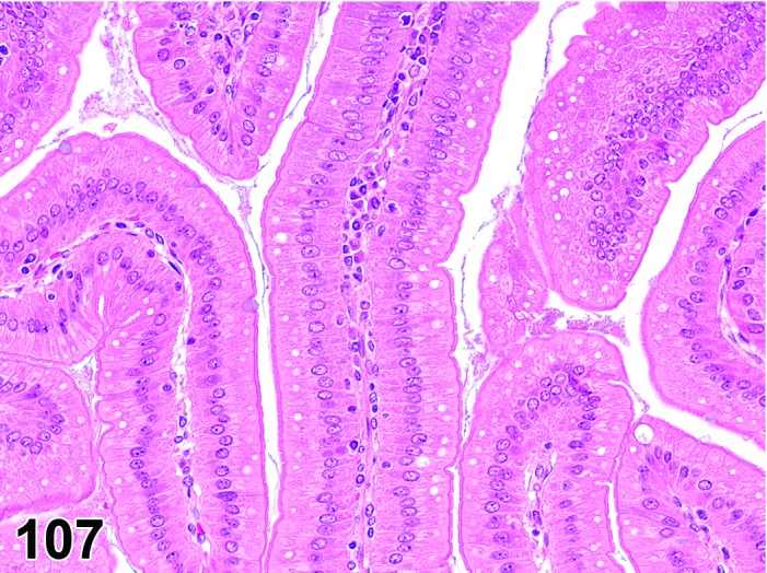
Rat, duodenum, vacuolation, mucosa (enterocytes) diffuse.
Figure 108.
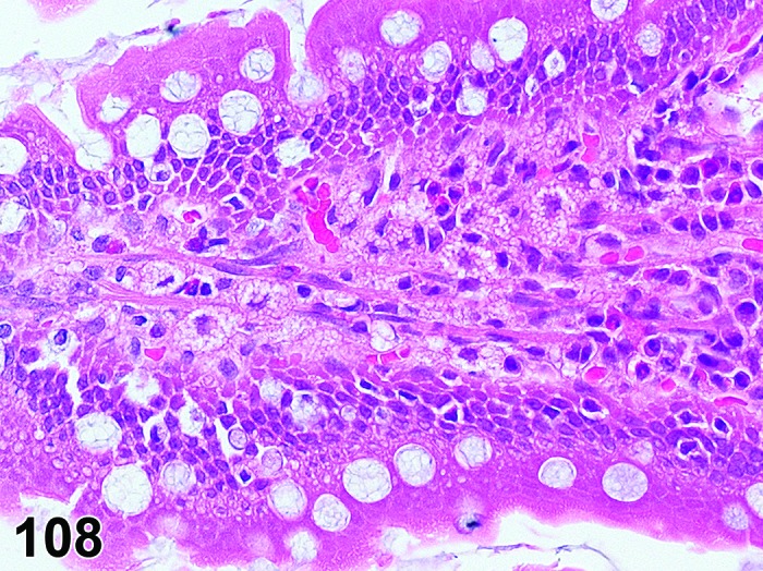
Rat, jejunum. Vacuolation, mucosa, foamy. Note affected cells in lamina propria.
Synonyms: Vacuolar degeneration, vesicular degeneration
Modifier: Foamy
Pathogenesis: Accumulation of substances of different character, including fluids, lipids, and phospholipids in epithelial cells of the villi and macrophages of the lamina propria in the absence of other signs of cell degeneration and necrosis.
Diagnostic features
Vacuoles in epithelial cells of the villi, particularly duodenum and jejunum.
Vacuoles are usually observed in the apical part of the epithelial cells in the upper third of the villi.
Vacuoles may also be observed in macrophages in the lamina propria and the mesenteric lymph nodes. Subsequent to rupture of macrophages, lipid droplets may be found free in the lamina propria. Occasional foreign body giant cells may be present in the lamina propria.
Foamy
Hypertrophy of cells with foamy cytoplasm
Differential diagnoses
Hypertrophy or hyperplasia, mucous cell: Presence of mucins can be confirmed by additional stains (PAS and alcian blue).
Necrosis: Cells swollen with pale eosinophilic cytoplasm; loss of nuclear basophilia, pyknosis and/or karyorrhexis that affects aggregates of cells.
Comment: The nature of the vacuolation can be further characterized by special techniques:
Vacuoles that contain neutral lipids stain positive with Oil Red O and Sudan Black.
Accumulation of intracytoplasmic lipid in villous epithelial cells has been reported with several xenobiotics, including glucose transport inhibitors, puromycin, ethionine and vanadium compounds which alter lipid synthesis or transport (Murgatroyd 1980; Greaves 2012; Imura et al. 2013). In addition, lipid droplets were described where an ester of erythromycin was administered and the compound appeared to be absorbed unhydrolysed and converted to chylomicron-like droplets which accumulated in macrophages (Visscher et al. 1980).
A fine cytoplasmic vacuolation, resulting in foamy appearance, may give the suspicion of phospholipidosis. In such cases, the modifier “foamy” should be used. If the change reflects phospholipidosis, increases in size and number of lysosomes can be identified by histochemistry (for alkaline phosphatase), immunostains (for lysosomal membrane proteins, e.g. LAMP-2) or by transmission electron microscopy. With the later technique, concentric multilaminated structures (myeloid bodies/lamellar bodies) are observed in the lysosomes. The term phospholipidosis should only be used when confirmation by one of these techniques has been achieved.
In the intestines, changes due to phospholipidosis are most commonly observed in macrophages, but epithelial cells, myocytes and neuronal ganglia can also be affected depending on the particular cellular distribution of the test article (Reasor et al. 2006). Affected cells will probably be present in other organs and tissues of the body, but the intestines are likely to be one of the more severely affected sites following oral administration of a phospholipidosis-inducing test article (Mazue et al. 1984). The intestines can also be affected following intravenous administration of such test articles depending on the tissue distribution of the test article in the body. The classical definitive diagnostic feature is the identification of lysosomal lamellar bodies by electron microscopy, but immunohistochemical staining for lysosomal membrane proteins can clearly distinguish lysosomes from neutral fat vacuole membranes stained immunohistochemically for adipophilin (Obert et al. 2007).
Degeneration/necrosis, Brunner’s glands
Figure 109.
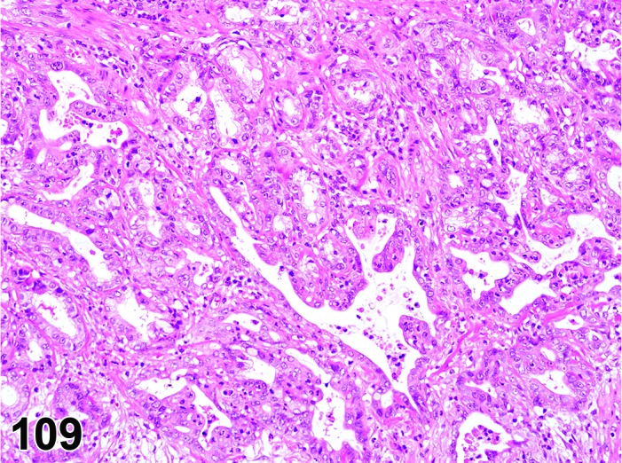
Degeneration/necrosis, Brunner’s glands.
Pathogenesis: Degeneration/necrosis of Brunner’s glands.
Diagnostic features
Degeneration and necrosis of epithelial cells.
Disorganisation of gland acini with disruption of basement membranes.
Glands may be dilated and lined with flattened, atrophic epithelial cells.
Accompanied by an inflammatory reaction.
May be accompanied by reactive hyperplasia of the duodenal epithelium.
Differential diagnosis
Atrophy, Brunner’s glands: Reduction in size and number of cells is present but necrosis and inflammation are absent.
Comment: Degeneration/necrosis of Brunner’s glands in rats has been described following administration of receptor tyrosine kinase (RTK) inhibitors directed against vascular endothelial growth factor (VEGF) (Inomata et al. 2014). Degeneration and necrosis of the glands is followed by disruption of the basement membrane leading to dilation of the glands and a neutrophilic inflammatory reaction. In the most severe lesions, inflammation extends into the surrounding mucosa and muscle layers. With more prolonged administration, dilated Brunner’s glands lined with flattened atrophic epithelium are observed and these may form cystic spaces which can extend into the muscle layers. A mixed neutrophilic and lymphocytic inflammatory reaction with fibrosis and reactive hyperplasia of neighbouring duodenal epithelium accompanies these Brunner’s gland changes after prolonged administration.
Lymphangiectasis
Figure 110.
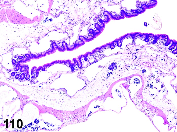
Mouse ileum. Lymphangiectasis and edema.
Synonyms: Lacteal dilatation
Pathogenesis: Lacteals are dilated with lipid.
Diagnostic features
Dilated lacteals are present in the villi.
Subcapsular sinuses of the mesenteric lymph node may also be dilated.
May be accompanied by minimal vacuolation of epithelial cells and/or macrophages in the lamina propria.
Differential diagnosis
None.
Comment: Lymphangiectasis may be primary, due to a metabolic alteration affecting the formation of chylomicrons or transit of lipid in the lacteals, or secondary to blockage or inflammatory conditions e.g. neoplasia, fibrosis, granulomatous infections. Lymphangiectasis in the small intestine due to the accumulation of large lipid droplets was described in rats administered indole-3-carbinol, possibly due to an impairment of lipid transport in the lacteals (Boyle et al. 2012). Although there were vacuolated macrophages and giant cells in the lamina propria, the epithelial cells were unaffected (Boyle et al. 2012).
Syncytia, epithelium
Figure 111.
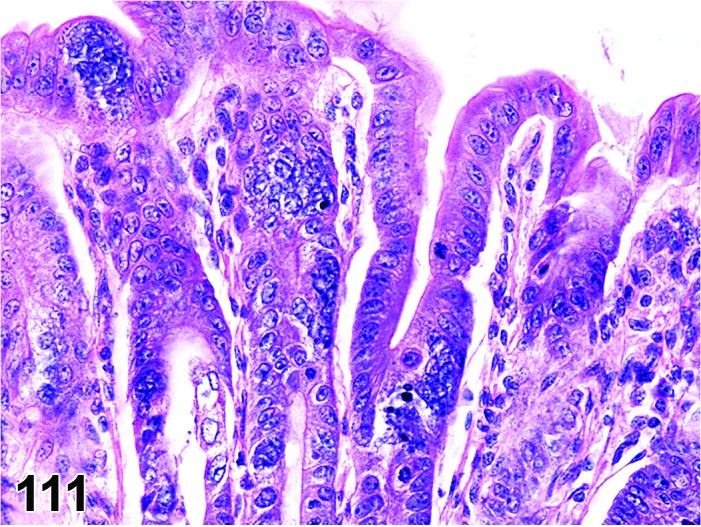
Mouse colon (Bouin’s fixed). Syncycia, epithelium.
Pathogenesis: Distrubance of cell division (cytokinesis), e.g. after Infection by enterotropic strains of mouse hepatitis virus (MHV), a coronavirus.
Diagnostic features
Formation of enterocyte syncytia (balloon cells occasionally containing eosinophilic intracytoplasmic inclusions).
Villous atrophy and mucosal necrosis also present, particularly in young mice.
Differential diagnosis
None.
Comment: Epithelial syncytia are common in MHV infection and MHV infection should be considered as an etiological agent when epithelial syncytia are observed.
With enterotropic MHV strains lesions are limited to the intestines and are generally located in ileum, cecum and proximal colon (Percy and Barthold 2007; Shackelford and Elwell 1999). Lesions are most severe in young mice and are limited to syncytia in adult mice. Compensatory hyperplasia of the intestinal epithelium develops in mice that survive the infection. With respiratory strains, enteric lesions are rare with infection largely restricted to GALT.
Apoptosis
Figure 112.
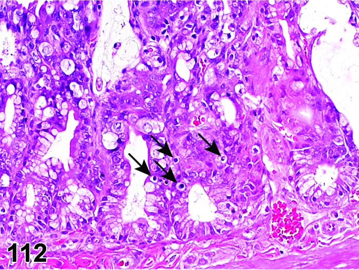
Rat jejunum. Apoptosis (arrows) in a severely atrophic mucosa.
Synonyms: Apoptotic cell death
Pathogenesis: Gene regulated, energy dependent process leading to formation of apoptotic bodies which are phagocytosed by adjacent cells.
Diagnostic features
Single cells or small clusters of cells.
Cell shrinkage.
Hypereosinophilic cytoplasm.
Nuclear shrinkage, pyknosis, karyorrhexis. Intact cell membrane.
Apoptotic bodies.
Cytoplasm retained in apoptotic bodies.
Phagocytosis of apoptotic bodies tissue macrophages or other adjacent cells.
No inflammation.
Differential diagnoses
Necrosis, mucosa: The morphologic features of cell death clearly fit with necrotic diagnostic criteria (cells and nuclei swollen, pale cytoplasm etc.).
Apoptosis/necrosis, mucosa: Both types of cell death are present and there is no requirement to record them separately or a combined term is preferred for statistical reasons. The term is also used when the type of cell death cannot be determined unequivocally.
Erosion/ulcer: Focal or focally extensive penetration of the mucosa, either partial (erosion) or full to the muscularis mucosa (ulcer).
Comment: The approach for the nomenclature and diagnostic criteria of cell death applied here is based on a draft recommendation of the INHAND Cell Death Nomenclature Working Group.
Apoptosis is not synonymous with necrosis. The main morphological differences between these two types of cell death are cell shrinkage with nuclear fragmentation and tingible body macrophages in apoptosis versus cell swelling, rupture and inflammation in necrosis; however, other morphologies (e.g. nucelar pyknosis and karyorrhexis) overlap. In routine H&E sections where the morphology clearly represents apoptosis or single cell necrosis, or whereby special procedures (e.g. TEM or IHC for caspases) prove one or the other, individual diagnoses may be used. However, because of the overlapping morphologies, necrosis and apoptosis are not always readily distinguishable via routine examination, and the two processes may occur sequentially and/or simultaneously depending on intensity and duration of the noxious agent (Zeiss 2003). This often makes it difficult and impractical to differentiate between both during routine light microscopic examination. Therefore, the complex term apoptosis/necrosis may be used in routine toxicity studies.
A differentiation between apoptosis and single cell necrosis may be required in the context of a given study, in particular if it aims for mechanistic investigations. Transmission electron microscopy is considered to represent the gold standard to confirm apoptosis. Other confirmatory techniques include DNA-laddering (easy to perform but insensitive), TUNEL (false positives from necrotic cells) or immunohistochemistry for caspases, in particular caspase 3. These techniques are reviewed in detail by Elmore (2007). Some of these confirmatory techniques detect early phases of apoptosis, in contrast to the evaluation of H&E stained sections, which detects late phases only; overall interpretation of findings should take into account these potential differences. Thus, low grades of apoptosis may remain unrecognized by evaluation of H&E stained slides only.
Certain studies may require the consideration of forms of programmed cell death other than apoptosis which requires specific confirmatory techniques (Galluzzi et al. 2012).
Necrosis, mucosa
Figure 113.
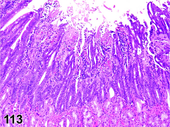
Rat duodenum. Necrosis, mucosa, focal.
Synonyms: Oncotic cell death; oncotic necrosis; necrosis
Modifiers: Single cell
Pathogenesis: Unregulated, energy independent, passive cell death with leakage of cytoplasm into surrounding tissue and subsequent inflammatory reaction; may be induced by direct contact with a test article after oral uptake/administration.
Diagnostic features
Cells swollen with pale eosinophilic cytoplasm.
Loss of nuclear basophilia, pyknosis and/or karyorrhexis that affects aggregates of cells.
In more severe cases, there may be detachment of the epithelium from the submucosa.
Typically, the presence of degenerative cells is a component of necrosis.
Minimal or slight inflammatory cell infiltrates may be present as a feature of necrosis.
Single cell
Only single cells affected.
Differential diagnoses
Apoptosis: The morphologic features of cell death clearly fit with apoptosis (cell and nucleus shrunken, hypereosinophilic cytoplasm, nuclear pyknosis etc.) and/or special techniques demonstrate apoptosis.
Apoptosis/necrosis, mucosa: Both types of cell death are present and there is no requirement to record them separately or a combined term is preferred for statistical reasons. The term is also used when the type of cell death cannot be determined unequivocally.
Atrophy, mucosa: Reduced thickness of all layers of the squamous epithelium or, in severe cases, absence of basal germinative layers; usually no cellular degeneration or necrosis.
Erosion/ulcer: Focal or focally extensive penetration of the mucosa, either partial (erosion) or full to the muscularis mucosa (ulcer).
Comments: Epithelial necrosis usually develops into erosion/ulcer and in the past has frequently been recorded as such. A separate recording of necrosis as the initial event in this process is recommended when the structure of the mucosa is still intact.
Apoptosis/necrosis, mucosa
Synonym: Cell death
Pathogenesis: Unregulated, energy independent, passive cell death with leakage of cytoplasm into surrounding tissue and subsequent inflammatory reaction (single cell necrosis) AND/OR gene regulated, energy dependent process leading to formation of apoptotic bodies which are phagocytosed by adjacent cells (apoptosis).
Diagnostic features
Both types of cell death are present and there is no requirement to record them separately or a combined term is preferred for statistical reasons.
The type of cell death cannot be determined unequivocally.
Differential diagnoses
Apoptosis: The morphologic features of cell death clearly fit with apoptosis (cell and nucleus shrunken, hypereosinophilic cytoplasm, nuclear pyknosis etc.) and/or special techniques demonstrate apoptosis and there is a requirement for recording apoptosis and necrosis separately.
Necrosis, mucosa: The morphologic features of cell death clearly fit with necrotic diagnostic criteria (cells and nuclei swollen, pale cytoplasm etc.) and there is a requirement for recording apoptosis and necrosis separately.
Erosion/ulcer: Focal or focally extensive penetration of the mucosa, either partial (erosion) or full to the muscularis mucosa (ulcer).
Comments: Necrosis and apoptosis are not always readily distinguishable via routine examination, and the two processes may occur sequentially and/or simultaneously depending on intensity and duration of the noxious agent (Zeiss 2003). This often makes it difficult and impractical to differentiate between both during routine light microscopic examination. In these cases, the combined term apoptosis/necrosis may be used. It is recommended to explain in detail the use of this combined term in the narrative part of the pathology report.
Erosion/ulcer
(Figure 114 and 124)
Figure 114.
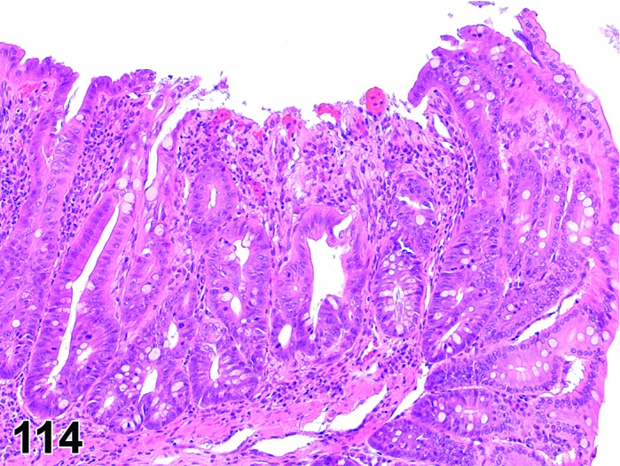
Mouse colon. Erosion with regenerative hyperplasia.
Figure 124.
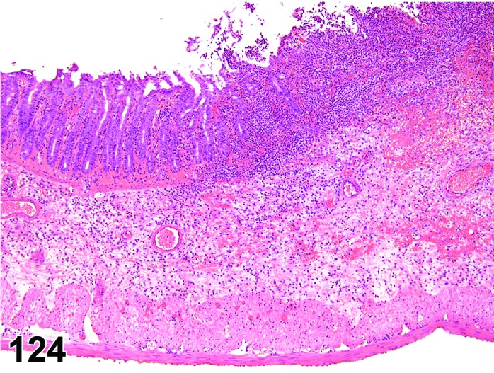
Rat cecum. Inflammation, mixed. Note mucosal erosion and submucosal edema.
Pathogenesis: Localized loss of mucosa with either partial mucosal penetration (erosion) or with full penetration of the mucosa to the muscularis mucosa (ulcer).
Diagnostic features
Epithelial cells are necrotic or absent but muscularis mucosa is intact (erosion).
Epithelial cell are necrotic or absent and muscularis mucosa has been destroyed (ulcer).
Submucosal edema may be present, particularly with ulceration.
Hemorrhage and hemosiderin may be present, particularly with ulcers.
Epithelial cells adjacent to an erosion or ulcer may be necrotic, cuboidal to flat or basophilic.
Inflammatory cell infiltration is present, except in recently formed lesions.
Chronic lesions will show chronic inflammatory cell infiltration, granulation tissue, fibrosis and regenerative epithelial hyperplasia which results in irregular, cystic glands in the mucosa and submucosa.
Differential diagnoses
Artifact: Mechanical loss of mucosa during necropsy; plane of section related to presentation of tissue in block.
Autolysis: Preferential destruction and loss of cells at the luminal surface; no inflammatory reaction.
Comment: The most likely cause of erosions and ulcers in immunocompetent barrier maintained rodents is the administration of xenobiotics (Greaves 2012). Acute erosion and ulceration of the distal small intestine has been described following administration of non-steroidal anti-inflammatory agents, the newer inhibitors of cyclooxygenase 2 (COX-2) and cytotoxic anticancer drugs, whereas ionizing radiation produces acute lesions followed by fibrosis and arteriolar sclerosis (Langberg and Hauer-Jensen 1996; Haschek et al. 2010). A variety of agents e.g. cysteamine, proprionitrile and 1-methy-4-pheny-1,2,3,6-tetrahydropyridine (MPTP) are capable of producing chronic ulcers in the duodenum possibly by altering gastric acid secretion, bicarbonate production and its delivery to the duodenum, changes in duodenal motility and/or dopamine inhibition (Szabo and Cho 1988). Some viral and bacterial infections may induce erosion and ulceration, particularly in immunodeficient mice or rats (Haines et al. 1998).
Erosion/ulcer usually is the result of epithelial necrosis. Because of its unique topography adjacent to the lumen and often involving multiple organ layers, which uniquely affects the progression and resolution of the lesion (e.g. margin hyperplasia, granulomatous tissue, inflammation), it is useful to record erosion/ulcer separately from epithelial necrosis.
Amyloid
Figure 115.
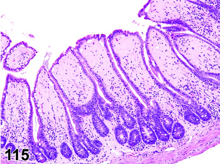
Mouse ileum. Amyloid in lamina propria of villi.
Pathogenesis: Extracellular deposits of polypeptide fragments of a chemically diverse group of glycoproteins, in various tissues including intestinal mucosa.
Diagnostic features
Deposition of pale amorphous, homogenous eosinophilic material.
Extracellular location in connective tissue and/or blood vessel walls.
Green birefringence with Congo red stain when viewed under polarized light.
Differential diagnoses
Hyaline connective tissue: Congo red negative.
Fibrinoid necrosis of blood vessels: Other evidence of tissue damage in section.
Comment: Amyloidosis is extremely rare in rats but common in some strains of mice in which the lamina propria of the intestinal villi is a common site for deposition of amyloid. In susceptible strains of mice a number of factors including sex, diet, housing conditions, stress, endocrine status and microbiological status can influence the occurrence of amyloidosis (Korenaga et al. 2004; Lipman et al. 1993). For as yet unidentified reasons the previously susceptible CD-1 strain in Europe now rarely develops amyloidosis.
The characteristic morphologic appearance and location of amyloid in H&E sections that is usually adequate to make this diagnosis. Confirmation of the deposits as amyloid can be accomplished with light microscopy using special stains such as Congo red. Amyloid appears apple green under polarized light with this stain.
The term “hyaline” should be used in case of uncertainties about the nature of extracellular deposits hyaline material in H&E sections and the unavailability of a Congo red stain. Hyaline is also the appropriate term for homogenously eosinophilic extracellular deposits that react negative with Congo red.
Mineralization
Figure 116.
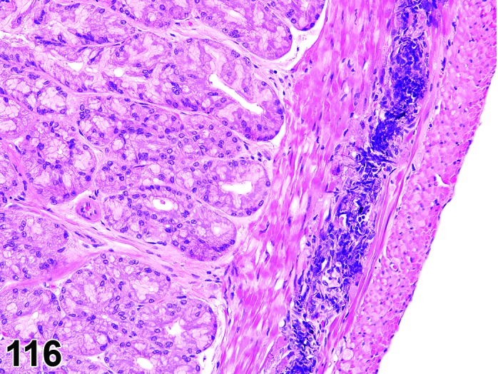
Rat duodenum. Mineralization of muscularis.
Synonym: Calcification
Pathogenesis: Deposition of mineral in muscle, fibrous or elastic tissues secondary to necrosis (dystrophic mineralization) or hypercalcemia (metastatic mineralization).
Diagnostic features
Mineral may be found in muscle layers, blood vessels, basement membranes and/or in the interstitium of mucosa and submucosa.
Amorphous basophilic material.
Mucosal mineralization can be limited to small focal deposits.
Differential diagnoses
Artifact: Hematoxylin stain precipitate.
Bacteria: Colonies of bacteria may be confused with mineral due to their basophilic staining. Ante mortem bacterial invasion will be accompanied by necrosis and usually inflammation; postmortem bacterial invasion will be accompanied by autolysis.
Osseous metaplasia: Osteoid present.
Comment: Presence of mineral can be confirmed with von Kossa or Alizarin red stains but these are rarely needed to make a diagnosis. Diffuse superficial mineralization of the colonic and rectal mucosa has been observed following administration of xenobiotics that decrease peristalsis and cause fecal retention (Gopinathet al. 1987). Multifocal or diffuse mineralization occurs secondary to chronic renal disease in rodents and in situations where serum calcium and/or phosphate balance are perturbed, but the intestines are not as susceptible as the glandular mucosa of the stomach.
Metaplasia, osseous
Figure 117.
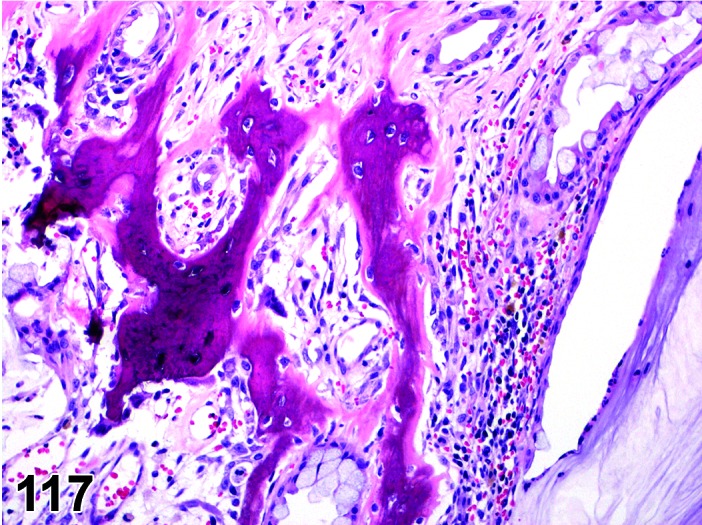
Rat duodenum. Metaplasia, osseous.
Pathogenesis: Presence of osteoid.
Diagnostic features
Presence of mineralized or non-mineralized bone matrix.
Differential diagnoses
Mineralization: Osteoid absent.
Sarcomas: Osseous metaplasia may be present in stroma of sarcomas.
Comment: Osseous metaplasia has been described in the submucosa and lamina propria of old rats in association with chronic inflammation, ulceration or regenerative hyperplasia (Elwell and McConnell 1990;Maekawa 1994; Bertram et al. 1996).
Dilatation
Figure 118.
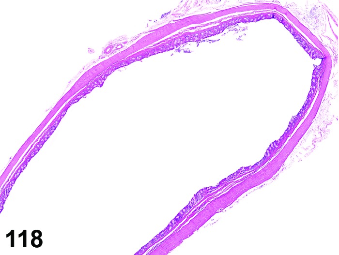
Rat rectum. Dilation.
Synonyms: dilation, megaileus; cecomegaly; megacolon
Pathogenesis: Dilatation of the lumen due to a defect in neuromuscular function.
Diagnostic features
Dilatation of the lumen is observable macroscopically.
Mucosa, submucosa and muscularis are thin due to stretching.
Degeneration of muscle fibers and ganglia cells in the nerve plexus may be present.
Differential diagnoses
Meteorism: Dilatation of the lumen due to the accumulation of air occurs as the result of mouth breathing and consequent aerophagia follows obstruction of nasal turbinates and/or nasopharynx in rodents as they are obligate nose breathers; the stomach will be more severely affected than the intestines.
Obstruction: Dilatation of the lumen due to simple mechanical obstruction e.g. proximal to a tumor.
Comment: Megaileus with inflammation (megaileitis) is often present in rats with Tyzzer’s disease (infection with Clostridium piliforme); the lesions are characterized by a necrotizing transmural ileitis with necrosis of the mucosa and muscularis, edema and infiltration primarily of mononuclear cells (Percy and Barthold 2007). Cecomegaly is observed following administration of some antibiotics, starches, polyols, lactose, fibers and vehicles which have an osmotic effect (Newberne et al. 1988). Megacolon as a result of constipation was induced in rats by a calcium channel antagonist; secondary mucosal necrosis, hemorrhage and inflammation were present in some rats (Nyska et al. 1994). Tribromoethanol (Avertin) administered intraperitoneally as an anesthetic has been associated with ileus in rodents (King and Russell 2006).
Intussusception
Figure 119.
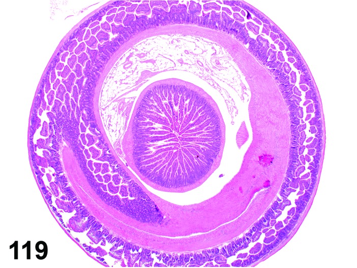
Mouse jejunum. Intussusception.
Pathogenesis: Invagination of a one portion of intestine into its immediate distal section (‘telescoping’).
Diagnostic features
Observable macroscopically.
Invagination of a portion of intestine into its immediate distal section.
Intestinal lumen proximal to the site of intussusception may be dilated whilst distally the lumen may be contracted and devoid of contents.
Histologically ischemic necrosis, inflammation, congestion and hemorrhage will often be present in the involved segments; the degree of such changes will depend on the length of time that the intussusception has been formed.
Adhesions may develop between the invaginating intussusceptum and the receiving intussuscipiens.
Associated with intestinal tumors, especially polyps.
Differential diagnosis
Artifacts, post mortem invagination of the intestine: No reactive changes present.
Comment: Intussusceptions are very rare in barrier maintained rodents but may occur in animals with intestinal tumors and rarely with high nematode infestations (Percy and Barthold 2007). Very occasionally an intussusception may be induced by a test article (Gopinath et al. 1987).
Although intussusception is primarily a gross pathological term, it is recommended to use it also microscopically, as an unequivocal macroscopic diagnosis is not always possible.
Prolapse
(Figures 120, 121 and 122)
Figure 120.
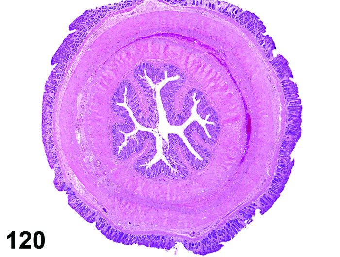
Rat rectum. Prolapse, cross section.
Figure 121.
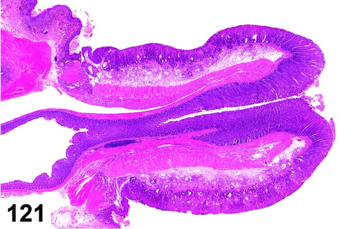
Mouse rectum (Bouin’s fixed). Polapse, longitudinal section.
Figure 122.
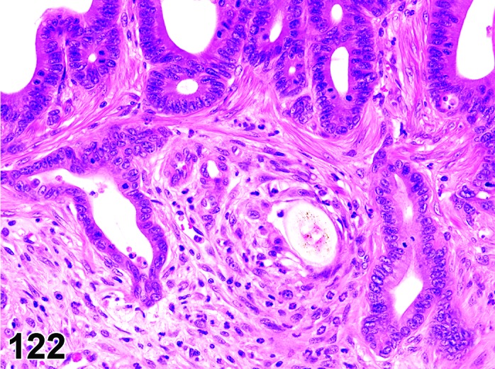
Mouse rectum. Prolapse. Higher magnification of Figure 121. Note diverticula in area of mixed inflammation with hair fragment.
Pathogenesis: Eversion of rectum through the anus.
Diagnostic features
Observable macroscopically.
Eversion of rectum through the anus.
Microscopic lesions of mucosal necrosis, hemorrhage, edema and inflammation.
Differential diagnosis
None.
Comment: Rectal prolapses are rare in barrier maintained rodents but may occur secondarily to inflammatory conditions in the colon and rectum such as occur with Citrobacter and Helicobacter infection in mice, weakening of anal sphincter and with a high nematode load (Ward et al. 1996; Percy and Barthold 2007; McInnes 2012; Miller et al. 2014).
The diagnosis of prolape is usually possible by gross pathological examination. Nevertheless it is recommended to confirm the gross observation histopathologically.
c. Inflammatory Lesions
Infiltrate
Figure 123.
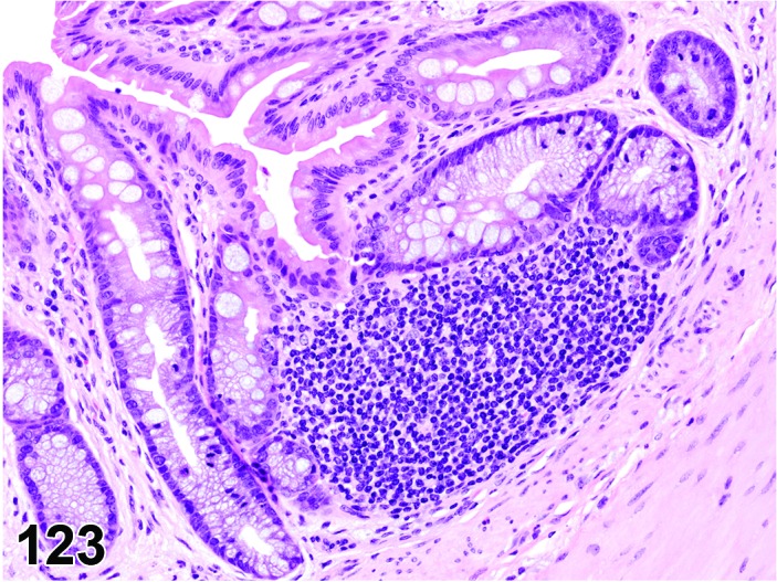
Mouse rectum. Infiltrate, mononuclear.
Synonyms: Infiltrate inflammatory; infiltrate inflammatory cell; infiltration (plus modifier); infiltration, inflammatory; infiltration, inflammatory cell
Modifiers: Type of inflammatory cell that represents the predominant cell type in the infiltrate
Pathogenesis: Infiltration of the lamina propria and/or submucosa with neutrophils (Infiltrate, neutrophil), eosinphils (Infiltrate, eosinophil) or mononuclear cells (Infiltrate, mononuclear cell) or a combination (Infiltrate, mixed) without other histological features of inflammation e.g. hemorrhage, edema, fibroplasia.
Diagnostic features
Focal, multifocal or diffuse infiltration of cells in the lamina propria and/or submucosa.
Presence of mononuclear or polymorphonuclear inflammatory cells but no other histological criteria of inflammation.
Differential diagnoses
Inflammation: Other morphologic features of inflammation such as edema, hemorrhage, necrosis and/or fibroplasia will be present.
Granulocytic leukemia: Infiltration of neutrophil precursors and/or abnormal neutrophils will probably be present along with mature neutrophils; infiltration of similar neoplastic cells is likely present in other organs.
Lymphoma: Infiltration of monomorphic population of lymphocytes (often with atypical, increased and/or abnormal mitoses); infiltration of similar neoplastic cells is likely present in other organs.
Comment: The number of mononuclear cells is the lamina propria of the large intestine (particularly cecum) is variable in healthy animals and care must be taken not to ‘over diagnose’ infiltration of inflammatory cells.
Inflammation
(Figures 124 and 125)
Figure 125.
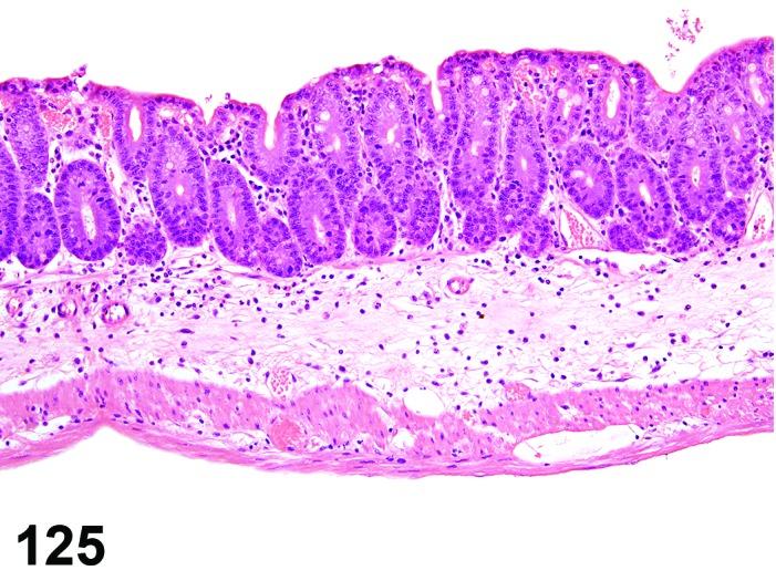
Mouse cecum. Inflammation, neutrophilic. Note submucosal edema.
Modifiers: Type of inflammatory cell that represents the predominant cell type in the inflammation
Pathogenesis: Infiltration of the lamina propria and/or submucosa with neutrophils (Inflammation, neutrophil) or mononuclear cell (Inflammation, mononuclear cell) or a combination (Inflammation, mixed) with additional histological features of inflammation e.g. hemorrhage, edema, fibroplasia.
Diagnostic features
Focal, multifocal or diffuse infiltration of cells in the lamina propria and/or submucosa.
Presence of other histological criteria of inflammation e.g. hemorrhage, edema, fibroplasia.
Differential diagnoses
Infiltrate, inflammatory cell: Other morphologic features of inflammation such as edema, hemorrhage, necrosis and/or fibroplasia will be absent.
Granulocytic leukemia: Infiltration of neutrophil precursors and/or abnormal neutrophils will probably be present along with mature neutrophils; infiltration of similar neoplastic cells is likely present in other organs.
Lymphoma: Infiltration of monomorphic population of lymphocytes (often with atypical, increased and/or abnormal mitoses); infiltration of similar neoplastic cells is likely present in other organs.
Comment: The most likely cause for inflammatory lesions in barrier maintained rodents is the administration of test articles, the best known examples being non-steroidal anti-inflammatory agents and anti-mitotic anticancer agents (Greaves 2012; Haschek et al. 2010). Hypersensitivity reactions may play a role in test article induced inflammatory reactions, for example trinitobenezensulfonic acid acts as hapten and induces a severe granulomatous reaction in the distal colon (Haschek et al. 2010). Because of the presence of numerous commensal bacteria in the intestine, there is the potential for secondary invasion of sites of mucosal injury and an exacerbation of any tissue injury and induction of an inflammatory response. Inflammatory lesions can be induced by pathogenic bacteria e.g. salmonella, Citrobacter freundii, Helicobacter spp or viruses (norovirus, mouse hepatitis virus) if there has been a barrier breakdown or in immunodeficient animals (Maggio-Price et al. 2005; Percy and Barthold 2007). Eaton et al. recently described a grading procedure for inflammation in the large intestine (Eaton et al. 2011).
d. Infectious Diseases
Features of specific bacterial and viral infections have been discussed in the previous sections. Nematode and protozoa are dealt with separately below.
Parasite
(Figures 126 and 127)
Figure 126.
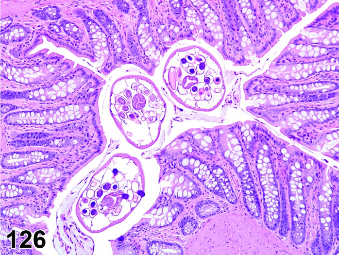
Rat rectum. Parasites.
Figure 127.
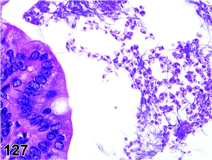
Mouse colon. Protozoa, probably Trichomonas muris.
Modifiers: Protozoa, nematode
Pathogenesis: Presence of protozoan or nematode parasitess in the intestinal lumen.
Diagnostic features
Protozoa
Presence of cross sections of protozoa in large intestinal lumen; morphology will depend on whether ameboid, ciliate or flagellates are present.
Inflammatory cell infiltration and/or edema may be present in lamina propria.
Nematodes
Presence of cross sections of parasites in large intestinal lumen.
Rarely submucosal granulomata may be present.
Differential diagnosis
None.
Comment: It is now uncommon for intestinal parasites to be present in barrier-reared and maintained rodents. Protozoa that have been reported in laboratory rodents include Spironucleus muris, Giadia muris and Cryptosporidia muris, but infections are often subclinical especially in adults of immunocompetent strains (Percy and Barthold 2007). The reader is referred to standard texts for a full list of intestinal protozoan that may be present in non-barrier maintained or wild rodents (Percy and Barthold 2007; Baker 2007).
The most likely nematodes to be seen in the event of a barrier break down in a rodent unit are the pinworms Syphacia muris, Syphacia obvelata and/or Aspicularis tetrapertera which inhabit the large intestine (Percy and Barthold 2007). The reader is referred to standard texts for a full list of intestinal parasites that may be present in non-barrier maintained or wild rodents (Percy and Barthold 2007; Baker 2007).
Neuronal degeneration, myenteric plexus
Figure 128.
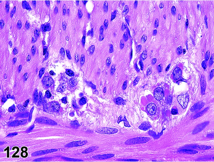
Mouse colon. Neuronal degeneration, myenteric plexus with formation of hyaline inclusions in ganglion cells (flavivirus infection).
Histogenesis: Degenerative changes in neurons of the myenteric/submucosal plexi.
Diagnostic features
Degenerative changes (hyaline, vacuolar) in neurons.
Intracytoplasmic inclusions (often hyaline) in neurons.
Differential diagnosis
None.
Comment: The degenerative changes and inclusions have only been seen with flaviviral infections (Ward, personal observation). Viral antigen is abundant in these ganglion cells.
e. Vascular Lesions
Edema and hemorrhage of the intestine are similar to those described for the upper digestive tract. Other vascular lesions in the intestine and mesentery are dealt with in the INHAND monograph on the cardiovascular system.
f. Non-neoplastic Proliferative Lesions
Diverticulum
(Figures 122, 129, 130 and 131)
Figure 129.
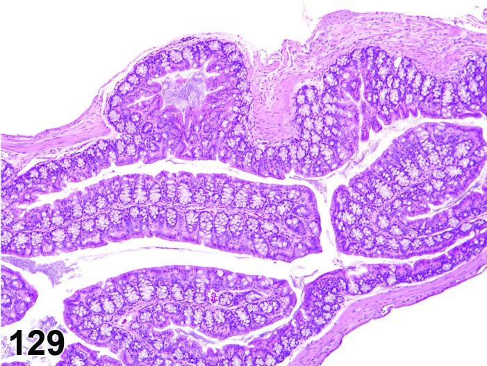
Mouse colon. Diverticulum, characterized by extension of intact mucosa into subserosal tissue.
Figure 130.
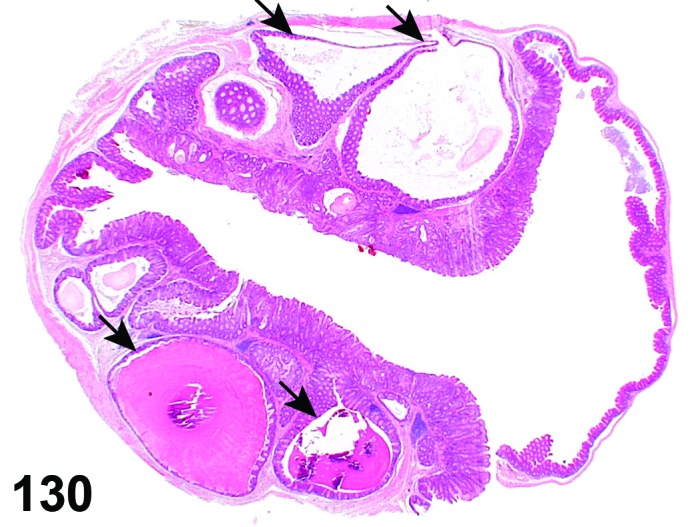
Mouse colon. Multiple diverticula, some of them cystic (arrows).
Figure 131.
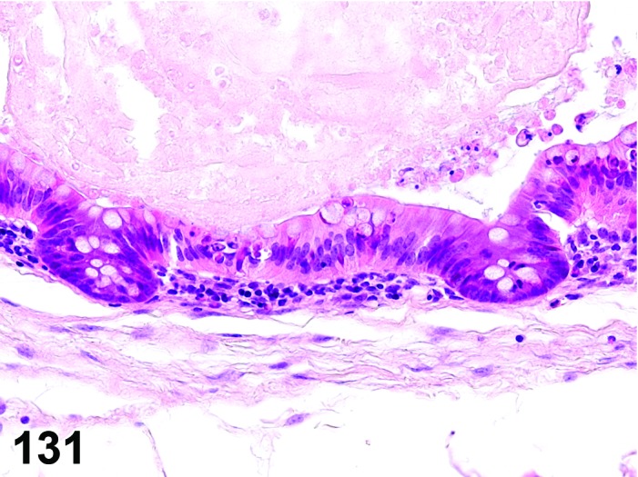
Mouse colon. Diverticulum, cystic. Higher magnification of Figure 130. Note flattened mucosa.
Synonyms: Cystic adenomatous hyperplasia; diverticulosis; herniated crypt; crypt herniation Diverticulum, atypical, may have been identified as: Atypical cystic hyperplasia; cystic hyperplasia with growth into the gastric wall; cystic adenomatous hyperplasia; herniation, atypical; “pseudoinvasion”, atypical
Modifiers: Cystic, atypical
Pathogenesis: Usually caused by mechanical or biochemical disruption of intestinal wall structures resulting in loss of extracellular matrix integrity and internal projection of surface lining into deeper layers; may alternatively represent a congenital abnormality.
Diagnostic features
Extension of crypts through muscularis mucosa, into submucosa and further in some cases.
Morphology of epithelial lining is variable, ranging from single layer cuboidal or columnar cells to complete mucosa.
Epithelium may show features of regeneration like increased basophilia, increased nucleus to cytoplasm ratio and gradual loss of polarity.
Basement membrane integrity always maintained.
Often accompanied by inflammation and regenerative / reparative processes.
May contain ingesta, hair, or other foreign material.
Cystic:
Rounded cystic structures in lamina propria or deeper layers.
Compression of surrounding structures, some degree of compression atrophy of the epithelium lining the cyst.
May contain mucus.
Atypical
Extension of atypical/dysplastic mucosal glands into or through muscularis mucosa, submucosa, and deeper in some cases.
Hyperplastic atypical (dysplastic) epithelial cells forming cystic glands that may appear to be “invading” into deeper layers of the intestine.
Basement membrane always intact.
Differential diagnoses
Adenoma: Proliferation beyond the confinements of the intestinal mucosa.
Adenocarcinoma: True infiltrative growth with loss of basement membrane integrity and often with a scirrhous stromal reaction.
Comment: These focal intestinal lesions are primarily formed by extensions of mucosal epithelium and do not involve evaginations of the other mural layers; as such they do not resemble diverticulosis in human colon, which may be focal, multifocal or diffuse. Diverticula do not appear to be neoplastic, although they do create an increased risk of rupture and septic peritonitis. These structures are typically lined by a simple cuboidal mucus epithelium.
It is not recommended to record “diverticulum” and its modifier as a separate finding, if it occurs in association with another proliferative process (e.g. hyperplasia, adenoma).
Small glandular diverticula lined by a single layer of cuboidal or columnar epithelium resemble “crypt herniation” of human gastrointestinal pathology (e.g. Rex et al. 2012). Such herniation of crypts through the muscularis mucosa is interpreted as “pseudoinvasive” or “inverted” growth pattern. Although the process-related term “crypt herniation” is common in literature (e.g. Betton et al. 2001), the term “diverticulum” is considered to better fit to the INHAND principle to use descriptive terms and to avoid process-related terms (Mann et al. 2012).
Spontaneous and induced intestinal diverticula occur more frequently in mice than in rats. They have been reported in the colon of Cdx-2 knockout mice due to a developmental defect (Tamai et al. 1999). In this model they appeared as polyps with herniation and due to the congenital etiology the authors termed it hamatoma.
Atypical diverticula must be distinguished from genuine neoplastic invasion (e.g. loss of basement membrane and other signs of malignancy in the latter). They are associated with preexisting mucosal atypical hyperplasia/dysplasia as seen in specific experimental conditions, tumor-associated intestinal toxins and genetically-engineered mice. Whereas they often do not progress to unequivocal adenocarcinoma, atypical glandular and cellular features are consistent with neoplastic potential.
Atypical diverticula may be associated with atypical hyperplasia and adenoma, but usually not with adenocarcinoma. As noted above for the simple diverticula, they should not be recorded as a separate diagnostic entity when associated with another proliferative process.
Hyperplasia, mucosa
(Figure 132, 133 and 134)
Figure 132.
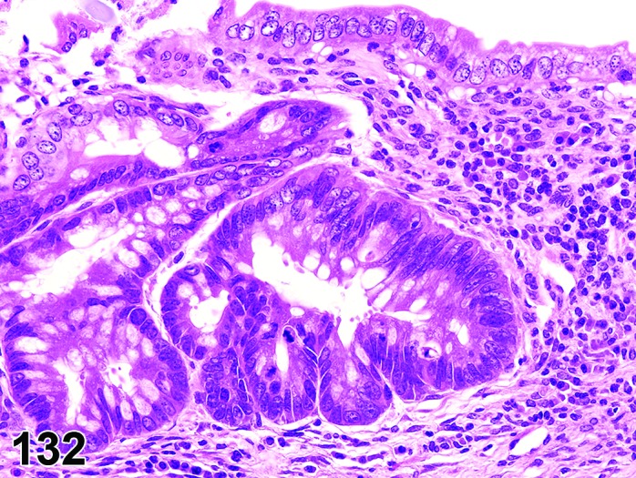
Mouse colon. Hyperplasia, mucosa, regenerative, at edge of ulcer.
Figure 133.
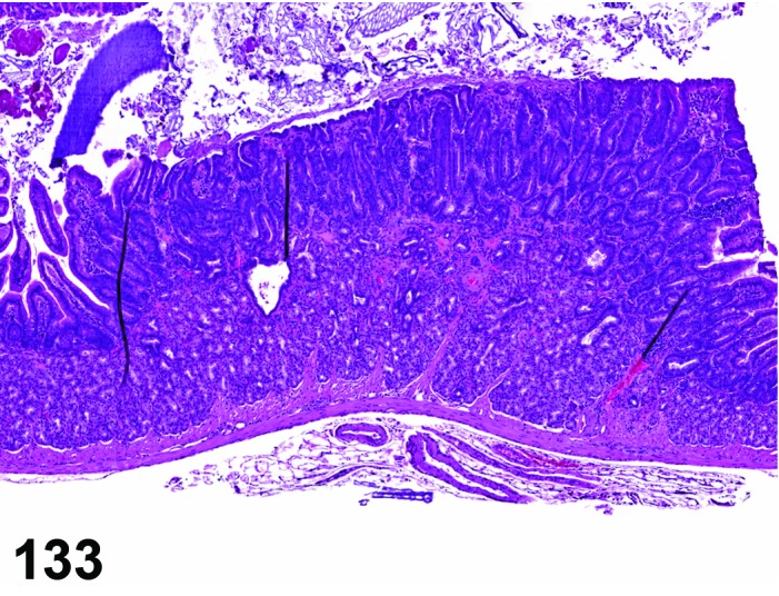
Mouse duodenum. Hyperplasia, avillous.
Figure 134.
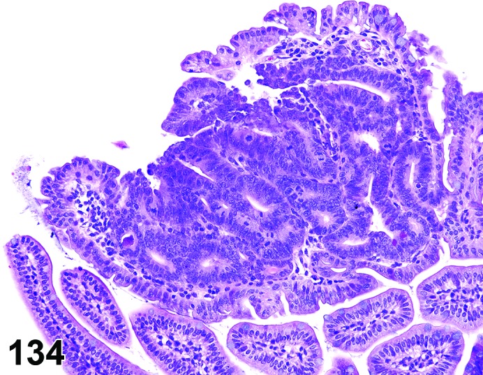
Mouse small intestine. Hyperplasia, focal, atypical. Note loss of regular architecture and cellular atypia.
Synonyms: Hyperplasia, regenerative
Synonyms for atypical hyperplasia may include dysplasia, gastrointestinal intraepithelial neoplasia (GIN, mice), early neoplastic lesion, atypical crypts, dysplastic crypts, dysplastic foci, or aberrant crypt foci.
Modifiers: Avillous, atypical, goblet cell
Histogenesis: Enterocytes of the intestinal mucosa.
Diagnostic features
Focal or diffuse process accompanying epithelial damage; no evidence of compression.
Villous and glandular architecture is not altered by the proliferative process itself but in regenerative hyperplasia may have been altered by the initiating event (degeneration and necrosis).
Focal penetration by glandular diverticula into lamina propria or deeper layers may be present but basement membrane is always intact.
No cellular or nuclear atypia.
Avillous:
Focal lesion of the duodenum in aging mice.
Smooth luminal surface lacking intestinal villi.
Hyperplastic crypts may often be interspersed between hyperplastic Brunner’s glands.
Goblet cells may be reduced or increased.
Crypt dilatation and diverticula may be present in larger lesions.
Frequently accompanied by submucosal edema and inflammatory cell infiltration.
Atypical:
Structure of the intestinal villi and crypts often abnormal.
Focal penetration by glandular diverticula into lamina propria or deeper layers may be present but basement membrane is always intact.
Cellular atypia and pleomorphism as evidenced by hyperchromatism with increased cytoplasmic basophilia and lost polarity, nuclear pleomorphism, increased N/C ratio, hyperchromasia, increased mitotic activity.
Goblet cell:
Increased proportion of goblet cells, predominantly in crypts; crypts exhibit normal length or are enlaged.
Differential diagnosis
Adenoma: Exophytic or endophytic growth pattern extending beyond the confinement of the mucosa; compression of adjacent tissue if growth pattern is not exclusively polypoid.
Comment: Reactive or reparative (regenerative) hyperplasias often follow injury due to chemicals, infectious agents and irradiation. Focal or diffuse reactive hyperplasia is seen in the cecum, colon and rectum of immuno-compromised mice following Helicobacter infection. Diffuse reactive hyperplasia is observed in the distal colon of mice infected with the gram negative bacterium Citrobacter freundii (transmissible murine colonic hyperplasia). Occasionally the transverse and ascending colon and the cecum are affected. In such cases of hyperplasia the surface epithelium is covered by myriads of cocobacillary bacteria. The lamina propria shows diffuse leukocytic infiltration.
Diffuse non-reactive hyperplasia of the mucosa with increased height of the villi and depth of the crypts is synonymous with hypertrophy, mucosa. Hypertrophy is the recommended term for such lesions.
An increased proportion of goblet cells has been reported in the literature as both goblet cell hyperplasia or metaplasia. We recommend the term hyperplasia, as goblet cells are normal constituents of the intestinal mucosa. Hyperplasia of goblet cells has been induced in the small and large intestine of rats by treatment with a gamma secretase inhibitor (Aguirre et al. 2014).
Focal hyperplasia of the proximal duodenum, adjacent to the pylorus, occurs in aged mice has been referred to in the literature as avillous hyperplasia (Facinni, et al. 1990; McInnes 2012). It may be visible macroscopically as a thickened plaque and is characterised by hyperplasia of all cells in the epithelium including Brunner’s glands, but with loss of villi. The epithelium can show erosion/ulcer formation with associated inflammation. In the more severe cases dilated mucosal glands can extend towards the submucosa and be interspersed with dilated Brunner’s glands.
Focal atypical hyperplasia is a frequent finding in mice treated with test articles which affect the intestinal mucosa or in genetically-engineered mice. Under such conditions they often occur in multiple foci. It has not been described in untreated wild type animals so far. The term GIN or dysplasia has been used in genetically-engineered mice. Other terms occasionally used but not preferred as synonyms are microadenoma, carcinoma in situ, intra-epithelial carcinoma. Dysplasia is often used for similar lesions in human colon (Bosman et al. 2010).
Colon crypts exhibiting focal atypia (“aberrant crypt foci”) can be demonstrated by the examination of methylene blue stained colon whole mounts with a dissecting microscope (Raju 2008). They are usually not seen grossly until 1 mm in size.
Atypical hyperplasia may be low-grade to high-grade, but this grading is usually not included in the diagnosis.
Under conditions of experimental intestinal carcinogenesis, focal atypical hyperplasia is considered as a preneoplastic lesion that may progress to an adenoma or adenocarcinoma.
Rectal prolapse often due to Helicobacter sp or Citrobacter sp infection, often shows severe degrees of simple hyperplasia, atypical hyperplasia and the appearance of a invasive-like growth pattern. These cases should be recorded as rectal prolapse.
Hyperplasia, Brunner’s glands
(Figures 135 and 136)
Figure 135.
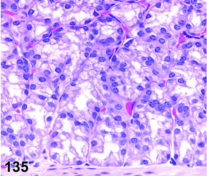
Mouse (hemizygous rasH2), duodenum by Normal Brunner’s glands.
Figure 136.
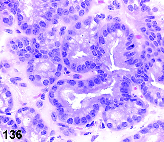
Mouse (hemizygous rasH2), duodenum by Hyperplasia Brunner’s glands. Note tortuous glandular structure with basophilic cytoplasmic staining.
Histogenesis: Cells of Brunner’s glands.
Diagnostic features
Brunner’s glands cells are often hypertrophic with more extensive cytoplasm, larger nuclei, or both.
Glands are more branched and coiled, compared with normal glandular structure.
Brunner’s glands cells and underlying stroma may form pedunculated structures within the spaces of cystic glands.
Cellular polarity is maintained.
Cells are tall columnar, with extensive, poorly-stained, or somewhat eosinophilic-stained cytoplasm with dense, basally-located nuclei.
Glands may expand into the underlying circular muscle layer, but basement membrane integrity always maintained.
Differential diagnosis
Diverticulum: Localized and discrete penetration of intestinal crypts into lamina propria or deeper layers with maintained basement membrane integrity and surrounded by pre-existing mature tissues lacking a desmoplastic stromal response.
Carcinoma, Brunner’s glands: Effacement of Brunner’s glands structures, prominent desmoplasia; invasive growth.
Comment: Brunner’s glands hyperplasia has been observed in transgenic mice (hemizygous rasH2) after dosing for one month with a receptor tyrosine kinase inhibitor that targets multiple cellular kinase receptors, including those of vascular endothelial growth factor (VEGF), platelet-derived growth factor (PDGF), stem cell factor receptor (KIT), FMS-like tyrosine kinase 3 (FLT-3) and glial cell line-derived neurotrophic factor rearranged during transfection (RET). Progression from preneoplastic hyperplasia to carcinoma/adenocarcinoma can occur.
To appropriately capture the area of interest in the rodent, it is important to retain the gastroduodenal junction in a manner that allows a comprehensive evaluation of the area of interest and prevents folding of the stomach and gastroduodenal region that can obscure Brunner’s glands. This can be accomplished by opening the stomach along the greater curvature, leaving the duodenum attached, and pinning the tissue flat for fixation.
Metaplasia, squamous cell
Figure 137.
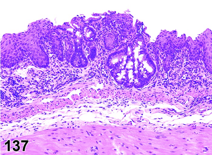
Mouse, rectum, metaplasia, squamous cell.
Synonyms: Squamous metaplasia.
Histogenesis: Colon or rectum epithelium.
Diagnostic features
Focal, multifocal or diffuse
Replacement of normal epithelium with squamous epithelium.
Frequently associated with chronic inflammation.
Differential diagnosis
Carcinoma, squamous cell: Invasion, areas with moderate to poor differentiation or anaplasia, ususally prominent mitoses.
Extension of anal mucosa into rectum: Usually in proximity to the anus and without concomitant inflammatory reaction.
Comment: Squamous metaplasia is often seen in chronic colitis models in mice, usually in the dextran sulfate model. Squamous cell carcinomas have not been reported to arise in the lesions (Seamons et al. 2013).
g. Neoplasms
Adenoma
(Figures 138, 139, 140, 141 and 142)
Figure 138.
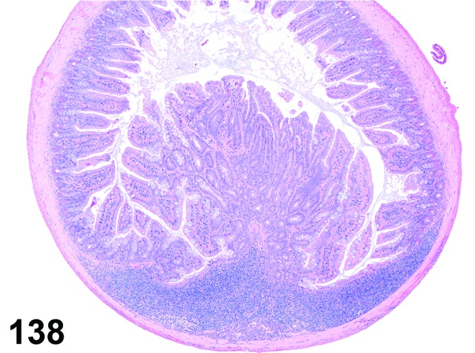
Mouse jejunum. Adenoma, polypoid.
Figure 139.
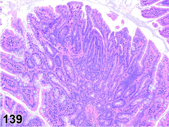
Mouse jejunum. Adenoma, polypoid. Higher magnification of Figure 138, demonstrating loss of regular mucosal architecture but low grade of atypia.
Figure 140.
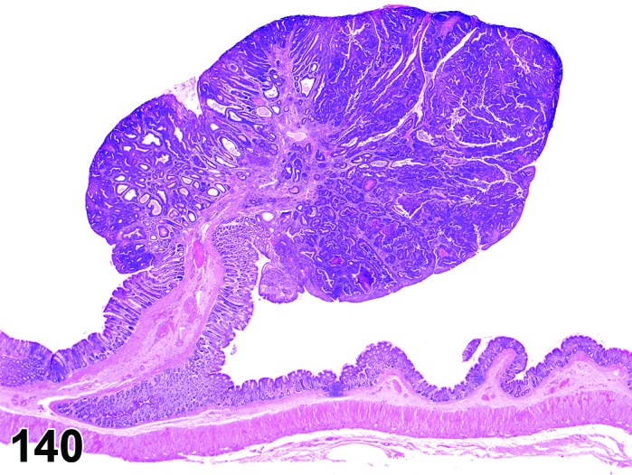
Rat colon. Adenoma, polypoid.
Figure 141.
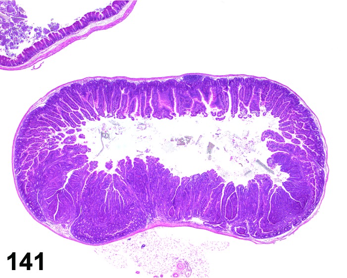
Rat jejunum. Adenoma, sessile; with diverticula, atypical. Note expansive growth beyond the confinement of the surrounding mucosa.
Figure 142.
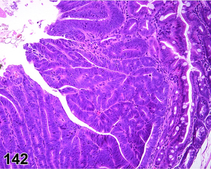
Rat jejunum. Adenoma, sessile. Higher magnification of Figure 141 demonstrating irregular growth pattern and moderate cellular atypia.
Synonyms: Polyp; adenomatous polyp
Histogenesis: Enterocytes of the intestinal mucosa.
Modifiers: Polypoid, papillary, sessile
Diagnostic features
Polypoid, sessile or papillary tumor projecting into intestinal lumen.
Sessile tumors have no stalk.
Compression of adjacent tissue if growth pattern is not exclusively polypoid.
Branching villi or tubular crypt proliferation can be present.
Squamous metaplasia and focal mineralization may be present.
May be associated with glandular or cystic diverticula, but basement membrane always intact.
One cell type is predominant and goblet cells are absent or reduced in number.
The epithelium may contain foci of varying degrees of dysplasia.
Signs of cellular atypia / dysplasia include cellular basophilia, loss of cellular polarity, hyperchromatic and pleomorphic nuclei, and nuclear stratification.
Differential diagnoses
Hyperplasia, atypical: Circumscribed proliferation with varying degree of atypia, but limited to the confinements of the intestinal mucosa.
Diverticulum: Extension of crypts through muscularis mucosa, into submucosa and further in some cases. Morphology of epithelial lining is variable, ranging from single layer cuboidal to columnar cells to complete mucosa. Epithelium may show features of regeneration like increased basophilia, increased nucleus to cytoplasm ratio and gradual loss of polarity. Often accompanied by inflammation and regenerative / reparative processes.
Adenocarcinoma: True invasion by individual, groups or areas of tumor cells, with or without a stromal response.
Comment: The term “polyp” was avoided, because of implied uncertainty about the neoplastic nature of this proliferative lesion. Benign neoplasms may develop dysplastic foci and evolve into malignant neoplasms. Chemicals causing benign intestinal tumors often also cause intestinal adenocarcinomas (Chandra et al. 2010; Pandiri et al. 2011; Ward 1974).
Adenomas, as well as atypical hyperplasia, exhibit some degree of dysplasia and distortion of pre-existing architecture. Macroscopic visibility has been suggested as a differential diagnostic criterion by some authors. This criterion is not used in the context of this description of histologic criteria. Any lesion exhibiting a papillary or polypoid growth pattern is an adenoma, irrespective whether it was grossly visible or not. An intra-epithelial small focus (compatible with atypical hyperplasia) with cytological characteristics seen in adenomas should NOT be diagnosed as an adenoma.
The frequency of spontaneous intestinal adenomas varies with the intestinal segment. Incidences up to 51% are reported for the duodenum and anterior part of the jejunum (Hare and Stewart 1956). In the other intestinal segments the incidence of adenomas is below 5%. However, the detection frequency is highly dependent on the mode of macroscopic examination of the intestines, e.g. random blocks, naked eye inspection of mucosal surface, transillumination or Swiss roll technique.
The determination of the malignancy status of a papillary or polypoid intestinal tumor requires a good fixation and trimming protocol, to obtain an optimal section through the middle of the tumor with its connecting area to the normal intestine. If there is no connection to the intestine, it is impossible to determine if invasion is present. The cytology of a non-invasive adenoma may include high grade dysplasia. In most rat and mouse chemically-induced or genetically-engineered tumor models, true invasion of the stalk is unusual but may occur commonly in other models.
Adenocarcinoma
(Figures 143, 144, 145 and 146)
Figure 143.
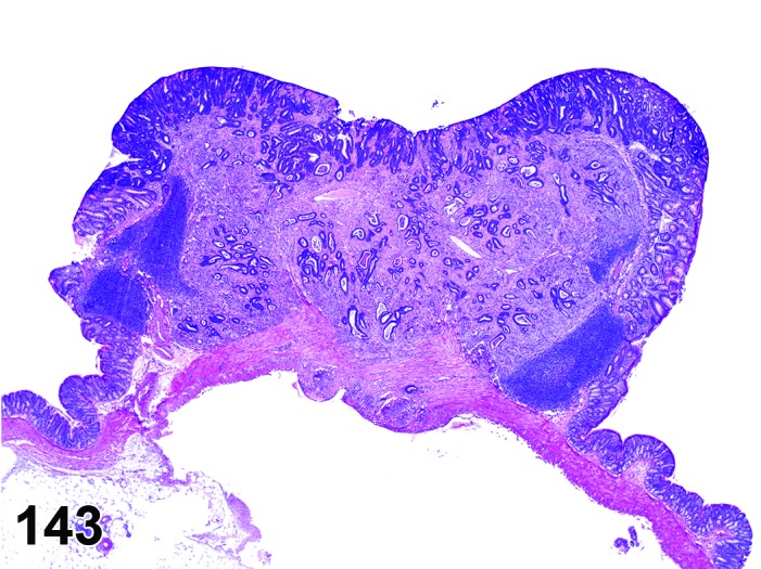
Rat colon. Adenocarcinoma, scirrhous.
Figure 144.
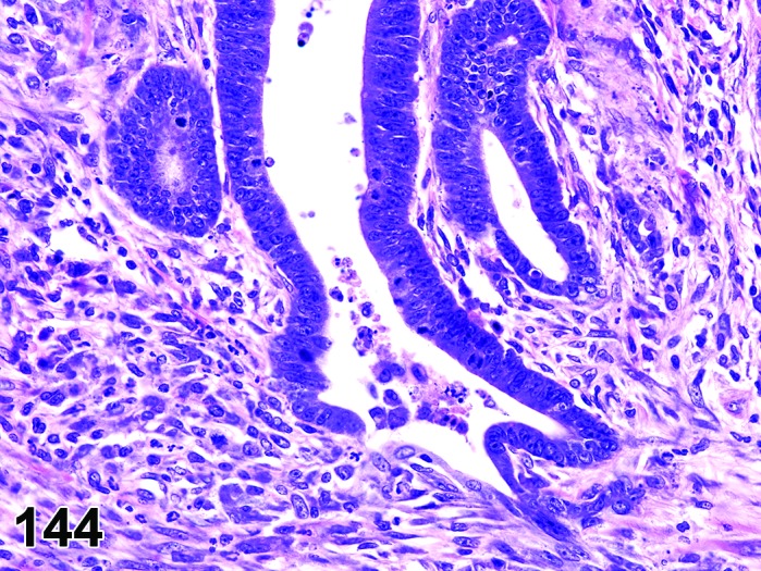
Rat colon. Adenocarcinoma, scirrhous. Higher magnification of Figure 143, demonstrating tubule structure of atypical tumor cell with lost basement membrane integrity, embedded in scirrhous stroma.
Figure 145.
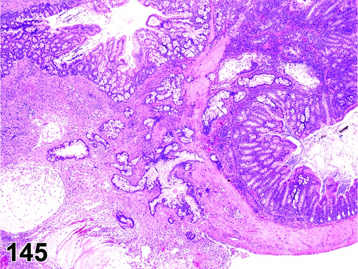
Mouse colon. Adenocarcinoma, mucinous.
Figure 146.
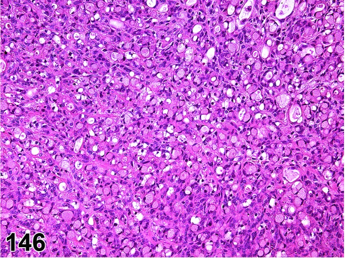
Mouse colon. Adenocarcinoma, signet ring type.
Synonym: Carcinoma
Histogenesis: Enterocytes of the intestinal mucosa.
Modifiers: Solid, papillary, cystic, scirrhous, mucinous, signet ring, mixed, undifferentiated
Diagnostic features
Mucosal architecture is distorted.
Growth patterns include tubular, papillary, tubulopapillary, mucinous, signet ring, cystic, scirrhous and combinations of all types; different etiologies may result in different growth patterns.
Mucus-producing cells may be interspersed between absorptive cells.
Mucus may be present intracellularly or extracellularly.
Signet ring cell type: intracellular accumulation of mucus, leading to the characteristic appearance of these cells; high invasive growth; therefore, single tumor cells and groups of cells are interspersed between pre-existing tissues.
Squamous metaplasia and focal mineralization are not present.
Varying degrees of cellular atypia.
Many mitotic figures.
Invasion through muscularis mucosa or deeper layers of the intestinal wall, stalk of an adenoma, or adjacent organs.
Often scirrhous response in areas of invasion.
Differential diagnoses
Diverticulum: Localized and discrete penetration into lamina propria or deeper layers with maintained basement membrane integrity and surrounded by pre-existing mature tissues lacking a scirrhotic stromal response.
Hyperplasia atypical and adenoma: May be associated with glandular or cystic diverticula, but basement membrane always intact.
Rectal prolapse (mice): Associated with Helicobacter sp or Citrobacter sp infection in mice; hyperplastic epithelium extending as diverticula through muscularis mucosa and into submucosa, with associated inflammation.
Comment: An intestinal adenocarcinoma may arise de novo from normal epithelium, but more commonly from a preneoplastic lesion such as atypical hyperplasia or from an adenoma. The histogenesis, morphology and biology of tumor progression often depend on the etiology, i.e. the chemical carcinogen or the kind of genetic change in a GEM model.
In contrast to humans, spontaneous rodent adenocarcinomas show a predilection for the small intestine, especially the jejunum and ileum, rather than the large intestine. The descriptions available indicate that the morphology of spontaneous lesions does obviously differ from the induced ones and that the etiology may determine the morphologic phenotype. In addition, induced adenocarcinomas have their preferred localization in the distal colon and show high invasiveness combined with a low tendency to metastasize. Helicobacter sp has been shown to be a colon co-carcinogen in some experimental mouse models involving GEM or immunodeficient mice (Erdman and Poutahidis 2010). Inflammatory mouse models of colon adenocarcinoma are often scirrhous and mucinous (Erdman and Poutahidis 2010; Kosa et al. 2012).
Metastases are often associated with a specific method of tumor induction. In rats, the mucinous adenocarcinoma induced by some carcinogens commonly metastasizes, predominantly to the lung. Mucinous intestinal adenocarcinomas are often the most malignant type of intestinal tumor in rats and mice (Ward et al. 1973).
Carcinoma, Brunner’s glands
Figure 147.
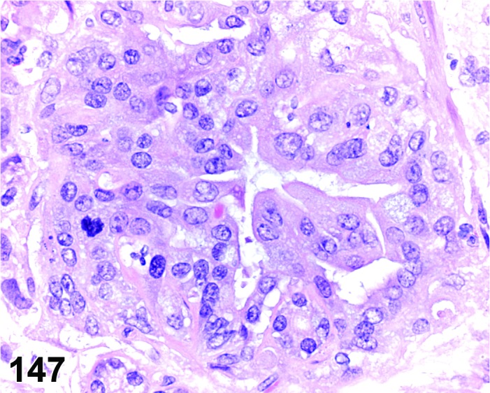
Mouse (hemizygous rasH2), duodenum. Carcinoma, Brunner’s glands.
Histogenesis: Cells of Brunner’s glands.
Diagnostic features
There is effacement of Brunner’s glandular structures and the gastroduodenal junction.
Desmoplasia is prominent and can be the most dominant feature.
Glandular structure is diminished or completely lost.
The mitotic index is usually low.
There is moderate pleomorphism of Brunner’s glandular cells.
Cells have well-stained cytoplasm that may be eosinophilic, amphophilic or basophilic.
Attenuated glandular cells may line cystic areas that contain mucus.
Invasion through the muscularis mucosa and/or through the circular and longitudinal muscles, as well as the serosa, can be extensive. The basement membrane of the glands is breached or not evident.
Differential diagnosis
Hyperplasia, Brunner’s glands: Glandular architecture may be slightly altered, but still recognizable; cell polarity maintained; glands may penetrate into the underlying circular muscle layer, but basement membrane maintained.
Diverticulum: Localized and discrete penetration of intestinal crypts into lamina propria or deeper layers with maintained basement membrane integrity and surrounded by pre-existing mature tissues lacking a desmoplastic stromal response.
Comment: Brunner’s glands carcinoma is a rare tumor, although a high occurrence can be induced with certain test articles. Brunner’s glands carcinoma has been observed in transgenic mice (CB6/F1/Jic Tg rasH2 hemizygous) and Sprague-Dawley rats after dosing for 6 and 24 months, respectively, with a receptor tyrosine kinase inhibitor that targets multiple cellular kinase receptors (see Brunner’s glands hyperplasia; Comment). The investigations in the transgenic model indicated that the rapid occurrence and high incidence of Brunner’s glands carcinoma is more consistent with altered transcriptional regulation rather than a genotoxic mechanism.
Knowledge of the site of the incipient lesion or the use of immunohistochemical methods are often required to diagnose the origin of the change because invasion of a gastric glandular cell or duodenal enterocyte carcinoma can have similar morphologic features to those of a malignant neoplasm of Brunner’s glands that has progressed extensively.
Leiomyoma
(Figures 148 and 149)
Figure 148.
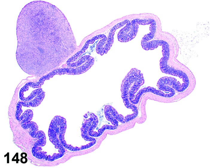
Mouse cecum. Leiomyoma.
Figure 149.
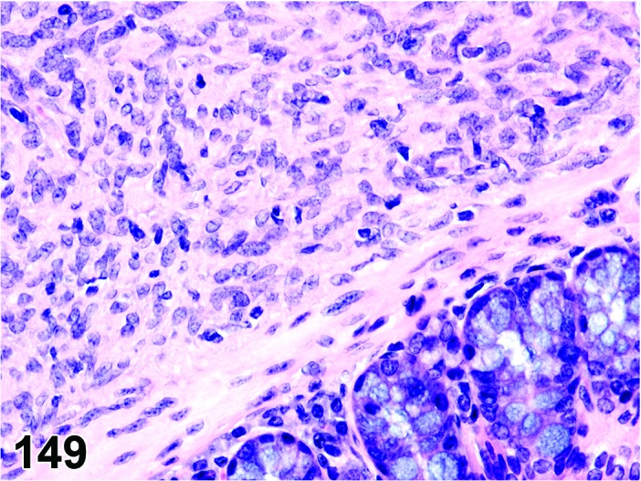
Mouse cecum. Leiomyoma. Higher magnification of Figure 148.
Histogenesis: Smooth muscle cells from tunica muscularis.
Diagnostic features
Intramural involvement.
Interlacing bundles and whorls of uniform spindle cells arranged in criss-cross patterns of bundles.
Nuclei typically blunt-ended or cigar shaped.
Eosinophilic cytoplasm contains longitudinal myofilaments and perinuclear clear space and show immunoreactivity for desmin.
Differential diagnoses
Leiomyosarcoma: Shows mitotic activity and cellular pleomorphism.
Gastrointestinal stromal tumor (GIST): Differentiation from other soft tissue tumors need only be made in cases of intergroup difference in the incidence of soft tissue tumors in a given study; unequivocal differentiation from other soft tissue tumors possible only by immunohistochemistry: Typically CD117 positive (cytokine receptor encoded by c-kit); spindle, epithelioid, or pleomorphic cell morphology. Indistinct cell borders and fibrillary cytoplasm; cells may be arranged in bundles with storiform architecture; spindle- or irregular-shaped nuclei.
Schwannoma: CD117 negative, S-100 positive; indistinct cell borders, elongate cells with eosinophilic cytoplasm; nuclei sometime arranged in palisades (i.e., Antoni A pattern).
Fibroma/sarcoma: CD 117 and S-100 negative; interlacing bundles of spindle cells; Masson’s trichrome-blue collagen.
Histiocytic sarcoma: Positive for histiocytic markers (ED-1 for rats, Factor 4/80 in mice), CD 117 and S-100 negative; infiltrative pattern, pleomorphic morphology.
Comment: Similar to those observed in other sites [see also INHAND-publication of soft tissue tumors (Greaves et al. 2013)].
Leiomyosarcoma
(Figures 150 and 151)
Figure 150.
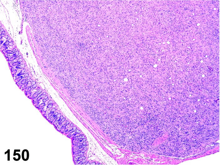
Mouse colon. Leiomyosarcoma.
Figure 151.
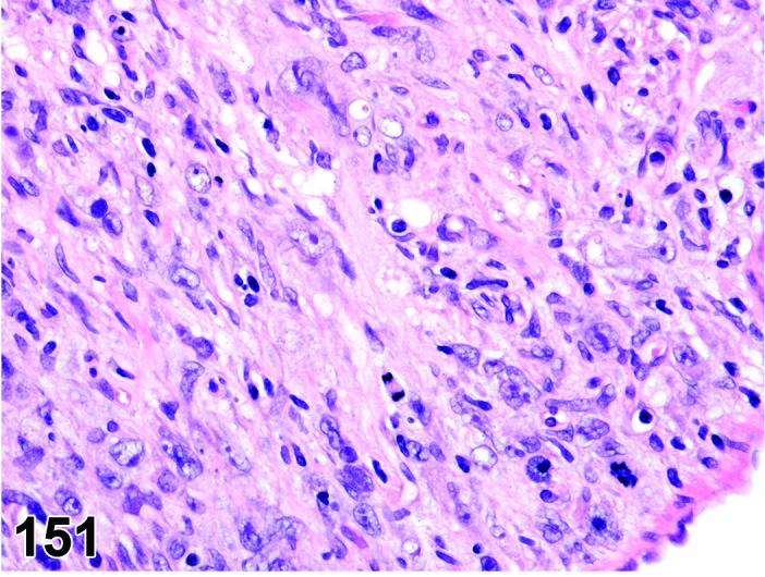
Mouse colon. Leiomyosarcoma; higher magnification of Figure 150.
Histogenesis: Smooth muscle cells from tunica muscularis.
Diagnostic features
Intramural involvement.
Similar to leiomyoma, but more pleomorphic and with mitotic activity.
Necrosis, cystic change and mineralization are common features.
Distant metastases.
Differential diagnoses
Leiomyoma: Shows little or no cellular pleomorphism or mitotic activity.
Gastrointestinal stromal tumor (GIST): Differentiation from other soft tissue tumors need only be made in cases of intergroup difference in the incidence of soft tissue tumors in a given study; unequivocal differentiation from other soft tissue tumors possible only by immunohistochemistry: Typically CD117 positive (cytokine receptor encoded by c-kit); spindle, epithelioid, or pleomorphic cell morphology. Indistinct cell borders and fibrillary cytoplasm; cells may be arranged in bundles with storiform architecture; spindle- or irregular-shaped nuclei.
Schwannoma: CD117 negative, S-100 positive; indistinct cell borders, elongate cells with eosinophilic cytoplasm; nuclei sometime arranged in palisades (i.e., Antoni A pattern).
Fibroma/sarcoma: CD 117 and S-100 negative; interlacing bundles of spindle cells; Masson’s trichrome-blue collagen.
Histiocytic sarcoma: Positive for histiocytic markers (ED-1 for rats, Factor 4/80 in mice), CD 117 and S-100 negative; infiltrative pattern, pleomorphic morphology.
Comment: Similar to those observed in other sites [see also INHAND-publication of soft tissue tumors (Greaves et al. 2013)].
Gastrointestinal stromal tumor (GIST), benign (Figures 152, 153 and 154)
Figure 152.
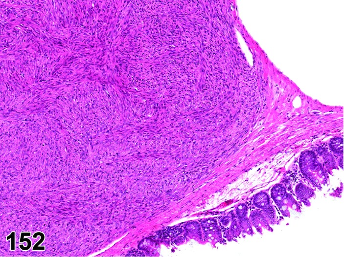
Mouse colon. Gastrointestinal stromal tumor, benign.
Figure 153.
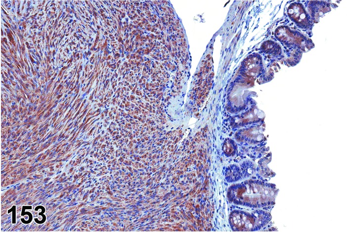
Mouse colon. Gastrointestinal stromal tumor, benign. Immunohistochemistry for CD117.
Figure 154.
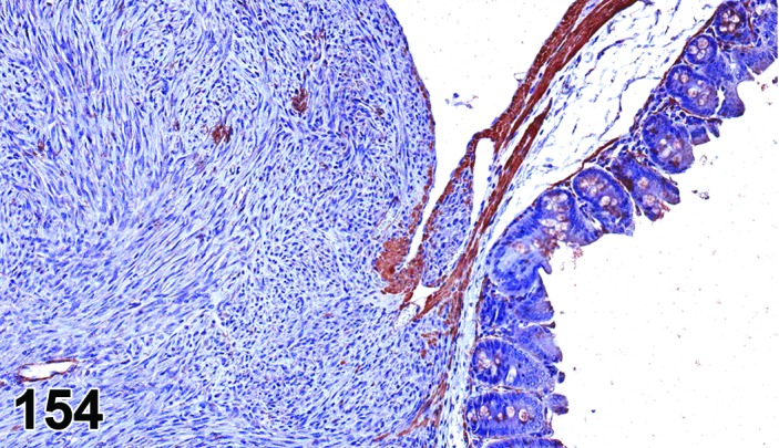
Mouse colon. Gastrointestinal stromal tumor, benign. Immunohistochemistry for smooth muscle actin.
Synonyms: Interstitial Cajal cell tumor, benign; gastrointestinal pacemaker cell tumor, benign
Histogenesis: Specialized smooth muscle cells (i.e., interstitial cells of Cajal) in the tunica muscularis or myenteric plexus.
Diagnostic features
Well demarcated tumor, expansive growth.
Cells may be arranged in bundles with storiform architecture, myxoid areas may be present.
Spindle, epithelioid, or pleomorphic cell morphology. Indistinct cell borders and fibrillary cytoplasm.
Spindle- or irregular-shaped nuclei.
Typically CD117 positive (cytokine receptor encoded by c-kit).
Differential diagnoses
Gastrointestinal stromal tumor, malignant: Infiltrative growth, distant metastases may be present.
Leiomyoma: CD117 negative, desmin positive; interlacing bundles and whorls of uniform spindle cells arranged in criss-cross pattern; blunt-ended, fusiform nuclei. Minimal nuclear pleomorphism; phosphotungstic acid-hematoxylin positive myofibrils.
Leiomyoscarcoma: CD117 negative, desmin positive; interlacing bundles and whorls of uniform spindle cells arranged in criss-cross pattern; blunt-ended, fusiform nuclei with hyperchromasia, pleomorphism, and high mitotic index; phosphotungstic acid-hematoxylin positive myofibrils.
Schwannoma: CD117 negative, S-100 positive; indistinct cell borders, elongate cells with eosinophilic cytoplasm; nuclei sometime arranged in palisades (i.e., Antoni A pattern).
Comment: GIST will generally not be easily differentiated from leiomyomas / leiomyosarcomas or other soft tissue tumors. However, immunohistochemical differentiation of the different tumor types, including GIST, needs to be performed in case of imbalances in the incidence of smooth muscle tumors in a given study.
Cecal GIST have been induced in GEM mice over-expressing a mutant KIT (Nakai et al. 2008). The question arises whether conventionally classified stromal tumors (e.g., leiomyoma) previously were actually GIST.
Gastrointestinal stromal tumor (GIST), malignant (Figure 155)
Figure 155.
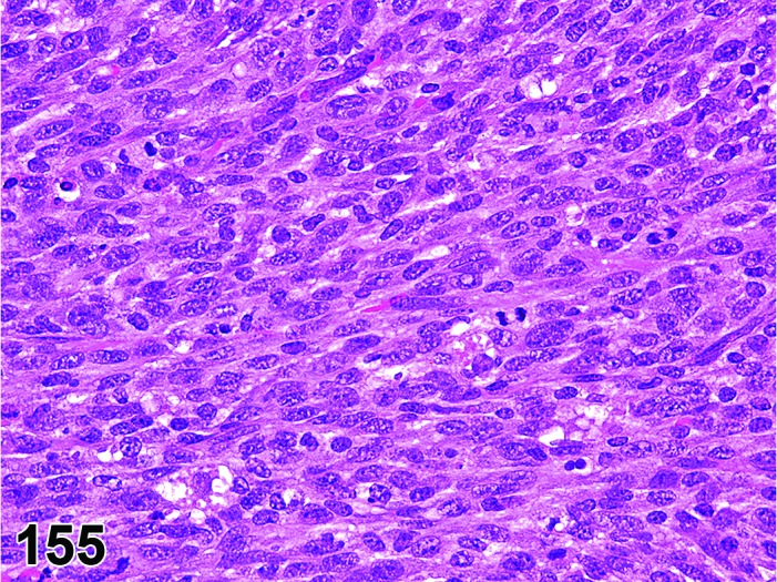
Mouse cecum. Gastrointestinal stromal tumor, malignant.
Synonyms: Interstitial Cajal cell tumor, malignant; gastrointestinal pacemaker cell tumor, malignant
Histogenesis: Specialized smooth muscle cells (i.e., interstitial cells of Cajal) in the tunica muscularis or myenteric plexus.
Diagnostic features
Locally infiltrative growth pattern.
Cells may be arranged in bundles with storiform architecture, myxoid areas may be present.
Spindle, epithelioid, or pleomorphic cell morphology. Indistinct cell borders and fibrillary cytoplasm.
Spindle- or irregular-shaped nuclei.
Typically CD117 positive (cytokine receptor encoded by c-kit).
Distant metastases may be present.
Differential diagnoses
Gastrointestinal stromal tumor, benign: Expansive growth pattern, no distant metastases.
Leiomyoma: CD117 negative, desmin positive; interlacing bundles and whorls of uniform spindle cells arranged in criss-cross pattern; blunt-ended, fusiform nuclei. Minimal nuclear pleomorphism; phosphotungstic acid-hematoxylin positive myofibrils.
Leiomyoscarcoma: CD117 negative, desmin positive; interlacing bundles and whorls of uniform spindle cells arranged in criss-cross pattern; blunt-ended, fusiform nuclei with hyperchromasia, pleomorphism, and high mitotic index; phosphotungstic acid-hematoxylin positive myofibrils.
Schwannoma: CD117 negative, S-100 positive; indistinct cell borders, elongate cells with eosinophilic cytoplasm; nuclei sometime arranged in palisades (i.e., Antoni A pattern).
Comment: GIST will generally not be easily differentiated from leiomyomas / leiomyosarcomas or other soft tissue tumors. However, immunohistochemical differentiation of the different tumor types, including GIST, needs to be performed in case of imbalances in the incidence of smooth muscle tumors in a given study.
Cecal GIST have been induced in GEM mice over-expressing a mutant KIT (Nakai et al. 2008). The question arises whether conventionally classified stromal tumors (e.g., leiomyoma) previously were actually GIST.
V. Salivary Glands
A. Morphology (Anatomy)
The salivary glands consist of major and minor glands and develop from the endodermal lining of the embryonal gut. The major salivary glands are grossly apparent and include the parotid, sublingual and submandibular glands. The minor salivary glands are microscopic and are distributed throughout the oral cavity encompassing the lingual (von Ebner’s glands around the vallate and foliate papillae, Weber’s glands at base of the tongue), sublingual, buccal, palatine, laryngeal and pharyngeal tissues. The submandibular gland matures with age and progress from rudimentary lobules of mesenchyme with ductular structures at birth to acinar development and complete maturation by about 30 days postnatally in rats (Proctor and Carpenter, 2007). The sublingual salivary glands are mature at birth unlike the parotid and submandibular salivary glands.
The major salivary glands are located on the ventral and lateral areas of the cervical region and extend to the base of the ear and are closely associated with the extraorbital lachrymal gland and mandibular lymph nodes. The largest of the three major salivary glands, the submandibular (mandibular or submaxillary) salivary gland is a tan, multilobular gland that is located in the ventral cervical region. It extends rostrally to the mandibular lymph nodes, caudally to the thoracic inlet, and is bordered bilaterally by the parotid glands. The sublingual glands consist of tan-brown bilateral single lobes located on the rostro-lateral surface of each of the submandibular glands and directly below the mandibular lymph nodes. The parotid glands are bilateral and consist of pink to off white, multiple flattened lobes extending from the submandibular gland ventrally, to the base of the ear dorsally, to the extraorbital lacrimal gland rostrally, and to the clavicle caudally. The parotid glands drain into the parotid duct while the submandibular and sublingual glands drain into the oral cavity via Wharton’s duct that terminates in small papillae near the incisors.
B. Morphology (Histology)
The submandibular gland is a seromucinous (mixed) compound tubuloacinar gland. In the mouse it is predominantly seromucinous, where as, in the rat it is mostly mucinous with a small serous component (Gresik, 1994; Ozono et al. 1991; Tucker, 2007)). The submandibular gland is comprised of several adenomeres with large pyramidal mucous cells that are flanked basally by variable numbers of cresentric serous demilunes. The large pyramidal mucous cells have abundant vesicular basophilic cytoplasm and basally located nuclei and the cresentric serous demilunes have scant eosinophilic cytoplasm and hyperchromatic nuclei. The acinar secretions from the adenomeres empty into intercalated ducts lined by low cuboidal to columnar epithelial cells. Few oncocytes are interspersed between the cuboidal epithelial cells of the intercalated ducts of aged rats. These cells are characterized by abundant granular eosinophilic cytoplasm (abundant mitochondria) and a central hyperchromatic nucleus. Thin fusiform myoepithelial cells are located between the basal lamina and epithelial cells of acini and intercalated ducts (Bogart, 10970; Sashima, 1986). Only in the submandibular gland, the intercalated ducts continue into granular (convoluted) ducts. The granular ducts are lined by tall columnar epithelial cells with abundant eosinophilic granular cytoplasm. The granulation of these ducts is much more prominent in sexually mature males when compared to females (Figures 156 and 157). These ducts continue into striated (intralobular) ducts that are lined by a single layer of tall columnar epithelial cells with marked basal infoldings (hence, striated cells) and central to apically located nuclei. Several of the striated (intralobular) ducts merge into interlobular excretory ducts that are lined by tall columnar epithelial cells with more apically located nuclei and prominent cytoplasmic striations. Several interlobular excretory ducts continue into one main excretory duct that expands into a diverticulum lined by simple columnar epithelium and end as squamous epithelium at the oral surface.
Figure 156.
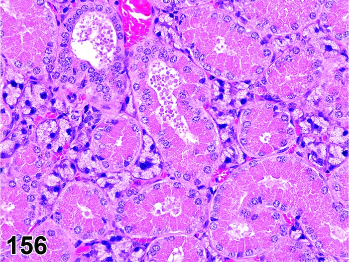
Mouse (male, 4-month old) submandibular gland. Note abundance of eosinophilic secretory granules in granular ducts.
Figure 157.
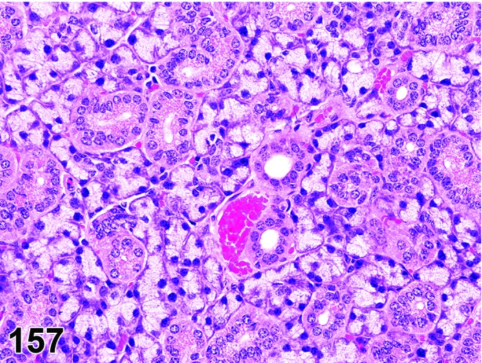
Mouse (female, 4-month old) submandibular gland. Note almost complete absence of eosinophilic secretory granules in granular ducts.
The sublingual gland is predominantly mucous secreting and has the largest acini. They are comprised of large pyramidal mucus cells with basally located nuclei and are infrequently capped by serous demilunes. The acinar secretions empty into intercalated ducts that in turn empty into striated (intralobular) ducts. The multiple striated ducts empty into the main excretory duct. The microscopic anatomy of the ducts is similar to their counterparts within the submandibular gland.
The parotid gland contains serous acini, which are morphologically very similar to the exocrine pancreatic acini (Wolff et al. 2002). The acinar cells are pyramidal with broad basophilic base and tapered apices. The eosinophilic granular cytoplasm contains zymogen granules, the nuclei are basally located. The duct system of the parotid gland is similar to the submandibular gland.
The minor salivary glands lack a true connective tissue capsules and are usually located within the submucosa or are interspaced between connective tissue stroma or muscle fibers. The glands are morphologically diverse and are organized into distinct serous (von Ebner’s glands), mucous (anterior and posterior buccal glands and minor sublingual glands) or mucous glands with serous demilunes (glossopalatal, palatal, and Weber’s glands, Figure 155) glands (Redman 2011). The acini are organized into lobule-like structures and collections of secretory end pieces and ducts open onto the mucosal surface.
C. Physiology
The salivary glands secrete a hypotonic sero-mucinous saliva containing electrolytes and enzymes that aid in lubrication and partial digestion of food. The serous secretions contain water, electrolytes (rich in K+ and HCO3– and poor in Na+ and Cl– compared to plasma), IgA and enzymes (ptyalin (amylase), lysozyme, lipase) and the mucinous secretions contain water, and glycoproteins. The minor salivary glands contribute about 14% protein and 1% amylase of the total salivary secretions, as determined by pilocarpine stimulation (Blazsek and Varga 1999).
The salivary secretions are tightly regulated by the sympathetic (adrenergic) and parasympathetic (cholinergic) nervous system (Garrett 1987). Most cell types within the submaxillary and parotid glands have both sympathetic and parasympathetic innervations while the sublingual glands have scant sympathetic innervation limited to striated ducts and blood vessels (Garrett et al. 1991). The parasympathetic system and the α-adrenergic sympathetic system regulate the water and inorganic ion phase of the primary secretory fluid, whereas the β-adrenergic sympathetic system effects primarily the macromolecular (enzymes) phase. Parasympathetic stimulation is mainly responsible for stimulation of the salivary acini resulting in copious secretion of saliva, however, the sympathetic stimulation does not inhibit salivary secretion but it modulates the saliva composition and causes salivary secretion to a lesser extent (Proctor and Carpenter 2007). The sympathetic and parasympathetic stimulation of the myoepithelial cells surrounding the acini results in contraction of acini and directing the saliva into the ducts (Tucker 2007). Surprisingly, it is the sympathetic stimulation but not parasympathetic stimulation that results in morphologically obvious depletion of storage granules within the acini (Garrett et al. 1991). β-adrenergic sympathomimetic agonists such as isoprenaline (Selye et al. 1961) and terbutaline (Sodicoff et al. 1980) first cause rapid depletion of secretory granules and later promotes re-synthesis of secretory material leading to acinar cell hypertrophy and hyperplasia. On the other hand, β-adrenergic antagonists like propranolol suppress salivary secretions and eventually cause acinar atrophy (Fukuda 1968).
The sexual dimorphism of the submandibular gland salivary glands is dependent on testosterone levels (Harvey 1952). The increased granularity in males (Figure 156) is accompanied by an about 10 times higher aminotransferase activity compared to females that have very little if any ductal secretory granules (Figure 156) (Hosoi et al. 1978).
D. Necropsy & Trimming
A typical approach for toxicological examination includes all the three grossly visible major salivary glands, i.e., submandibular, sublingual, and parotid gland. At necropsy, all three major salivary glands are removed in one piece, usually together with the mandibular lymph nodes. Trimming of the salivary glands is then undertaken as specified in the RITA trimming guide (website//reni.item.fraunhofer.de/reni/trimming): The salivary glands are trimmed longitudinally and through the largest surface. All three salivary glands should be present in the section.
The typical approach may be varied according to study protocol requirements, e.g. determination of organ weights and tissues required for pathology examination.
The microscopic minor lingual glands are routinely examined if the entire tongue, including its base, is removed at necropsy and trimmed longitudinally. The other minor salivary glands located within the oral cavity and pharynx are examined only occasionally during microscopic evaluation of these tissues.
E. Nomenclature, Diagnostic Criteria, and Differential Diagnoses
a. Congenital Lesions
Ectopic tissue
(Figures 158 and 159)
Figure 158.
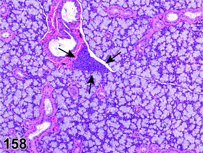
Rat sublingual gland. Ectopic tissue, parotid gland (arrow).
Figure 159.
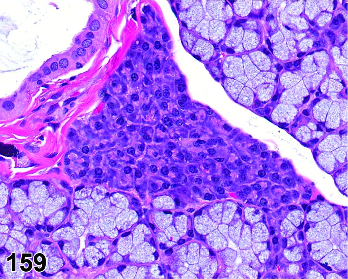
Rat sublingual gland. Ectopic tissue, parotid gland; higher magnification of Figure 158.
Pathogenesis: remnant of embryonic cell rests.
Modifiers: Parotid gland, submandibular gland, sublingual gland
Diagnostic features
Focal.
Circumscribed normal parotid, submandibular or sublingual gland acini within a heterotopic salivary gland.
Differential diagnosis:
None.
Comment: Care should be taken to differentiate sectioning artifacts (“floaters”) from the ectopic parotid gland in the sublingual glands.
b. Cellular Degeneration, Injury and Death
Vacuolation
Figure 160.
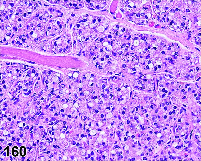
Rat linugal von Ebner’s gland. Vacuolation and secretory depletion.
Synonyms: hydropic change, cloudy swelling, hydropic degeneration, fatty degeneration, lipidosis, lipid accumulation
Modifier: Foamy
Pathogenesis: Accumulation of substances of different character, including fluids, , lipids, phospholipids, and glycoproteins within cells.
Diagnostic features
Distribution may be focal or multifocal and involve several lobules or a diffuse change involving all the lobes.
Cells may be swollen with pale eosinophilic cytoplasm and have intracytoplasmic vacuoles with variably margins.
The size of vacuoles may vary from small (microvesicular) to large when multiple small vacuoles coalesce together (macrovesicular) often resulting in nuclear displacement.
Variable loss of zymogen granules or mucinous secretions within the affected cells
Lobular architecture of gland is retained.
Foamy
Hypertrophic cells with foamy cytoplasm characterized by clear vesicles with eosinophilic fine granules
Differential diagnoses
Artifact: Medium-sized clear vesicles within the cytoplasm of acinar cells, especially at the periphery. No positive staining by fat stains.
Comments: Based solely on light microscopic evaluation of hematoxylin and eosin stained tissue sections, it is not possible to conclusively characterize the nature of the intracytoplasmic vacuoles. Hence, vacuolation, cytoplasmic is the preferred terminology of the lesion that was previously diagnosed as fatty change, hydropic degeneration, cloudy swelling etc. Any chemical/factor affecting the integrity of the cell membranes can potentially cause poorly defined intracytoplasmic vacuoles that was previously diagnosed as hydropic degeneration (Sela et al. 1977; Simson, 1972). Care should be taken to discriminate artifactual vacuolation induced by processing from the lesion induced by treatment.
Some metabolic insults cause disruption of normal lipid transport and metabolism and subsequently cause accumulation of lipid within the cytoplasm. In the streptozotocin-diabetes rat model, the parotid is the major salivary gland most susceptible to cytoplasmic vacuolation (Anderson 1998). Cytoplasmic vacuolation within the parotid was observed in doxylamine treated F344 rats (Jackson and Blackwell 1988). Cytoplasmic vacuolation within the parotid but not submandibular glands was noted in aged Wistar rats (Andrew 1949). Oil Red O and Sudan Black stains on frozen sections help to identify the neutral lipids within the micro/macro vesicles and to differentiate fatty change from vacuoles of different nature, in particular hydropic degeneration.
A fine cytoplasmic vacuolation, resulting from foamy appearance, may give the suspicion of phospholipidosis. In such cases, the modifier “foamy” should be used. If the change reflects phospholipidosis, increases in size and number of lysosomes can be identified by histochemistry (for alkaline phosphatase), immunostains (for lysosomal membrane proteins, e.g. LAMP-2) or by transmission electron microscopy. With the later technique, multilamellated structures (myeloid bodies/lamellar bodies) are observed in the lysosomes. The term phospholipidosis should only be used when confirmation by one of these techniques has been achieved.
Accumulation, adipocytes
Figure 161.
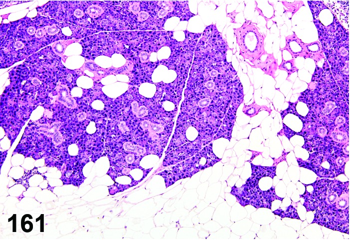
Mouse submandibular gland. Accumulation, adipocytes.
Synonyms: Adipocyte accumulation; lipomatosis
Pathogenesis: unknown.
Diagnostic features
Multifocal to coalescing accumulations of adipocytes within the interstitium.
Compression of the adjacent acini, ducts and lobes.
Moderate replacement (loss) of acini in rare cases.
Disruption of lobular structure.
Differential diagnosis
Vacuolation due to storage of lipids: Moderate to abundant intracytoplasmic lipid vacuoles; acinar and ductular architecture is retained.
Atrophy: Reduction in the number/size of acini is the predominant finding.
Comment: The accumulation of adipocytes within salivary glands is an uncommon lesion and it may be an age related change. It occurs predominantly in parotid and less commonly in submandibular and sublingual salivary glands (Andrew 1949).
Apoptosis
Figure 162.
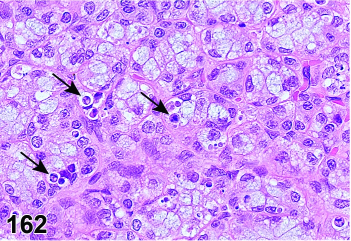
Rat sublingual gland. Single cell necrosis / apoptosis. Note numerous apoptotic bodies, some indicated by arrows.
Synonyms: Apoptotic cell death
Pathogenesis: Gene regulated, energy dependent process leading to formation of apoptotic bodies which are phagocytosed by adjacent cells.
Diagnostic features
Single cells or small clusters of cells.
Cell shrinkage.
Hypereosinophilic cytoplasm.
Nuclear shrinkage, pyknosis, karyorrhexis. Intact cell membrane.
Apoptotic bodies.
Cytoplasm retained in apoptotic bodies.
Phagocytosis of apoptotic bodies tissue macrophages or other adjacent cells.
No inflammation.
Differential diagnoses
Necrosis: The morphologic features of cell death clearly fit with necrotic diagnostic criteria (cells and nuclei swollen, pale cytoplasm etc.).
Apoptosis/necrosis: Both types of cell death are present and there is no requirement to record them separately or a combined term is preferred for statistical reasons. The term is also used when the type of cell death cannot be determined unequivocally.
Comment: The approach for the nomenclature and diagnostic criteria of cell death applied here is based on a draft recommendation of the INHAND Cell Death Nomenclature Working Group.
Apoptosis is not synonymous with necrosis. The main morphological differences between these two types of cell death are cell shrinkage with nuclear fragmentation and tingible body macrophages in apoptosis versus cell swelling, rupture and inflammation in necrosis; however, other morphologies (e.g. nucelar pyknosis and karyorrhexis) overlap. In routine H&E sections where the morphology clearly represents apoptosis or single cell necrosis, or whereby special procedures (e.g. TEM or IHC for caspases) prove one or the other, individual diagnoses may be used. However, because of the overlapping morphologies, necrosis and apoptosis are not always readily distinguishable via routine examination, and the two processes may occur sequentially and/or simultaneously depending on intensity and duration of the noxious agent (Zeiss 2003). This often makes it difficult and impractical to differentiate between both during routine light microscopic examination. Therefore, the complex term apoptosis/necrosis may be used in routine toxicity studies.
A differentiation between apoptosis and single cell necrosis may be required in the context of a given study, in particular if it aims for mechanistic investigations. Transmission electron microscopy is considered to represent the gold standard to confirm apoptosis. Other confirmatory techniques include DNA-laddering (easy to perform but insensitive), TUNEL (false positives from necrotic cells) or immunohistochemistry for caspases, in particular caspase 3. These techniques are reviewed in detail by Elmore (Elmore 2007). Some of these confirmatory techniques detect early phases of apoptosis, in contrast to the evaluation of H&E stained sections, which detects late phases only; overall interpretation of findings should take into account these potential differences. Thus, low grades of apoptosis may remain unrecognized by evaluation of H&E stained slides only.
Certain studies may require the consideration of forms of programmed cell death other than apoptosis which requires specific confirmatory techniques (Galluzzi et al. 2012).
Necrosis
Figure 163.
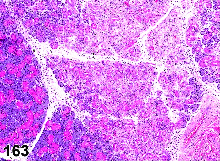
Rat submandibular gland. Necrosis, accompanied by neutrophilic inflammation with edema.
Synonyms: Oncotic cell death; oncotic necrosis; necrosis
Modifiers: Single cell
Pathogenesis: Unregulated, energy independent, passive cell death with leakage of cytoplasm into surrounding tissue and subsequent inflammatory reaction; may be induced by direct contact with a test article after oral uptake/administration.
Diagnostic features
Focal affecting cell groups, lobular, diffuse.
Cells swollen with pale eosinophilic cytoplasm.
Loss of nuclear basophilia, pyknosis and/or karyorrhexis that affects aggregates of cells.
In more severe cases, there may be detachment of the epithelium from the submucosa.
Typically, the presence of degenerative cells is a component of necrosis.
Minimal or slight inflammatory cell infiltrates, edema and fibrin may be present as a feature of necrosis.
Single cell
Only single cells affected.
Differential diagnoses
Apoptosis: The morphologic features of cell death clearly fit with apoptosis (cell and nucleus shrunken, hypereosinophilic cytoplasm, nuclear pyknosis etc.) and/or special techniques demonstrate apoptosis.
Apoptosis/necrosis: Both types of cell death are present and there is no requirement to record them separately or a combined term is preferred for statistical reasons. The term is also used when the type of cell death cannot be determined unequivocally.
Inflammation, acute: Infiltrates of inflammatory cells, edema, and fibrin predominate; necrosis may be present, but is a minor component.
Comments: Necrosis is usually accompanied by inflammatory cell infiltrate and is usually a significant component of acute inflammation (Levin et al. 1999). Necrosis of salivary glands may be due to chemical toxicity or emboli.
Apoptosis/necrosis
Synonym: Cell death
Pathogenesis: Unregulated, energy independent, passive cell death with leakage of cytoplasm into surrounding tissue and subsequent inflammatory reaction (single cell necrosis) and/or gene regulated, energy dependent process leading to formation of apoptotic bodies which are phagocytosed by adjacent cells (apoptosis).
Diagnostic features
Both types of cell death are present and there is no requirement to record them separately or a combined term is preferred for statistical reasons.
The type of cell death cannot be determined unequivocally.
Differential diagnoses
Apoptosis: The morphologic features of cell death clearly fit with apoptosis (cell and nucleus shrunken, hypereosinophilic cytoplasm, nuclear pyknosis etc.) and/or special techniques demonstrate apoptosis and there is a requirement for recording apoptosis and necrosis separately.
Necrosis: The morphologic features of cell death clearly fit with necrotic diagnostic criteria (cells and nuclei swollen, pale cytoplasm etc.) and there is a requirement for recording apoptosis and necrosis separately.
Comments: Necrosis and apoptosis are not always readily distinguishable via routine examination, and the two processes may occur sequentially and/or simultaneously depending on intensity and duration of the noxious agent (Zeiss 2003). This often makes it difficult and impractical to differentiate between both during routine light microscopic examination. In these cases, the combined term apoptosis/necrosis may be used. It is recommended to explain in detail the use of this combined term in the narrative part of the pathology report.
Secretory depletion, acinar cell
(Figures 164 and 165)
Figure 164.
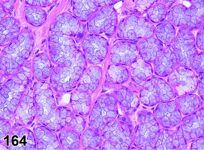
Rat lingual Weber’s gland. Control (for comparison to Figure 165).
Figure 165.
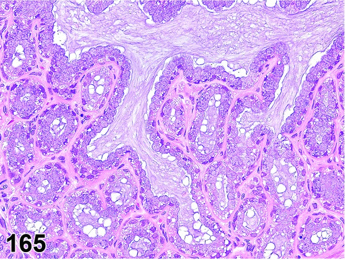
Rat lingual Weber’s gland. Secretory depletion, acinar cell. Same magnification as Figure 164.
Synonym: Degranulation, acinar cell
Pathogenesis: Decrease of acinar cell zymogen granules or mucin leading to shrunken acinar cells.
Diagnostic features
Focal, lobular or diffuse.
Reduced acinar diameter.
Partial or complete loss of acinar cell zymogen granules or mucin leading to a reduced cell size and increased basophilia.
Lack of fibrosis or adipocyte infiltration.
Differential diagnosis
Atrophy, acinar cell: Complete degranulation of acini may be accompanied by fibrosis, mononuclear cell infiltrates, prominent intra- and inter-lobular ducts.
Comment: The morphology of the acini may be altered due to several physiological conditions, such as anorexia/inanition. Degranulation of the acini may also be noted in rodents treated with sympathomimetic agents like isoprenaline (Selye et al. 1961) and terbutaline (Sodicoff et al. 1980).
Secretory depletion, granular duct (submandibular gland) (Figure 166)
Figure 166.
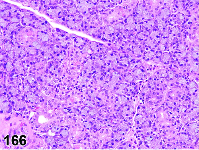
Mouse (male) submandibular gland. Secretory depletion, granular ducts; compare to Control – Figure 156.
Synonym: Feminization of the granular duct; degranulation granular duct
Pathogenesis: Decrease of granulated duct cell serous granules leading to shrunken granulated duct cells.
Diagnostic features
May be focal or multifocal, but usually diffuse.
Affects males, not females.
Partial or complete loss of serous granules leading to a reduced cell size.
Reduced cell diameter of the granulated duct cell.
Lack of fibrosis or adipocyte infiltration.
May be accompanied by degranulation of the acinar cells.
Differential diagnosis
None.
Comment: The morphology of the granular ducts is androgen dependent. Secretory depletion and subsequent atrophy of granular ducts may be observed in male rats and mice with decreased androgen levels. It is also observed in the streptozotocin-induced chronic diabetes rat model. The acinar cells are less susceptible compared to the granular ducts. The atrophy of the granular ducts in chronic diabetic rats may be due to the hypogonadal effect of diabetes. In cases of sympathetic but not parasympathetic nerve stimulation, extensive degranulation may be observed in both acinar (beta-adrenergic) and granular duct (alpha-adrenergic) cells.
Granules increased, granular duct (submandibular gland) (Figure 167)
Figure 167.
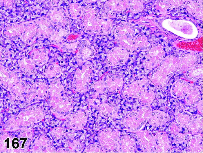
Mouse (female) submandibular gland. Granulation increased, granular duct. Compare to Control – Figure 157.
Synonym: Masculinization of the granular duct
Pathogenesis: Increased androgenic stimulus in females.
Diagnostic features
Diffuse, limited to the submandibular gland of females.
Prominent large granular ducts with increased eosinophilic granules within female animals.
Increased granularity of the granular duct.
Increased diameter of the granular cells.
Except for the change in the granular duct, rest of the histomorphology is normal.
Differential diagnosis
None.
Comment: The submandibular salivary gland is sexually dimorphic and shows increased granularity of the convoluted (granular) ducts in males compared to females. When female rats or mice are treated with androgenic test articles (e.g. androstenedione or oxymetholone) or have increased endogenous androgen levels, their granular ducts of the submandibular glands acquires male morphology.
Atrophy
(Figures 168, 169 and 170)
Figure 168.
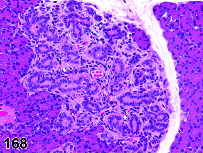
Rat parotid gland. Atrophy, focal.
Figure 169.
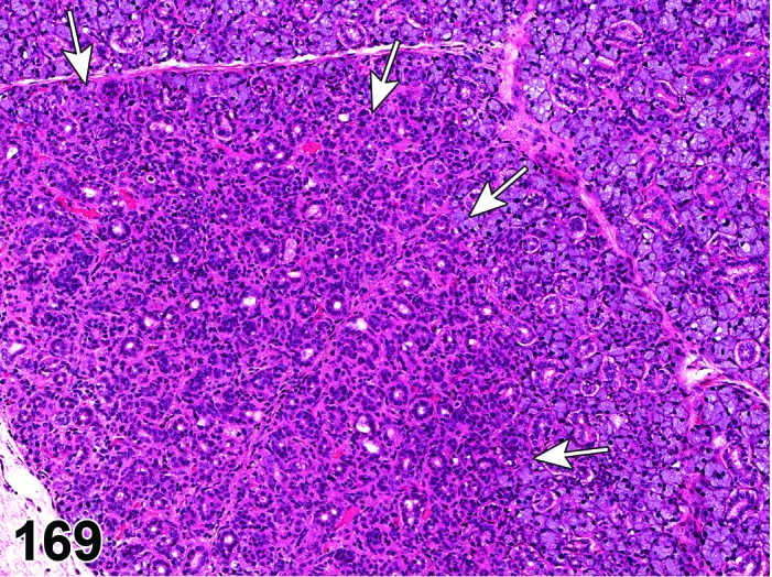
Rat submandibular gland. Atrophy, acinar cell. Area most severely affected indicated by arrows.
Figure 170.
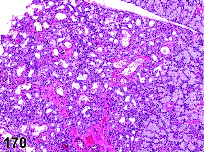
Rat sublingual gland. Atrophy, acinar cell. Normal tissue limited to upper and lower right corner.
Pathogenesis: Spontaneous or experimentally induced.
Diagnostic features
Focal, lobular or diffuse.
Variable reduction in the number and/or size of acini to total loss of acini.
Degranulation (parotid), reduced mucin (submandibular and sublingual).
Relatively more numerous, prominent, occasionally dilated, intra- and inter-lobular ducts lined by cuboidal or flattened epithelial cells.
Pyknotic and karyorrhectic nuclei, apoptotic bodies may be present,.
Variable degree of interstitial fibrosis with/without lymphocytes and plasma cells.
Scattered to coalescing adipocytes may be present within the interstitium.
May be accompanied by mineralization of luminal contents, salivary calculus, ductular foreign bodies.
Differential diagnosis
Secretory depletion, acinar cell: Variable loss of serous or mucinous granules resulting in reduced cellular size, lack of fibrosis or adipocyte infiltration, or lack of chronic inflammatory cell infiltrate.
Accumulation, adipocytes: Accumulation of adipocytes is the predominant finding.
Comment: Atrophy more commonly affects the submandibular and parotid glands than the sublingual gland. It may be an age related lesion or may be induced experimentally, usually as a secondary response after obstruction of excretory ducts or after alteration of trophic factors. Examples for obstruction of excretory ducts include ligation (Bhaskar et al. 1956; Standish and Shafer 1957), sialoliths and foreign bodies. Testosterone, adrenocorticoids, and sympathetic stimulation are important trophic factors for the salivary glands. Any experimental condition that acts on their homeostasis may potentially induce diffuse salivary gland atrophy. This has been shown for hypophysectomy (Koerker 1967), adrenalectomy, denervation and α- and β-adrenergic antagonists. Also decreased food consumption, and protein starvation lead to salivary gland atrophy. Atrophy of salivary glands due to liquid diet affects parotid and submandibular glands but the sublingual glands are apparently spared (Hall and Schneyer 1964; Scott and Gunn 1991). Also, the sublingual glands lack sympathetic innervation and thus are spared in situations where the chemicals act on the sympathetic nervous system. Salivary gland atrophy was noted in F344 rats treated with sodium dichromate dihydrate (NTP, 2008). Often, salivary atrophy is accompanied by mild chronic inflammation and mild interstitial fibrosis.
Mineralization
Synonym: Calcification
Pathogenesis: Deposition of mineral either secondary to cellular necrosis (dystrophic mineralization) or secondary to hypercalcemia (metastatic mineralization).
Diagnostic features
Focal, lobular, diffuse.
Affected structures are ducts and arteries (metastatic mineralization) or areas of degeneration / necrosis (dystrophic mineralization).
Dark basophilic granular material (on H&E) replacing acini, ducts or interstitium.
Differential diagnoses
Artifacts: Hematoxylin stain deposits (distribution unrelated to tissue structures), acid hematin (birefringent in polarized light).
Pigment: Lipofusin and porphyrin stain brown to golden brown on H&E and may further be differentiated from mineralization by special stains.
Calculi (concretions, sialoliths): Localization in the lumen of ductules or ducts.
Comment: Dystrophic calcification occurs in necrotic/injured tissue even in animals with reference range serum calcium levels, whereas metastatic calcification occurs throughout the body, in hypercalcemic animals, especially within interstitial tissues and blood vessel walls. Special stains like Alizarin red S and von Kossa can help identify calcium salts within tissue sections.
Pigment
Figure 171.
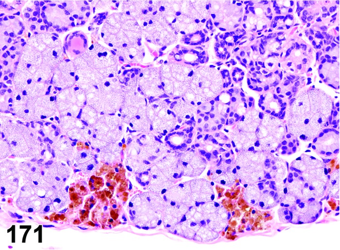
Mouse sublingual gland. Pigment.
Synonyms: Pigmentation; pigment deposition; endogenous pigmentation
Pathogenesis: diverse, dependent on the nature of pigment.
Diagnostic features
Focal, lobular or diffuse.
Yellow to brown pigmentation within the interstitium, acini and ducts.
Differential diagnoses
Mineralization: Dark basophilic appearance in hematoxylin & eosin stained sections; Special stains like Alizarin red S and von Kossa can help identify calcium salts.
Artifacts: Hematoxylin stain deposits (distribution unrelated to tissue structures), acid hematin (birefringent in polarized light).
Comment: The distribution of the pigment will differ dependent on its nature. For example, lipofuscin may be found mainly within epithelial cells of the intralobular ducts (Buchner and David 1978); hemosiderin may be found within the interstitium and macrophages and acid hematin may be present diffusely within the section. The type of pigment can be identified by special stains like Schmorl’s (late lipofuscin, melanin), Fontana-Masson (melanin), and Perl’s (ferric iron-hemosiderin). Formalin-heme pigment (acid-hematin) is observed in tissues fixed in unbuffered formalin. Polarization can help differentiate some pigments like lipofuscin (strong orange autofluorescence on unstained FFPE sections) and acid-hematin (birefringence on H&E sections).
Amyloid
Figure 172.
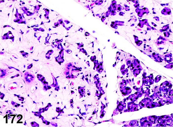
Mouse parotid gland. Amyloid, accompanied by atrophy.
Synonyms: Amyloidosis, amyloid deposition
Pathogenesis: Extracellular deposits of chemically diverse complex insoluble polypeptide fragments.
Diagnostic features
Light eosinophilic amorphous extracellular material within the tunica media of blood vessels, within the interstitium and basement membrane.
Variable degree of atrophy and loss of intervening acini and ducts may occur, if amyloid deposition is prominent.
Green birefringence using polarized light with Congo red stain.
Differential diagnoses
Necrosis: loss of cellular detail, but without extracellular Congo red positive material.
Fibrinoid change (necrosis) in vessel walls: Deep eosinophilic homogenization of vascular media.
Comment: Amyloid and amyloid-like material has been observed in various tissues, including salivary glands of aged rodents, especially mice. Rats are much more resistant to develop amyloidosis than mice.
The characteristic morphologic appearance and location of amyloid in H&E sections that is usually adequate to make this diagnosis. Confirmation of the deposits as amyloid can be accomplished with light microscopy using special stains such as Congo red. Amyloid appears apple green under polarized light with this stain.
The term “hyaline” should be used in case of uncertainties about the nature of extracellular deposits hyaline material in H&E sections and the unavailability of a Congo red stain. Hyaline is also the appropriate term for homogenously eosinophilic extracellular deposits that react negative with Congo red.
c. Inflammatory Lesions
Infiltrate
Figure 173.
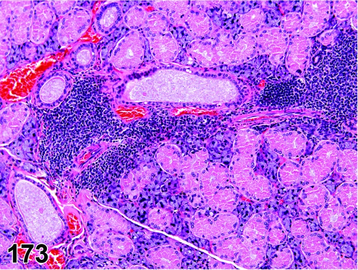
Mouse submandibular gland. Infiltrate, mononuclear cell.
Synonyms: Infiltrate inflammatory; infiltrate inflammatory cell; infiltration (plus modifier); infiltration, inflammatory; infiltration, inflammatory cell
Modifiers: Type of inflammatory cell that represents the predominant cell type in the infiltrate
Pathogenesis: Infiltration with neutrophils (Infiltrate, neutrophil), eosinophils (Infiltrate, eosinophil), mononuclear cells (Infiltrate, mononuclear cell) or a combination of more than one type (Infiltrate, mixed) present without other morphological features of inflammation, e.g. hemorrhage, edema, fibroplasia.
Diagnostic features
Focal, multifocal or diffuse.
Presence of mononuclear or polymorphonuclear leukocytes but without other histological features of inflammation like edema, congestion or necrosis.
Mild atrophy of surrounding cells may be present.
Usually no acinar cell degranulation or mucus depletion.
Differential diagnoses
Inflammation: An inflammatory cell infiltrate associated with additional morphologic features of inflammation such as edema, hemorrhage, necrosis, and/or fibroplasia.
Granulocytic leukemia: Infiltration of neutrophil precursors and/or abnormal neutrophils will probably be present along with mature neutrophils; infiltration of similar neoplastic cells is likely present in other organs.
Lymphoma: Infiltration of monomorphic population of lymphocytes (often with atypical, increased and/or abnormal mitoses); infiltration of similar neoplastic cells is likely present in other organs.
Comment: In toxicity studies, inflammatory cell infiltrates are encountered more commonly than inflammation (sialadenitis). The term inflammation (or sialadenitis) should not be confused with or used in lieu of inflammatory cell infiltrate. The term inflammatory cell infiltrate should be used rather than sialadenitis (or inflammation) when there is no tissue injury/reaction associated with the inflammatory cell infiltrate. Also, these inflammatory cell infiltrates should be qualified with the type of inflammatory cells that are predominant, such as mononuclear, lymphocyte, neutrophil, etc. The use of terminology summarizing the nature of the infiltrates as acute or suppurative (in lieu of neutrophilic), granulomatous (in lieu of histiocytic), and chronic (in lieu of lymphocytic, plasmacytic, mononuclear, etc.) is not recommended since these terms may imply inflammation. The term inflammation (or sialadenitis) should be reserved for lesions where the inflammatory cell infiltrate is associated with obvious tissue injury/reaction associated with inflammation such as edema, congestion, acinar (parotid) cell degranulation, hemorrhage or necrosis.
Inflammation
(Figures 174, 175, 176 and 177)
Figure 174.
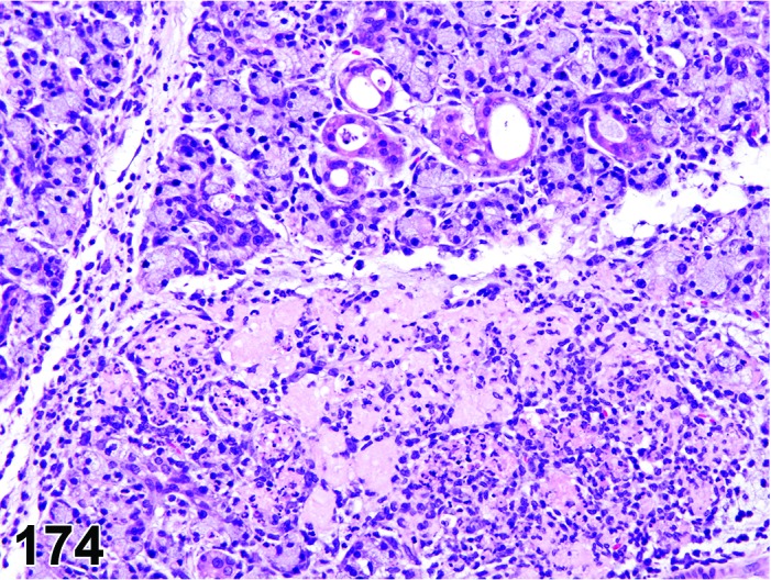
Rat submandibular gland. Inflammation, neutrophil. The necrosis is part of the inflammation and covered by this term.
Figure 175.
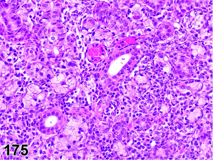
Mouse submandibular gland. Inflammation, mononuclear cell. The vacuolation of acinar cells and other degenerative processes are covered by this term; viral etiology.
Figure 176.
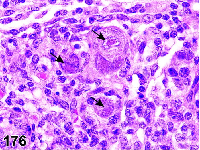
Mouse parotid gland. Inflammation, mononuclear cell. Intranuclear inclusion bodies, some of them indicated by arrows (cytomegaly virus).
Figure 177.
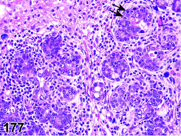
Rat parotid gland. Inflammation, mixed; acompanied by regenerative hyperplasia with atypia. Intranuclear inclusion bodies, some of them indicated by arrows (rat papilloma virus).
Synonym: Sialadenitis
Modifiers: Type of inflammatory cell that represents the predominant cell type in the inflammation
Pathogenesis: Infiltration of the lamina propria and/or submucosa with neutrophils (Inflammation, neutrophil) or mononuclear cell (Inflammation, mononuclear cell) or a combination (Inflammation, mixed) with additional histological features of inflammation e.g. hemorrhage, edema, fibroplasia.
Diagnostic features
Focal, multifocal, or diffuse.
Infiltrate of mononuclear or polymorphonuclear leukocytes into the gland parenchyma.
Presence of other histological criteria of inflammation, e.g. hemorrhage, edema, fibroplasia
Inflammation, neutrophil
Infiltrate predominantly neutrophilic; frequently accompanied by edema and congestion, parotid acinar cell degranulation, acinar and ductular degeneration and necrosis.
Inflammation, mononuclear cell
Infiltrate predominantly mononuclear cell; frequently accompanied by acinar cell atrophy and mild acinar cell degeneration, ductal proliferation, interstitial fibrosis; mineralization or nonkeratinizing squamous metaplasia of acini and ducts may be present.
Differential diagnoses
Infiltrate, inflammatory cell: Other morphologic features associated with inflammation such as edema, hemorrhage, necrosis, and/or fibroplasias will be absent.
Granulocytic leukemia: Infiltration of neutrophil precursors and/or abnormal neutrophils will probably be present along with mature neutrophils; infiltration of similar neoplastic cells is likely present in other organs.
Lymphoma: Infiltration of monomorphic population of lymphocytes (often with atypical, increased and/or abnormal mitoses); infiltration of similar neoplastic cells is likely present in other organs.
Comment: When a mononuclear or mixed cell infiltrate is considered to represent an inflammatory process (inflammation, mononuclear cell or inflammation, mixed) and other morphologic features (fibrosis, mineralization) associated with the infiltrate suggest an ongoing/continuing inflammatory process, the modifier ‘chronic’ may be included in the diagnosis (i.e. inflammation, mononuclear cell, chronic) to better characterize the finding.
Inflammation, neutrophilic of the salivary glands is uncommon in toxicological evaluation of tissues. Infection with murine cytomegalovirus (MCMV) and sialodacryoadenitis virus (SDAV) were once common causes of acute sialodacryoadenitis in mice and rats (Jacoby et al. 1975), respectively, but are now rare in barrier bred and maintained colonies. MCMV predominantly affects the submandibular glands and in rare cases the parotid glands. SDAV has a tropism for tubuloalveolar glandular tissue of serous (parotid) or mucous/serous (submandibular) glands. The sublingual gland is spared in SDAV infections in rats. Sialadenitis may be differentiated by their respective pathognomonic clinical signs and lesions. Bacterial infections due to Klebsiella aerogenes and Staphylococcus aureus may also cause severe acute necrotizing sialadenitis as well as abscesses.
Spontaneous autoimmune sialadenitis is seen in several strains of mice (NOD, NZB/NZW, SL/Ni, BDF females) and rats (Percy and Barthold 2007). The infiltrating lymphocytic population is predominantly CD4+ T cells and less than 10% cells are C8+ T cells. Variable degree of chronic inflammation of the salivary glands may be seen occasionally in aged animals of other strains. Chronic infections of MCMV and SDAV or regenerative lesions from acute MCMV and SDAV infections may appear morphologically similar to chronic inflammatory lesions (Ohyama et al. 2006). Rats treated with sympathomimetics like isoproterenol present with mild chronic inflammation in the submandibular salivary gland (Cohen et al. 1992).
d. Vascular Lesions
Refer also to the INHAND monograph on the cardiovascular system.
Edema
Figure 178.
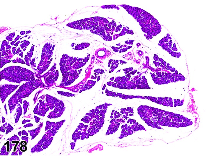
Mouse parotid gland. Edema.
Pathogenesis: Accumulation of fluid in salivary interstitium resulting from increased vascular permeability due to damage to capillaries and blood vessels, increased hydrostatic pressure in vasculature, decreased oncotic pressure, or impaired lymphatic drainage.
Diagnostic features
Diffuse.
Increased clear spaces between acini and interstitium.
Increase in total lobe volume.
Rare inflammatory cell infiltrate secondary to diapedesis may be present.
Differential diagnosis
Inflammation, neutrophil: Tissue and capillary damage accompanied by exudative edema, granulocytic infiltration, fibrin, congestion and hemorrhage.
Comment: Edema within the salivary glands is very uncommon. It may be exudative (inflammatory edema due to capillary injury secondary to leakage of enzymes) or transudative (hydrodynamic derangement secondary to increased hydrostatic osmotic pressure and decreased oncotic pressure). The diagnosis of edema should be reserved for a transudative type of edema since exudative edema is usually accompanied by abundant inflammatory cell infiltrate and possibly tissue injury. Hence, the primary diagnosis in cases of exudative edema should be inflammation, neutrophil. Chemical induced salivary gland edema has been noted in mice treated with P-Nitrophenol and rats administered D & C Yellow No. 11 in US-NTP studies.
Hemorrhage
Pathogenesis: Increased vascular permeability (diapedesis) or rupture of blood vessels.
Diagnostic features
Presence of erythrocytes in parenchymal or interstitial tissue or lumina of ductules and ducts.
Differential diagnoses
Angiectasis: Blood present within dilated vascular lumina.
Artifact: Erythrocytes on the surface of tissues as a result of necropsy procedures.
e. Miscellaneous Lesions
Calculus, ductular
Figure 179.
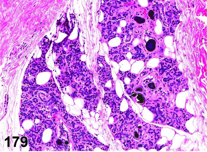
Rat parotid gland. Calculi, ductular, accompanied by atrophy, accumulation adipocytes and fibrosis.
Synonyms: Sialoliths; sialolithiasis; salivary concretion; duct concretion; salivary calculus; inspissated material
Pathogenesis: Precipitation of secretory components after reabsorption of the water component by the ductular cells.
Diagnostic features
Basophilic or eosinophilic inspissated material within the ductular lumen.
Ductular epithelium may be low to flattened cuboidal epithelium.
May be accompanied by mild periductular mononuclear inflammatory cell infiltrate.
Differential diagnoses
Ectasia, duct: Salivary calculi are absent.
Foreign body: Foreign body within the duct is usually associated with some tissue damage and elicits an inflammatory response.
Comment: A diagnosis of ductular ectasia should not be used in the presence of the ductular calculi, even though they share other morphologic features.
Ectasia, duct
Figure 180.
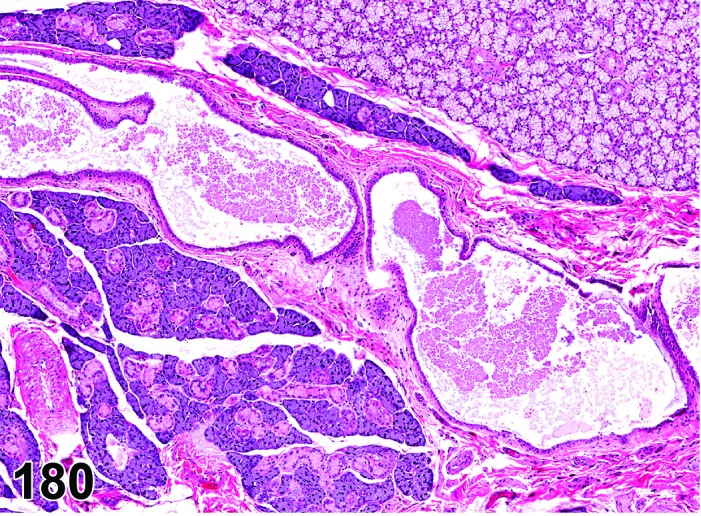
Rat submandibular gland. Ectasia, duct.
Synonyms: Dilatation; luminal distension
Pathogenesis/cell of origin: Luminal dilatation due to obliteration of the intralobular or interlobular ducts.
Diagnostic features
Solitary or multiple tortuous irregularly enlarged lumina that may or may not contain gland-specific secretory material.
Lined by flat cuboidal epithelial cells.
May be accompanied by mild interstitial infiltration of lymphocytes and plasma cells.
Differential diagnosis
Calculus, ductular: Salivary calculi are present.
Comment: Usually ectasia results from blockage of inter- and intra-lobular ducts either proximally or distally due to rare inspissated secretory material (sialolithiasis), sialodacryoadenitis tumors, or foreign bodies. However, a diagnosis of ectasia is used only in the absence of the calculi or the foreign body within the histologic section.
Fibrosis
Figure 181.
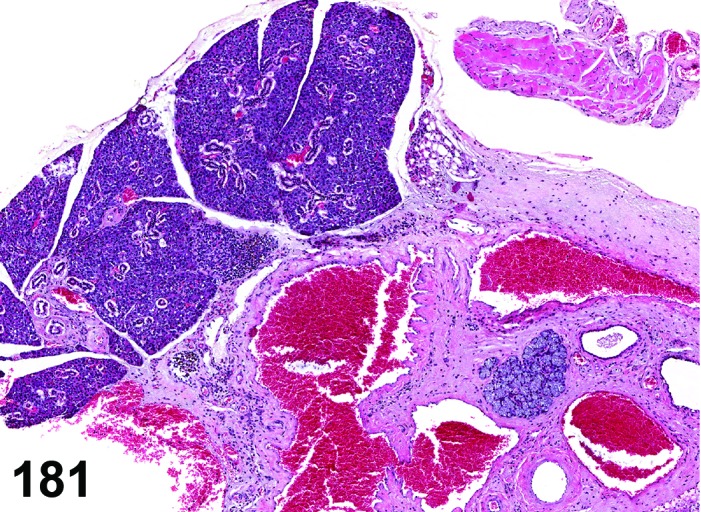
Mouse sublingual gland. Fibrosis, accompanied by atrophy and angiectasis.
Synonym: Interstitial fibrosis
Pathogenesis: Collagen deposition by fibroblasts following inflammation, necrosis or hemorrhage.
Diagnostic features
Focal, lobular, or diffuse.
Increase in the interstitial tissue due to collagen deposition and fibroblast cell proliferation.
Differential diagnoses
Amyloid: There is abundant extracellular eosinophilic amorphous ground substance that exhibits apple green birefringence in Congo red stained sections under polarized light and there is an absence of fibroblast cell proliferation.
Inflammation, mixed cell, chronic: Fibrosis associated with inflammatory cell infiltrates and degenerative parenchymal changes.
Comment: Fibrosis may be minimal to marked depending on the severity and duration of insult. It is usually a significant component of chronic inflammation and may be a component of healing after acute toxic insults. Mature collagen in H&E sections is birefringent under polarized light.
Hypertrophy, acinar cell
Synonym: Cytomegaly
Pathogenesis: Usually secondary to increased functional demand or due to sympathomimetic stimulation.
Diagnostic features
Focal, multifocal or diffuse.
Acinar structure not altered, no encapsulation, no compression.
Enlarged cells with abundant cytoplasm of normal staining pattern.
Large nuclei with euchromatin and prominent nucleoli.
Relative reduction in the number of secretory ducts due to separation by enlarged acini.
Differential diagnoses
Basophilic hypertrophic focus (parotid gland): Discrete non-compressing foci of hypertrophic cells with increased basophilia within discrete non-compressing foci.
Hyperplasia, non-regenerative: May be associated with hypertrophy, but always increase in cell number; minimal compression or distortion of architecture may be present.
Hyperplasia, regenerative: May be associated with hypertrophy, but always increase in cell number and cytoplasmic basophilia; minimal compression or distortion of architecture may be present.
Comment: Acinar hyperplasia is often associated with hypertrophy. In these cases, it is recommended to record hyperplasia, but not hypertrophy.
Hypertrophy (and hyperplasia) of the parotid and submandibular glands has been observed after administration of certain drugs such as isoproterenol (Selye et al. 1961, Brenner and Stanton, 1970), methoxamine and pilocarpine (Inanaga et al. 1988), terbutaline (Sodicoff et al. 1980), methacholine, epinephrine, phenylephrine, reserpine, furosemide (Scarlett et al. 1988), alloxan (Sagstrom et al. 1987), thyroxine, and dexamethasone (Sagulin and Roomans 1989). Single or repeated mandibular incisor tooth amputation also resulted in enlargement of submandibular glands, presumably due to neural regulation (Wells 1963). This enlargement was due to abundant intracytoplasmic accumulation of mucus along with calcium, amylase and other protein.
Focus, hypertrophic, basophilic (parotid gland) (Figures 182 and 183)
Figure 182.
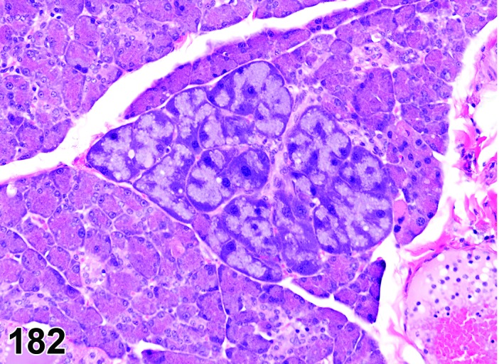
Rat parotid gland. Focus, hypertrophic, basophilic.
Figure 183.
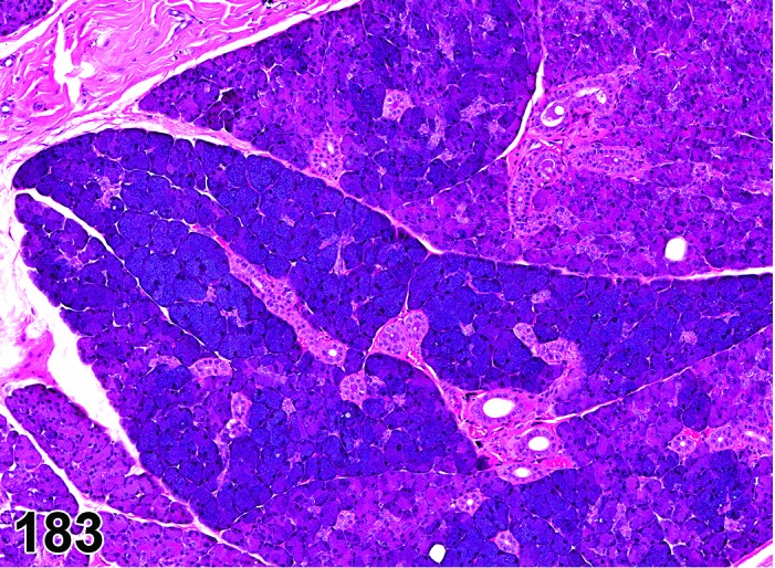
Rat parotid gland. Focus, hypertrophic, basophilic, exaggerated lesion.
Synonyms: Focus, basophilic; basophilic focus; basophilic hypertrophic focus; focus, basophilic, hypertrophic; focus of cellular alteration;
Pathogenesis/cell of origin: Unknown.
Diagnostic features
Focal, multifocal or diffuse.
Discrete unencapsulated, noncompressing foci involving one or multiple acini.
Enlarged cells with increased cytoplasm and occasionally enlarged nuclei.
Apical regions of the acinar cells have an eosinophilic granular or fine vesicular cytoplasm on H&E stain.
Basal regions of the acinar cells have intensely basophilic cytoplasm with large hyperchromatic nuclei.
The enlarged cells can be diffusely basophilic in extreme cases where most of the parotid is affected.
Pyknotic nuclei or mitoses may be observed.
In diffusely affected cases, the acinar enlargement is not as prominent as seen in smaller focal basophilic foci.
Relative reduction in the number of secretory ducts due to separation by enlarged acini.
Differential diagnoses
Hyperplasia, non-regenerative OR Hyperplasia, regenerative: Increase in cell number that may or may not be associated with hypertrophy; minimal compression or distortion of architecture may be present.
Hypertrophy, acinar cell: Cells in single or multiple acini (foci) enlarged without increased cytoplasmic basophilia.
Comment: In untreated animals, basophilic hypertrophic foci are more common in rats than in mice (Chiu and Chen 1986). The incidence of basophilic hypertrophic foci slightly increases with age. In addition, several chemicals such as doxylamine (Jackson and Blackwell 1993), triprolidine (Greenman et al. 1995), glyphosate (NTP, 1992a), methyleugenol (NTP, 2000), and diethanolamine (NTP, 1992b) induce basophilic hypertrophic foci in rodents. These foci are considered adaptive hypertrophic lesions rather than precursors of neoplasia.
Metaplasia, acinar cell
Figure 184.
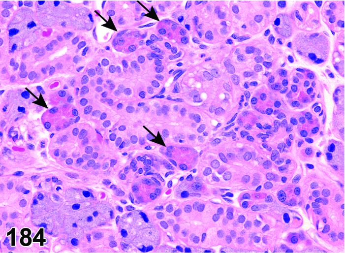
Rat submandibular gland. Metaplasia, acinar cell (arrows) aquiring the morphology of parotid acinar cells.
Pathogenesis: Transdifferentiation of one fully differentiated salivary gland epithelium into a different type of fully differentiated salivary gland epithelium.
Diagnostic features
Focal to multifocal scattered distribution.
Usually accompanied by prior tissue injury.
Presence of fully differentiated epithelial cells that are not native to the tissue, e.g. presence of parotid gland epithelial cells within the submandibular gland.
Differential diagnosis
Ectopic tissue: Usually focal, circumscribed and not associated with prior tissue injury.
Comment: Documenting prior injury and the corresponding chronic inflammation as well as the multifocal nature of the lesion supports the diagnosis of metaplasia instead of ectopic tissue.
Metaplasia, squamous cell
(Figures 185 and 186)
Figure 185.
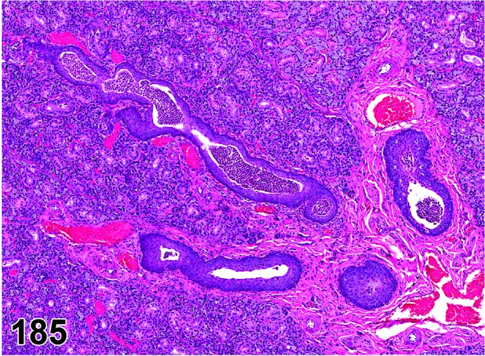
Rat submandibular gland. Metaplasia squamous cell.
Figure 186.
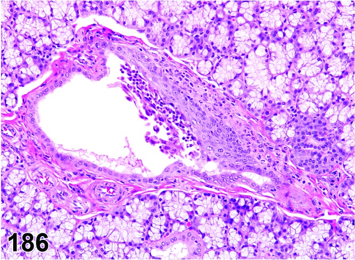
Rat sublingual gland. Metaplasia, squamous cell.
Synonyms: Squamous metaplasia; ductal squamous metaplasia
Histogenesis: Ductular epithelial structures of the salivary glands.
Diagnostic features
Cuboidal ductular epithelium replaced by squamous epithelium.
Squamous epithelial cells may be uni- or multilayered.
Keratinization may be present.
May be accompanied by duct hyperplasia.
Differential diagnoses
Carcinoma, squamous cell: Local invasion of adjacent tissue, distant metastasis, or marked cellular atypia.
Hyperplasia: Hyperplastic cells cuboidal to columnar in shape and unilayered.
Hyperplasia, atypical: Multiple layers of cuboidal to columnar epithelial cells with lost polarity and increased basophilia; no horizontal flattening as in squamous metaplasia.
Comment: Ductal squamous cell metaplasia is usually seen as a part of the regenerative response following necrosis of the ductal epithelium. In rats, a common cause of this lesion in parotid and submandibular glands is due to sialodacryoadenitis viral (SDAV) infection. The sublingual glands are reportedly spared from pathologic changes in SDAV infections (Jacoby 1985). However, the sublingual excretory ducts may have ductal squamous metaplasia in the absence of SDAV infections. Vitamin A deficient diet induced squamous metaplasia in the ducts of parotid, submandibular and sublingual glands in Sprague-Dawley rats (Horn et al. 1996). Squamous cell metaplasia in the submandibular gland was described in mice after treatment with DMBA and was proven to be of ductal segment origin by the presence of keratin proteins (Takai et al. 1986).
Light and electron microscopy studies have demonstrated that the principal portion of salivary gland tissue undergoing squamous metaplasia is the acinar-intercalated duct cell complex (Dardick et al. 1985). Though, squamous cell metaplasia of the ductular epithelium has been considered as a preneoplastic condition progressing to neoplasia (Neuenschwander and Elwell 1990), the potential for progression of hyperplasia and squamous metaplasia of the ductular epithelium is unclear. These changes in the sublingual gland of Wistar rats, often associated with inflammation/fibrosis, were not associated with neoplasia at that site (Van Esch et al. 1986). In addition, treatment-related squamous cell metaplasia of the salivary gland in a chronic F344 rat study with iodinated glycerol was not associated with neoplastic findings in the salivary gland after two years (NTP, 1990). Squamous cell metaplasia was induced with chlorodibromomethane in F344 rats but not in B6C3F1 mice after 13-week treatment, but no tumor progression was noted after 2 years treatment (Dunnick et al. 1985). On the other hand, transition of squamous metaplasia into squamous cell carcinoma in the submandibular gland via a non-genotoxic, proliferation-dependent mechanism was postulated after 2-year treatment with potassium iodide in F344 rats (Takegawa et al. 1998). In addition, in experiments with implanted sponge pellets containing DMBA inducing squamous cell carcinomas in Sprague-Dawley rats, the authors concluded that ductal segments undergoing squamous cell metaplasia may have participated in the genesis of neoplasia during experimental carcinogenesis (Cao et al. 2000, Sumitomo et al. 1996).
f. Non-neoplastic Proliferative Lesions
Hyperplasia
Figure 187.
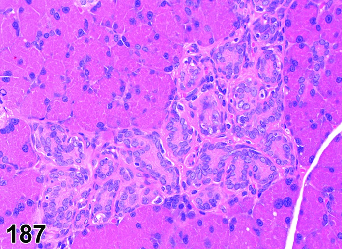
Rat parotid gland. Hyperplasia ductal.
Modifiers: Acinar, ductal, reactive, atypical
Histogenesis: Ductal and/or acinar epithelium of the salivary glands.
Diagnostic features
Focal, multifocal or diffuse.
Glandular architecture is preserved or only minimally altered.
Minimal compression of the surrounding tissue may be present.
Often rounded circumference, but maintenance of normal acinar or ductal architecture.
No capsule.
Acinar:
Acinar cells with atypia may occur.
Ductal:
Ducts may show dilated lumina lined by flattened epithelium.
Reactive:
Acinar or ductular proliferation with cytoplasmic basophilia; evidence of an underlying process, e.g. inflammation, fibroplasias, ductal obstruction or foreign material.
Atypical:
Focal or multifocal, not diffuse.
Multiple layers of acinar / ductular epithelial cells.
Gradual loss of cellular polarity and differentiation, cytoplasm may be amphophilic, increased nucleus to cytoplasm ratio.
Differential diagnoses
Adenoma: Loss or distortion of the normal acinar/ductular structure; well-demarcated and distinct compression of the surrounding tissue.
Ectopic parotid gland: Foci of serous acini between mucous acini of the sublingual gland.
Hypertrophy: Enlarged acinar cells without increase in cell number; no compression, no alteration of glandular architecture.
Focus, hypertrophic, basophilic: Enlarged acinar structures with increased basophilia; no compression.
Comment: Hyperplastic or preneoplastic changes associated with spontaneously occurring neoplasms have not been identified in F344 rats (Neuenschwander and Elwell. 1990). Spontaneous hyperplastic and metaplastic duct epithelium was described in the sublingual glands of Wistar rats (Van Esch et al. 1986). Hyperplasia of the salivary ducts is a common feature of many inflammatory and reactive conditions in the salivary glands of rodents (Greaves 2012). Multifocal ductal cell hyperplasia was induced in the intercalated duct cells of the submandibular salivary glands of Wistar rats after chronic treatment with a steroidal test article comprising prostagenic and estrogenic properties (De Rijk et al. 2003).
Spontaneous hyperplastic changes of the salivary gland have not been observed in B6C3F1 mice (Botts et al. 1999). Transgenic mice models were reported to show hyperplasia of the submandibular glands in serous acinar cells expressing a retinoid acid receptor (Bérard et al. 1994) and ductular cells expressing simian virus 40 T antigen (Ewald et al. 1996), respectively.
g. Neoplasms
Spontaneous and primary tumors of the rodent major salivary glands are rare (Elwell and Leininger 1990; Frith and Heath 1985; Greaves 2012). Salivary gland adenomas in rats and mice may arise from either acinar or ductular components (Neuenschwander and Elwell 1990; Botts et al. 1999). Few carcinomas and adenocarcinomas have been reported in mice and rats (Botts et al. 1999; Hosokawa et al. 2000; Nishikawa et al. 2010; Tsunenari et al. 1997). Malignant tumors in the salivary glands may be induced with chemical substances in rats (Neuenschwander and Elwell 1990;Sumitomo et al.1996; Cao et al. 1999; Zaman et al. 1996) and mice (Botts et al. 1999; Takegawa et al. 1998; Takai et al. 1986; Yura et al. 1995) or in various transgenic animal models (Dardick et al. 2000; Declercq et al. 2005; Nielsen et al. 1991; Nielsen et al. 1995).
Adenoma
(Figures 188, 189, 190, 191 and 192)
Figure 188.
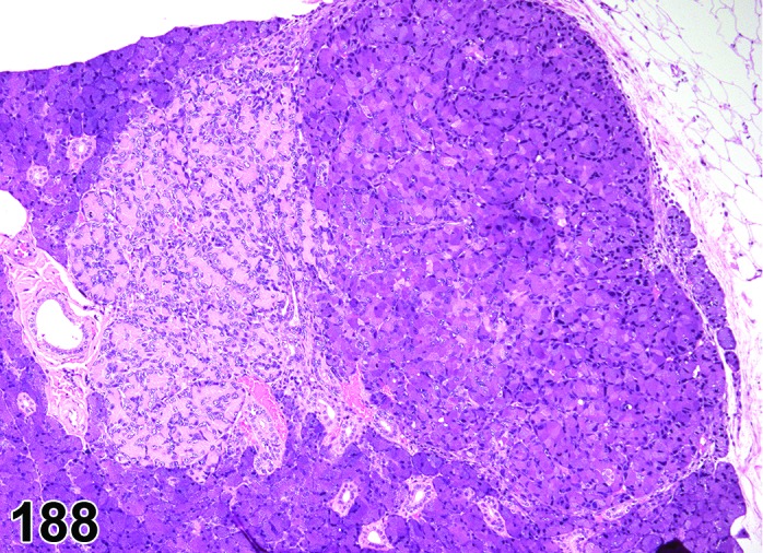
Rat paroid gland. Adenoma, tubular (left) and acinar (right).
Figure 189.
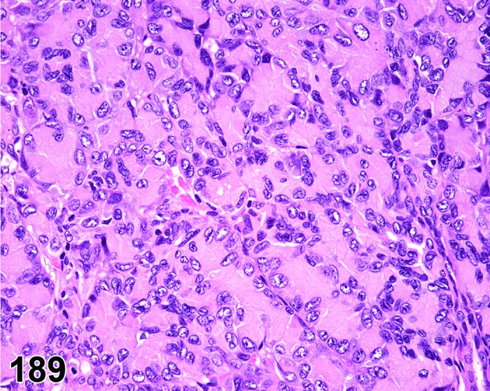
Rat parotid gland. Adenoma, tubular. Higher magnification of Figure 188.
Figure 190.
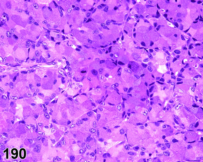
Rat parotid gland. Adenoma, acinar. Higher magnification of Figure 188.
Figure 191.
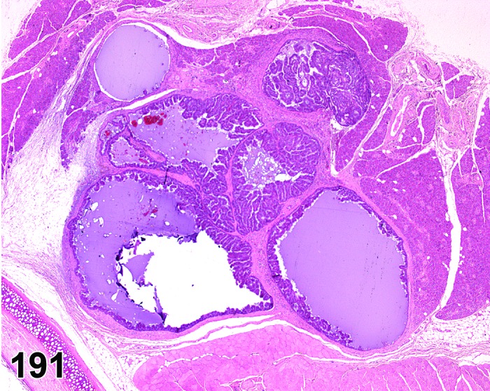
Rat parotid gland. Adenoma, papillary.
Figure 192.
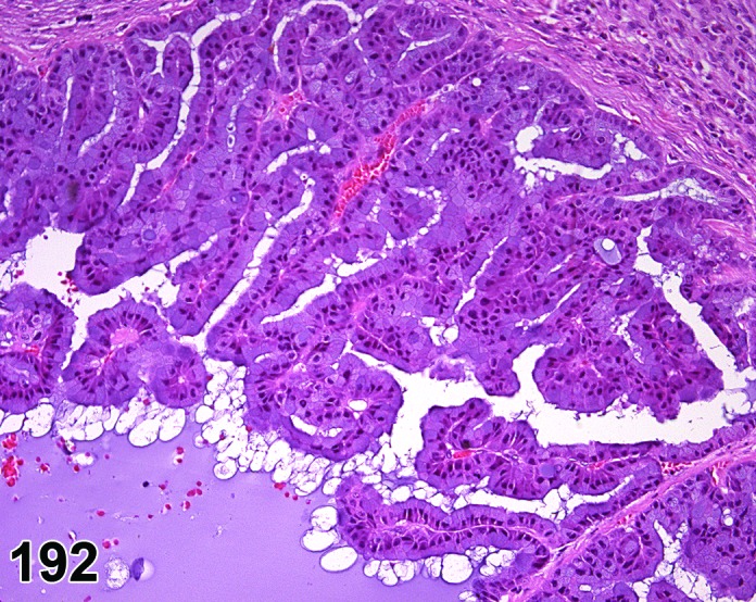
Rat parotid gland. Adenoma, papillary. Higher magnification of Figure 191.
Modifiers: Acinar, tubular, papillary, solid, mixed acinar/tubular
Histogenesis: Ductal or acinar epithelium of salivary glands.
Diagnostic features
Well demarcated.
Compression of the adjacent parenchyma or connective tissue.
Alteration of normal acinar or ductular architecture.
May be surrounded partially or completely by a thin capsule.
Nuclei are hyperchromatic and basally oriented when cytoplasmic mucus present.
Mitotic figures are rare.
Growth pattern may be acinar, tubular, papillary, solid, or mixed.
Acinar:
Show some signs of secretory activity. Minor areas with solid growth pattern may be present; excretory ducts are usually absent.
Tubular:
Consists of lobules or aggregates of tubules lined by squamous to cuboidal or columnar epithelium, separated by a variable amount of fibrous stroma. Some ducts may have dilated lumina.
Papillary:
Papillary projections dominate and cystic dilatation may be present.
Solid:
Diffuse sheet of epithelial cells without acinar or ductular structure.
Mixed acinar/tubular:
A mixture of acinar and tubular forms is not infrequent.
Differential diagnoses
Hyperplasia: Acinar structure preserved or only minimally altered; no or only minimal compression, no capsule; in case of reactive hyperplasia evidence of an underlying process, e.g. inflammation, fibroplasia, ductal obstruction or foreign material.
Hyperplasia, atypical: Focal proliferation of acinar or tubular epithelium with no or only minimal compression, with multiple epithelial layers and cellular atypia; no capsule.
Adenocarcinoma: Cellular pleomorphism and cytoplasmic amphophilia; numerous mitotic figures, invasive growth.
Tumor, mixed, benign: Consists of two proliferative cell types, one myoepithelium and the other glandular acinar epithelium.
Adenoma of the mammary gland (anterior part): Glandular structure is not as compact; staining characteristics of epithelium different.
Comment: In mice, only acinar adenomas of the salivary glands have been described, while rats may develop the different growth patterns listed as Modifiers.
Multinodular acinar adenomas may be difficult to distinguish from well-differentiated adenocarcinomas. Salivary gland adenomas in rats may arise from either acinar or ductular components (Neuenschwander and Elwell 1990). Few adenomas have been observed in mice and it is unclear if they arise from ducts or acini (Botts et al. 1999). Therefore it is recommended to use the modifiers acinar and tubular not in the sense of histogenesis but as a description of the predominant growth pattern.
Mice transgenic for the pleomorphic adenoma gene (PLAG1 or PLAG2) developed a high incidence of submandibular gland adenomas. These tumors developed pleomorphic characteristics, similar to human pleomorphic adenomas of the salivary glands. They were composed of epithelial and myoepithelial structures. The epithelial component showed various growth patterns, including keratinization. The spindle-shaped myoepithelial cells were embedded in a myxoid stroma (Declercq et al. 2005).
Adenocarcinoma
(Figures 193, 194, 195 and 196)
Figure 193.
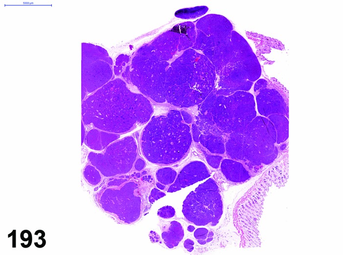
Rat parotid gland. Adenocarcinoma, acinar.
Figure 194.
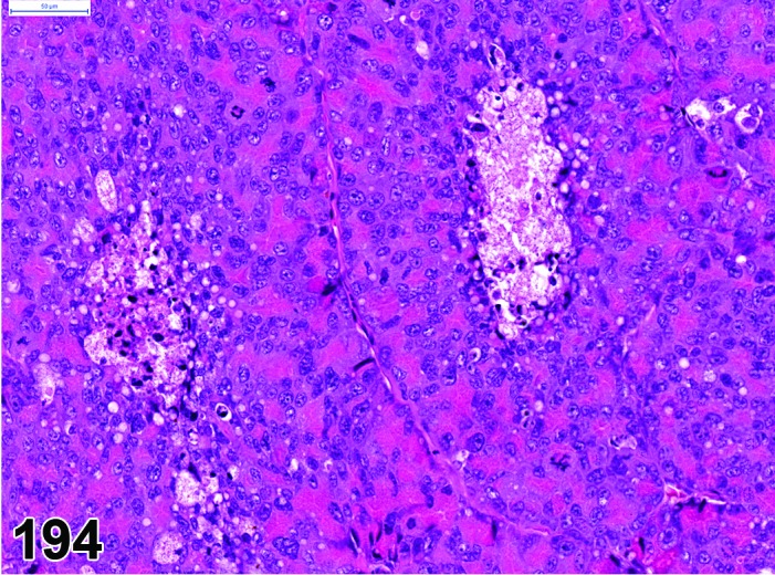
Rat parotid gland. Adenocarcinoma, acinar. Higher magnification of Figure 193.
Figure 195.
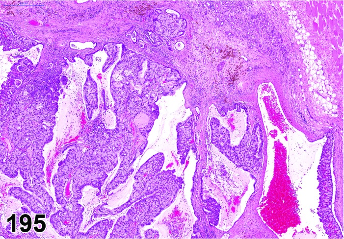
Rat sublingual gland. Adenocarcinoma, mixed.
Figure 196.
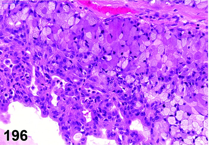
Rat sublingual gland. Adenocarcinoma, mixed. Higher magnification of Figure 195.
Modifiers: Acinar, tubular, papillary, solid, mixed acinar/tubular/solid
Histogenesis: Ductal or acinar epithelium of salivary glands.
Diagnostic features
Grows in whorls or spindle pattern.
Necrosis frequently present.
Areas of squamous differentiation may be present.
Cells are large, pleomorphic and polyhedral with amphophilic cytoplasm.
Nucleus to cytoplasm ratio high.
Nuclei are large vesicular and have multiple nucleoli.
Mitotic figures numerous.
Invasion of the surrounding tissue present.
Metastasis to the lung may be present.
Growth pattern may be acinar, tubular, papillary, solid, or mixed.
Acinar:
Show some signs of secretory activity.
Minor areas with solid growth pattern may be present, excretory ducts are usually absent.
Tubular:
Differentiation pattern variable, ranging from well-formed tubular structures to poorly formed tubules or nodular masses of anaplastic epithelial cells.
Some ducts may have dilated lumina.
Solid:
Diffuse sheet of epithelial cells without acinar or ductular structure.
Mixed acinar/tubular/solid:
May have different patterns in one tumor: acinar, tubular and solid.
Papillary:
Papillary projections dominate and cystic dilatation may be present.
Differential diagnoses
Hyperplasia: No evidence of invasion, glandular structure preserved or only slightly altered (acinar hyperplasia; in case of reactive hyperplasia evidence of an underlying process like inflammation, fibroplasia, ductal obstruction or foreign material.
Hyperplasia, atypical: Focal proliferation of acinar or tubular epithelium with multiple layers and cellular atypia; no invasion of surrounding tissue.
Adenoma: Minimal cellular pleomorphism; acinar cells well differentiated with secretory granules; no or only minimal pleomorphism; non-invasive; Mitotic figures rare.
Adenocarcinoma of the mammary gland: Cytoplasmic staining differences and lipid vacuolation may be present.
Comment: Multinodular acinar adenomas may be difficult to distinguish from well-differentiated adenocarcinomas. Spontaneous tumors of the major salivary glands are rare in the rat (Elwell and Leininger 1990; Glucksmann and Cherry 1973). Poorly differentiated carcinomas of the parotid gland of young Sprague-Dawley rats (Tsunenari, et al. 1997; Nishikawa et al. 2010) and papillary cystadenocarcinoma in the parotid gland of a F344 rat (Hosokawa et al. 2000) were reported. Induction of adenocarcinomas in the submandibular gland was reported after intraglandular injection of DMBA in female Wistar rats (Zaman et al. 1996) and female mice (Yura et al. 1995).
Adenocarcinomas have rarely been reported in mice (Botts et al. 1999). A naturally occurring mucoepidermoid carcinoma in the salivary gland associated with both myoepithelium and duct epithelium was described in two mice (Ishikawa et al. 1998).
Submandibular gland adenocarcinomas of intercalated duct origin were induced in Smgb-TAG mice (Dardick et al. 2000). In these mice the oncogene SV40 T antigen is expressed from the neonatal submandibular gland secretory protein b (Smgb) gene promoter. Development of pleomorphic adenoma with malignant characteristics and lung metastasis was described in the submandibular gland of transgenic mice with PLAG1 proto-oncogene over expression (Declercq et al. 2005). Transgenic mice expressing a human Ha-ras oncogene developed adenosquamous carcinomas arising from the serous areas from the submandibular gland (Nielsen et al. 1991). In wap-ras transgenic mice adenocarcinomas arising from the submandibular gland were reported to develop spontaneously by one year of age (Nielsen et al.1995).
Carcinoma, squamous cell
(Figures 197, 198 and 199)
Figure 197.
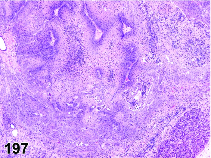
Mouse parotid gland. Carcinoma, squamous cell.
Figure 198.
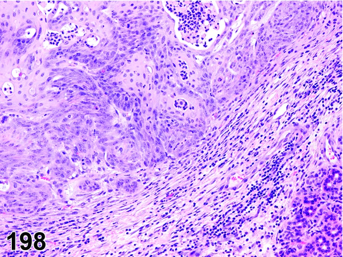
Mouse parotid gland. Carcinoma, squamous cell. Higher magnification of Figure 197.
Figure 199.
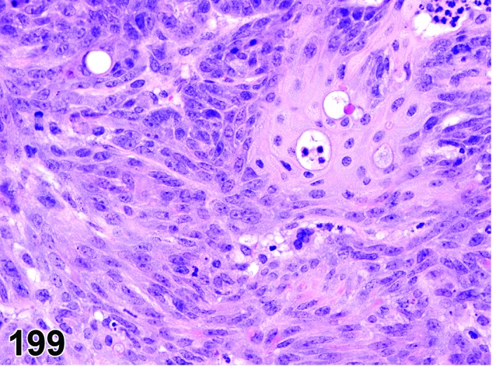
Mouse parotid gland. Carcinoma, squamous cell. Higher magnification of Figure 197.
Synonym: Carcinoma, epidermoid
Histogenesis: Probably ductular epithelial structures of the salivary glands.
Diagnostic features
Well-differentiated islands and cords with keratin formation.
Variable mitotic activity.
Invasion of adjacent tissues or metastasis.
High grades of anaplasia may occur.
Tumors may be extensively keratinized.
Differential diagnoses
Adenocarcinoma: Consisting of acinar, tubular, mixed, solid or papillary components; squamous differentiation may occur, but is not predominant.
Carcinoma, squamous cell (Zymbal’s gland): Squamous cell carcinomas (often with a sebaceous component) located at the base of ear pinna should be ascribed to the Zymbal’s gland, if origin from other sites (mammary, salivary glands) is not evident.
Adenosquamous carcinoma (mammary gland): With adenomatous component.
Metaplasia, squamous cell: Lack of invasion, well differentiated with retention of the ductular structure.
Myoepithelioma, malignant: Tumor cells with epithelioid differentiation or palisading around blood vessels; areas of densely packed elongated to spindeloid tumor cells, separated by a delicate and inactive tumor stroma.
Comment: Spontaneous squamous cell carcinomas in rodents are rare, but have been induced with chemicals in rats (Neuenschwander and Elwell 1990; Sumitomo et al. 1996; Cao et al. 1999) and mice (Botts et al. 1999; Takegawa et al.1998; Takai et al. 1986), and occur spontaneously in mice with the v-Ha-ras oncogene (Cardiff et al. 1993). Transgenic mice expressing a human Ha-ras oncogene developed adenosquamous carcinomas arising from the serous areas of the submandibular gland (Nielsen et al.1991). Also adenosquamous carcinomas of the mammary gland, which are not uncommon in mice, need to be considered as a differential diagnosis. Squamous cell carcinomas of the salivary glands should be distinguished from invasive Zymbal’s gland carcinomas with squamous differentiation by their location in situ (Neuenschwander and Elwell 1990).
Tumor, mixed, benign
Synonym: Tumor, composite, benign
Histogenesis: Glandular epithelial, myoepithelial, and mesenchymal components of the salivary glands.
Diagnostic features
Well circumscribed.
Features of both a benign mesenchymal tumor and an adenoma are present.
Neoplastic epithelial structures often intermixed with neoplastic myxomatous/fibrous tissue.
Cellular pleomorphism is not present.
No evidence of invasion.
Differential diagnoses
Tumor, mixed, malignant: Features of both a sarcoma and a carcinoma present; cellular pleomorphism present.
Adenoma: Lack of a neoplastic mesenchymal component.
Fibroma: Lack of a neoplastic epithelial component.
Fibroadenoma of the anterior mammary glands: Epithelial component with cytoplasmic staining characteristics of mammary gland fibroadenomas; according to well-established diagnostic criteria, “fibroadenomas” are separately classified, although they are benign mixed tumors.
Comment: Mouse polyoma virus causes salivary gland tumors including a mixed epithelial-mesenchymal type (Botts et al. 1999; Frith and Heath 1994; Dawe 1979).
Tumor, mixed, malignant
Figure 200.
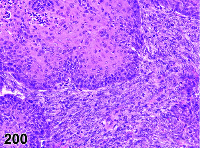
Mouse submandibular gland. Tumor mixed, malignant.
Synonym: Tumor, composite, malignant
Histogenesis: Glandular epithelial and myoepithelial / mesenchymal cells.
Diagnostic features
Features of both a sarcoma and a carcinoma are present.
Cellular and nuclear pleomorphism.
Invasion is present.
Differential diagnoses
Tumor, mixed, benign: Features of both a benign mesenchymal tumor and an adenoma; no cellular pleomorphism, no local invasion.
Myoepithelioma, malignant: Tumor cells with epithelioid differentiation or palisading around blood vessels; areas of densely packed elongated to spindeloid tumor cells, separated by a delicate and inactive tumor stroma.
Sarcoma, not otherwise specified: Lack malignant epithelial component.
Schwannoma, malignant: Lack malignant epithelial component.
Adenocarcinoma, OR carcinoma, squamous cell: Lack malignant mesenchymal component.
Comment: Mouse polyoma virus causes salivary gland tumors including a mixed epithelial-mesenchymal type (Botts et al. 1999; Frith and Heath 1994).
The differentiation between the mixed malignant tumor and the malignant myoepithelioma with epithelioid and spindeloid growth pattern may be difficult based on hematoxylin and eosin stained slides. Confirmatory immunohistochemistry may be required which in case of the mixed tumor would demonstrate two different tissue compartments that show either epithelial or mesenchymal immunophenotype, but usually not a mixed one. In contrast, mixed epithelial and mesenchymal antigenic expression would be demonstrated in case of the myoepithelioma.
Myoepithelioma, malignant
Figure 201.
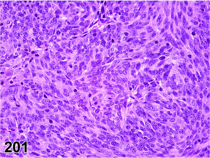
Mouse submandibular gland. Myoepithelioma, malignant.
Histogenesis: Myoepithelial cells, extraglandular ductal origin, salivary glands (possibly other glandular tissues).
Diagnostic features
Unencapsulated tumor; may show invasive cords of solid tissue extending into adjacent structures.
In larger tumors, central degeneration and necrosis may form pseudo-cysts, which may be filled by brown, mucoid material or cell debris.
Tumor parenchyma solid without acinar or tubular architecture.
Mixed tumor morphology with confluent regions of basal, stratified squamous, and spindle cell patterns. May contain tubular structures. Adjacent to blood vessels, tumor cells may show palisading basal cell pattern or align in epithelioid fashion.
Squamous appearance without keratinization or pearl formation may occur in some areas.
Invasion into surrounding tissue or vessels may be present; large tumors may metastasize to the lung.
Infiltrates of lymphocytes and plasma cells are not present (unlike polyoma virus neoplasms).
Differential diagnoses
Carcinoma, squamous cell: True epithelial differentiation without vascular orientation; spindle shaped tumor cells in case of scirrhous or anaplastic growth pattern only; keratinization in well differentiated tumors.
Tumor mixed, malignant: Presence of true epithelial and true mesenchymal tumor cells.
Comment: Myoepitheliomas of the salivary glands have not been described in rats. They are rare in most strains of mice but occur more often in the BALB/c, A/J, and C58 strains (mainly females). The most frequent locations are the parotid and submandibular glands (Frith and Ward 1988), but they are not limited to salivary glands or the cervical region. Neoplastic myoepithelial cells have been reported to stain positive for cytokeratin and for keratins K5 and K14 and vimentin. Also PTAH staining will aid in confirmation of the intracytoplasmic fibrils within the myoepithelial cells (Botts et al. 1999; Sundberg et al. 1991). They are negative for smooth muscle cell alpha-actin immunohistochemistry, in contrast to normal myoepithelial cells; furthermore, Sundberg et al. has suggested that the neoplasms arise from a subset of extraglandular ductal myoepithelial cells that are negative for smooth muscle cell alpha-actin (Sundberg et al. 1991). Such confirmatory stains may be required for differentiation of myoepitheliomas from scirrhous/anaplastic squamous cell carcinomas or mixed malignant tumors.
Myoepitheliomas in mice preferably occur in the ventral neck region (Sundberg et al. 1991). Although most of these tumors have been ascribed to the salivary glands, it needs to be taken into account that they may occur from any other glandular tissue, e.g. the mammary gland or extraorbital lacrimal gland. The frequent poor encapsulation, pleomorphic morphology, and central necrosis are compatible with a malignant interpretation; however, Sundberg et al. suggested few are malignant based on stringent criteria of metastasis to the lung or infiltration of underlying bone (Sundberg et al. 1991). The morphology of tumors with confirmed metastases is not unique, as compared to those without, and the clinical appearance of the neoplasms may lead to culling prior to development of readily defined metastases, so it is recommended that they should be regarded as potentially malignant.
VI. Pancreas (Exocrine)
A. Morphology (Anatomy)
During embryonic development the pancreas is formed by fusion of its dorsal and ventral buds derived from the septum transversum at the embryonic foregut/midgut junction. The pancreas consists of both exocrine and endocrine components. The exocrine pancreas forms the bulk of the pancreas and produces numerous enzymes (in proenzyme form) that aid in digestion.
The pancreas of rat and mouse is classified as mesenteric type (versus compact type) since discrete pancreatic tissues are diffusely distributed in the mesentery of the duodenal loop, transverse colon, and greater omentum adjacent to stomach and spleen. The large interlobular ducts open into a common hepatic (biliary)/pancreatic duct that enters the duodenum. In addition, there are some ducts that open directly into the duodenum (Eustis et al. 1990; Boorman and Sills 1999).
B. Morphology (Histology)
The exocrine pancreas is classified as a compound tubuloalveolar or compound acinar gland. The pancreatic acinus consists of a single layer of pyramidal acinar cells arranged concentrically around a lumen. The apex of the pyramidal cells facing the lumen contains eosinophilic zymogen granules and the base of the pyramidal cells is basophilic due to abundant rough endoplasmic reticulum and also contains the nucleus. Each pancreatic acinus is surrounded by a thin basal lamina, scant reticular stroma, and pancreatic stellate cells (akin to the hepatic stellate cells). Located centrally within the acinus, the centroacinar cells form an interface between the acinus and the intercalated duct. The intercalated duct continues into intralobular ducts formed by the ductular cells. The intralobular ducts fuse to form the interlobular ducts that finally open into the pancreato-hepatic (biliary) duct (Eustis et al. 1990; Boorman and Sills 1999).
The exocrine pancreas is physiologically and morphologically compartmentalized into peri- and tele-insular regions. The peri-insular acinar cells are in the immediate proximity of the islets of Langerhans and are larger due to more abundant cytoplasm, larger zymogen granules and larger nuclei than the tele-insular acinar cells that are farther from the islets. Due to differences in sizes of the peri- and tele-insular acinar cells, at lower magnification, the islets of Langerhans appear to be surrounded by “halos” (peri-insular halos) (Greaves 2012).
The histologic constituents of the pancreatic tissue such as the exocrine acini, centro-acinar cells, ductal cells and the endocrine cells within the islets have a unique ability to de-differentiate and transdifferentiate into other pancreatic histologic cell types based on the severity and type of injury, distinct transcriptional and protein alterations and epigenetic factors (Stanger and Hebrok 2013). As a result, tissue injury in exocrine pancreas may resolve by forming a histologic constituent that may be similar or different from the damaged tissue type. After certain types of pancreatic tissue injury, the exocrine acini undergo de-differentiation and may attain a ductular morphology and in some cases even attain an endocrine phenotype. Notch, Wnt and Hedgehog signaling pathways play important roles in these de-differentiation and transdifferentiation pathways (Stanger and Hebrok 2013).
C. Physiology
The pancreas is a dual function gland with both exocrine (acini) and endocrine (islets of Langerhans) functions.
The acinar cells store the digestive enzymes in heterogeneously composed vacuoles and the release of these enzymes occurs in a cyclic and secretagogue-specific fashion. The exocrine acinar secretions collect within the acinar lumen and then accumulate within the interlobular ducts that subsequently drain into the duodenum via the pancreato-biliary duct. Hormones like gastrin (from G-cells in the stomach) and cholecystokinin (from I-cells in the duodenum) stimulate the exocrine pancreas to secrete inactive proenzymes (zymogens like trypsinogen, chymotrypsinogen, elastase and carboxypeptidase) as well as active enzymes such as lipase, amylase and nuclease. In addition, the duodenal hormone secretin stimulates the centroacinar cells (in addition to the Brunner’s glands) to secrete bicarbonate ions that aid in neutralizing acidic chyme from the stomach and stabilizing the enzymes. The intestinal enteropeptidases activate the trypsinogen to trypsin that in turn cleaves the rest of trypsinogen and chymotrypsinogen to their active forms resulting in protein digestion. Lipase and amylase aid in the digestion of lipids and carbohydrates, respectively (Greaves 2012).
The exocrine pancreas has great reserve capacity and no clinical evidence of exocrine pancreatic insufficiency is noted with up to 90% destruction of exocrine pancreas. Exocrine pancreatic function deficits > 90% results in improper digestion of food and malabsorption of nutrients (Hotz et al. 1973).
D. Necropsy & Trimming
At necropsy, the whole left lobe of the pancreas (located in the omentum close to the spleen) is removed for trimming. For microscopic evaluation, a large part of the left lobe is cut in longitudinally in the horizontal plane, making the cut surface as large as possible. The right lobe is removed together with the adjacent small intestine (Ruehl-Fehlert et al. 2003).
E. Nomenclature, Diagnostic Criteria, and Differential Diagnoses
a. Congenital Lesions
Ectopic tissue
Figure 202.
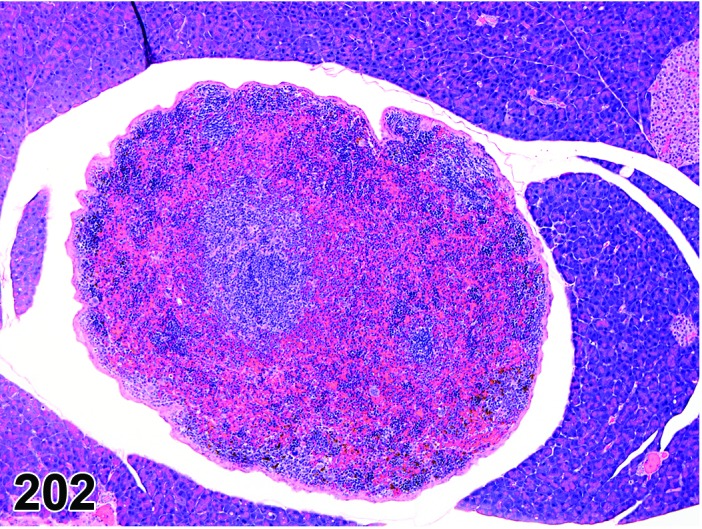
Mouse pancreas. Ectopic tissue, spleen.
Synonym: Heterotopia
Pathogenesis: Congenital abnormalities resulting in splenic or liver tissues in pancreas.
Diagnostic features
Usually focal distribution.
Fully differentiated non-pancreatic tissue in the pancreas.
Usually circumscribed and not wholly integrated into the ‘host’ tissue.
No evidence of dysplasia or history of tissue injury.
Differential diagnosis
Metaplasia: The metaplastic tissue is usually integrated into the histological pattern of the host tissue and is usually a result of tissue reaction and adaptation.
Comment: Ectopic tissues are usually a rare finding in toxicity studies and care should be taken to properly interpret them and not be confused with metaplasia or neoplasia.
b. Cellular Degeneration, Injury and Death
Vacuolation, acinar cell
Figure 203.
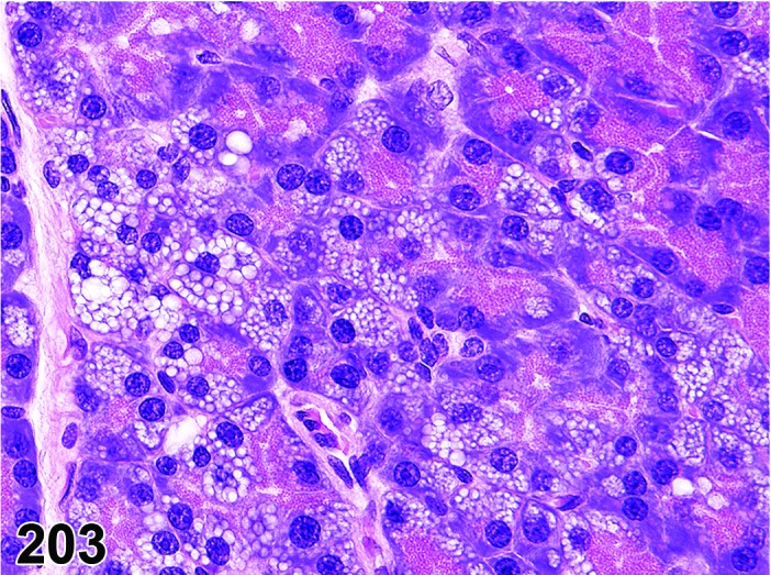
Rat pancreas. Vacuolation, acinar cell.
Synonyms: Hydropic change; cloudy swelling; hydropic degeneration; fatty degeneration; lipidosis; lipid accumulation, vacuolation epithelial
Modifier: Foamy
Pathogenesis: Degenerative processes that lead to accumulation of substances of different character, including fluids, lipids, phospholipids and glycoproteins within acinar cells.
Diagnostic features
Focal, multifocal or diffuse.
Moderate to total loss of zymogen granules within the affected pyramidal acinar cells.
The cells may be swollen with pale eosinophilic cytoplasm and have intracytoplasmic vacuoles with variably margins.
The size of vacuoles may vary from small (microvesicular) to large when multiple small vacuoles coalesce together (macrovesicular) often resulting in nuclear displacement.
Retained lobular architecture.
Islets of Langerhans usually unaffected.
Foamy
Hypertrophy of cells with foamy cytoplasm characterized by clear vesicles with eosinophilic fine granules.
Differential diagnoses
Artifact: Medium sized clear vesicles within the cytoplasm of acinar cells, especially at the periphery. No positive staining by fat stains.
Comments: Based solely on light microscopic evaluation of hematoxylin and eosin stained tissue sections, it is not possible to conclusively characterize the nature of the intracytoplasmic vacuoles. Hence, vacuolation is the preferred terminology of the lesion that was previously diagnosed as fatty change, hydropic degeneration, cloudy swelling etc. Acinar cell vacuolation is a reversible change and is usually a reflection of hypoxia and metabolic insults resulting in damage to mitochondria, endoplasmic reticulum, protein machinery, and plasma membrane. Oil Red O and Sudan Black stains on frozen sections help to identify the neutral lipids within the micro/macro vesicles. It is useful to differentiate from cytoplasmic vacuoles due to water accumulation (hydropic change) resulting from cell injury due to hypoxia. Marked vacuolation of the exocrine pancreatic acini has been documented in female Sprague-Dawley rats exposed to dioxins and dioxin-like compounds (Yoshizawa et al. 2005).
A fine cytoplasmic vacuolation, resulting from foamy appearance, may give the suspicion of phospholipidosis. In such cases, the modifier “foamy” should be used. If the change reflects phospholipidosis, increases in size and number of lysosomes can be identified by histochemistry (for alkaline phosphatase), immunostains (for lysosomal membrane proteins, e.g. LAMP-2) or by transmission electron microscopy. With the later technique, multilamellated structures (myeloid bodies/lamellar bodies) are observed in the lysosomes. The term phospholipidosis should only be used when confirmation by one of these techniques has been achieved.
Accumulation adipocytes
Figure 204.
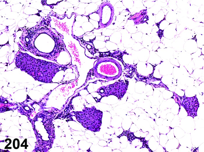
Mouse pancreas. Accumulation adipocytes after severe atrophy, acinar cell.
Synonyms: Adipocyte accumulation; lipomatosis; fatty infiltrate
Pathogenesis: Replacement of exocrine parenchyma after age-related or chemically-induced atrophy or in case of obesity.
Diagnostic features
Multifocal to coalescing adipocytes within the interstitium.
Compression of the adjacent exocrine tissue only occasionally.
Multifocal to diffuse atrophy of exocrine acini and replacement by adipose tissue.
Islets of Langerhans unaffected.
Differential diagnosis
Vacuolation, acinar cell: Moderate to abundant intracytoplasmic vesicles with a multifocal to diffuse distribution. The exocrine acinar architecture is retained.
Comments: Fatty infiltration of exocrine pancreas is an uncommon lesion which may be age related. The pathogenesis is not usually apparent but may be secondary to atrophy or is the cause of atrophy.
Eosinophilic globules
(Figures 205 and 206)
Figure 205.
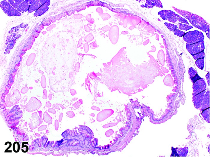
Mouse pancreas. Ectasia duct, with eosinophilic globules.
Figure 206.
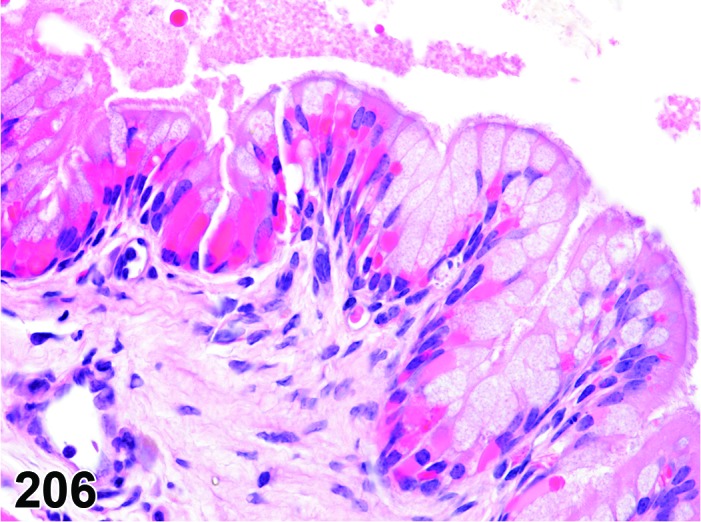
Mouse panceas. Detail of Figure 205 showing osinophilic globules in the epithelium of an ectatic duct.
Synonyms: Hyalinosis; eosinophilic change
Pathogenesis: Storage of hyaline material within the cytoplasm of ductal cells.
Diagnostic features
Focal or multifocal.
Intensely pink-red cytoplasmic droplets and/or crystals in ductal cells.
Cells are usually hypertrophic.
Crystals, if present, may be intracellular or extracellular.
Differential diagnosis
None.
Comments: Aged mice of the C57BL/6, 129 and B6,129 strains develop a high incidence of eosinophilic globules in various epithelia, including glandular stomach, respiratory tract, bile duct, gall bladder and pancreatic duct (Ward et al. 2001). The protein isolated from the characteristic eosinophilic lesions has been identified as Ym1/Ym2 a member of the chitinase family that may be produced in response to mucosal irritation (Ward et al. 2001; Rogers and Houghton 2009). The use of the term hyalinosis for this lesion will potentially cause confusion in safety assessment as the same term may also be used for a distinctly different clinical disorder in humans and can be used to describe vascular and renal glomerular changes.
Autophagic vacuoles, acinar cell
Figure 207.
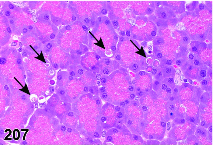
Rat pancreas. Autophagic vacuoles, some of them indicated by arrows)
Synonyms: Autophagy, acinar cell
Pathogenesis: Increased autophagocytosis by segregation of cytoplasmic organelles or cytoplasmic contents in sublethally injured acinar cells.
Diagnostic features
Multifocal.
Retained lobular architecture.
Intracytoplasmic hypereosinophilic to basophilic globules surrounded by a thin clear halo.
Differential diagnoses
Apoptosis: Apoptotic bodies also have a hypereosinophilic cytoplasm and are surrounded by a clear halo but they always contain basophilic condensed nuclear fragments. Apoptosis may be differentiated from autophagy by using TUNEL staining that marks the nuclear fragments (Zhang et al. 2014).
Vacuolation, acinar cell: Swollen cells with intracytoplasmic vacuoles with no intraluminal contents
Artifact: Medium sized clear vesicles within the cytoplasm of acinar cells, especially at the periphery. No positive staining by fat stains.
Foam cells (Phospholipidosis): The cells have foamy cytoplasm characterized by clear vesicles with eosinophilic fine granules. Ultrastructurally, it consists of large lysosomal inclusions composed of densely packed concentric membranes with a fingerprint pattern.
Comments: Autophagy is a homeostatic mechanism that plays a major role in the catabolism by lysosomal digestion of organelles and/or aged cytoplasmic contents. Autophagy occurs at basal levels in all tissues to maintain cellular homeostasis. However, it is rapidly increased during energy depletion, hypoxia, endoplasmic reticulum stress, high temperature, hormonal stimulation, or cellular remodeling to combat oxidative stress. Alterations in autophagy may lead to disruption of cellular homeostasis and can result in pancreatitis and cell death (Helin et al. 1980; Gukovskaya and Gukovsky 2012). Increased autophagic vacuoles in a treated group should always be considered in the context of the control group.
Autophagy and apoptosis are not necessarily mutually exclusive entities. There is evidence that apoptosis can occur simultaneously with autophagy (Maiuri et al. 2007).
The exocrine acini are very sensitive to agents affecting autophagic flux such as vinblastine (enhances autophagic segregation) or cycloheximide (regression of the autophagic compartment). Autophagy may be detected by electron microscopy, by the fluorescent localization pattern of LC3 puncta (intracytoplasmic granular fluorescent material) or by immunohistochemistry using antibodies against LC3-II (processed LC3). The autophagosomes within the cytoplasm, under light microscopy, appear as discrete vacuoles with some dark eosinophilic or amphophilic contents and under electron microscopy, remnants of mitochondria or endoplasmic reticulum may be observed. It is important to note that accumulation of autophagosomes is not always indicative of autophagy induction since it could be the result of blockage of autophagosomal maturation or increased generation of autophagosomes. Autophagic flux assays may measure LC3 turnover, levels of autophagic substrates, mRFP-GFP-LC3 color change, free GFP generated from GFP-LC3, and lysosome-dependent long-lived protein degradation (Iovanna and Vaccaro 2010).
Apoptosis
Figure 208.
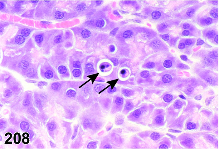
Rat pancreas. Apoptosis: apoptotic bodies (arrows).
Synonyms: Apoptotic cell death
Pathogenesis: Gene regulated, energy dependent process leading to formation of apoptotic bodies which are phagocytosed by adjacent cells.
Diagnostic features
Single cells or small clusters of cells.
Cell shrinkage.
Hypereosinophilic cytoplasm.
Nuclear shrinkage, pyknosis, karyorrhexis. Intact cell membrane.
Apoptotic bodies.
Cytoplasm retained in apoptotic bodies.
Phagocytosis of apoptotic bodies tissue macrophages or other adjacent cells.
No inflammation.
Differential diagnoses
Necrosis: The morphologic features of cell death clearly fit with necrotic diagnostic criteria (cells and nuclei swollen, pale cytoplasm etc.).
Apoptosis/necrosis: Both types of cell death are present and there is no requirement to record them separately or a combined term is preferred for statistical reasons. The term is also used when the type of cell death cannot be determined unequivocally.
Comment: The approach for the nomenclature and diagnostic criteria of cell death applied here is based on a draft recommendation of the INHAND Cell Death Nomenclature Working Group.
Apoptosis is not synonymous with necrosis. The main morphological differences between these two types of cell death are cell shrinkage with nuclear fragmentation and tingible body macrophages in apoptosis versus cell swelling, rupture and inflammation in necrosis; however, other morphologies (e.g. nucelar pyknosis and karyorrhexis) overlap. In routine H&E sections where the morphology clearly represents apoptosis or single cell necrosis, or whereby special procedures (e.g. TEM or IHC for caspases) prove one or the other, individual diagnoses may be used. However, because of the overlapping morphologies, necrosis and apoptosis are not always readily distinguishable via routine examination, and the two processes may occur sequentially and/or simultaneously depending on intensity and duration of the noxious agent (Zeiss 2003). This often makes it difficult and impractical to differentiate between both during routine light microscopic examination. Therefore, the complex term apoptosis/necrosis may be used in routine toxicity studies.
A differentiation between apoptosis and single cell necrosis may be required in the context of a given study, in particular if it aims for mechanistic investigations. Transmission electron microscopy is considered to represent the gold standard to confirm apoptosis. Other confirmatory techniques include DNA-laddering (easy to perform but insensitive), TUNEL (false positives from necrotic cells) or immunohistochemistry for caspases, in particular caspase 3. These techniques are reviewed in detail by Elmore (Elmore 2007). Some of these confirmatory techniques detect early phases of apoptosis, in contrast to the evaluation of H&E stained sections, which detects late phases only; overall interpretation of findings should take into account these potential differences. Thus, low grades of apoptosis may remain unrecognized by evaluation of H&E stained slides only.
Certain studies may require the consideration of forms of programmed cell death other than apoptosis which requires specific confirmatory techniques (Galluzzi et al. 2012).
Acinar cell apoptosis is classically manifested in many cases of xenobiotic-induced injury. It is considered a “preferred” response to injury, as it does not lead to subsequent inflammation. An inverse relationship between apoptosis and necrosis in acinar cell injury has been described for various experimental models. Stimulation of apoptosis seems to protect against acute necrotizing reactions, while inhibition of apoptosis leads to necrosis and acute inflammation (Wallig and Sullivan 2013).
Exocrine pancreatic acinar apoptosis is observed in rodents treated with synthetic cholecystokinin analog caerulein (Reid and Walker 1999), ethionine (Fitzgerald and Alvizouri 1952; Walker et al. 1993), physical blockage of the pancreas by ductal ligation (Abe and Watanabe 1995; Doi et al. 1997), duct obstruction involution following soy flour-induced hyperplasia (Oates et al. 1986) and zinc toxicants (Kazacos and van Vleet 1989), copper-deficient diet supplemented with a copper-chelating agent (Rao et al. 1993), Azaserine (Woutersen 1996), lipopolysaccharide (Laine et al. 1996) and pan CDK inhibitor (Ramiro-Ibáñez et al. 2005). In addition, exocrine pancreatic apoptosis is also observed in some genetically modified mice such as Serpini2-deficient mouse model for pancreatic insufficiency (Loftus et al. 2005). Non-treatment associated sporadic apoptosis may be noted in pancreas from rodents and this is part of the normal tissue homeostasis and is considered as a background change. Acinar cell apoptosis may be increased in fasted or chronically anorectic animals. The degree of apoptosis is in these cases lower than that of acute pancreatic injury.
Necrosis
Figure 209.
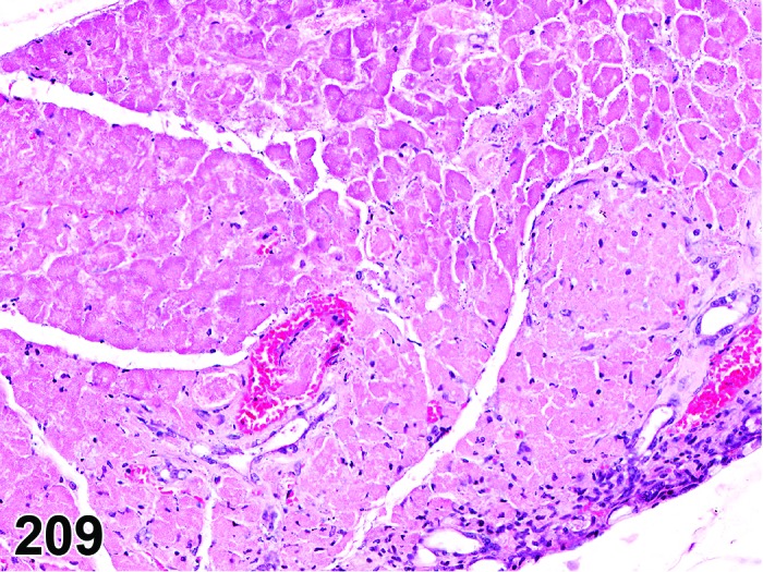
Mouse pancreas. Necrosis.
Synonyms: Oncotic cell death; oncotic necrosis; necrosis
Modifiers: Single cell
Pathogenesis: Unregulated, energy independent, passive cell death with leakage of cytoplasm into surrounding tissue and subsequent inflammatory reaction; may be induced by direct contact with a test article after oral uptake/administration.
Diagnostic features
Focal affecting cell groups, lobular, diffuse.
Cells swollen with pale eosinophilic cytoplasm.
Loss of nuclear basophilia, pyknosis and/or karyorrhexis that affects aggregates of cells.
In more severe cases, there may be detachment of the epithelium from the submucosa.
Typically, the presence of degenerative cells is a component of necrosis.
Minimal or slight inflammatory cell infiltrates, edema and fibrin may be present as a feature of necrosis.
Single cell
Only single cells affected.
Differential diagnoses
Apoptosis: The morphologic features of cell death clearly fit with apoptosis (cell and nucleus shrunken, hypereosinophilic cytoplasm, nuclear pyknosis etc.) and/or special techniques demonstrate apoptosis.
Apoptosis/necrosis: Both types of cell death are present and there is no requirement to record them separately or a combined term is preferred for statistical reasons. The term is also used when the type of cell death cannot be determined unequivocally.
Inflammation, acute: Infiltrates of inflammatory cells, edema, and fibrin predominate; necrosis may be present, but is a minor component.
Comments: Necrosis is usually accompanied by an inflammatory cell infiltrate and may be a significant component of acute inflammation. Usually, either necrosis or acute inflammation will be recorded. However, in the context of a specific study it may be more appropriate to record both processes separately.
In the pancreas, dead cells quickly pass through the stage of coagulation necrosis to liquefaction because of the high content of hydrolytic enzymes activated and released into the interstitial tissue. Necrosis of acinar tissue develops very rapidly and is best recognized within 12–48 hours after the insult (Wallig and Sullivan 2013).
Apoptosis/necrosis
Synonym: Cell death
Pathogenesis: Unregulated, energy independent, passive cell death with leakage of cytoplasm into surrounding tissue and subsequent inflammatory reaction (single cell necrosis) AND/OR gene regulated, energy dependent process leading to formation of apoptotic bodies which are phagocytosed by adjacent cells (apoptosis).
Diagnostic features
Both types of cell death are present and there is no requirement to record them separately or a combined term is preferred for statistical reasons OR
The type of cell death cannot be determined unequivocally.
Differential diagnoses
Apoptosis: The morphologic features of cell death clearly fit with apoptosis (cell and nucleus shrunken, hypereosinophilic cytoplasm, nuclear pyknosis etc.) and/or special techniques demonstrate apoptosis and there is a requirement for recording apoptosis and necrosis separately.
Necrosis: The morphologic features of cell death clearly fit with necrotic diagnostic criteria (cells and nuclei swollen, pale cytoplasm etc.) and there is a requirement for recording apoptosis and necrosis separately.
Comments: Necrosis and apoptosis are not always readily distinguishable via routine examination, and the two processes may occur sequentially and/or simultaneously depending on intensity and duration of the noxious agent (Zeiss 2003). This often makes it difficult and impractical to differentiate between both during routine light microscopic examination. In these cases, the combined term apoptosis/necrosis may be used. It is recommended to explain in detail the use of this combined term in the narrative part of the pathology report.
Secretory depletion, acinar cell
Figure 210.
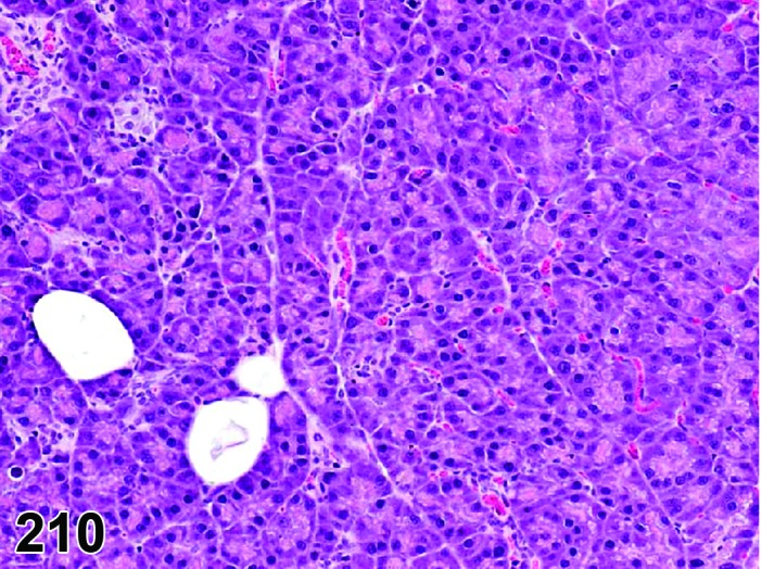
Rat pancreas. Secretory depletion, acinar cell.
Pathogenesis: Decrease of acinar cell zymogen granules leading to shrunken acinar cells with increased basophilia.
Diagnostic features
Focal, lobular, or diffuse lesion.
Reduced acinar diameter.
Partial or complete loss of acinar cell zymogen granules leading to a reduced cell size and increased basophilia.
Nuclei of acinar cells inactive
Lack of fibrosis or adipocyte infiltration.
Islets of Langerhans are unaffected.
Differential diagnoses:
Atrophy, acinar cell: Loss of acinar cell basophilia and decreased zymogen granules resulting in small acini lined by small columnar cells almost completely devoid of cytoplasm and with a small and inactive nucleus; may be accompanied by fibrosis and minimal mononuclear cell infiltrates.
Focus, basophilic: Basophilic cells of normal size and commonly slightly enlarged; nulcei slightly enlarged and with prominent nucleoli.
Peri-insular halos: Tele-insular acinar cells have relatively less zymogen granules when compared to peri-insular acinar cells.
Comment: The morphology of the exocrine pancreatic acini may be altered due to several physiological conditions, such as anorexia/inanition. The volume of the zymogen granules and RER is reciprocally related. During protein synthesis, RER is increased with a proportionate decrease in zymogen granules. In moribund and/or anorexic animals with protein deficiency, the zymogen granules are decreased and the cells are shrunken with apparently increased basophilia due to relative predominance of the basally located basophilic cytoplasm (Longnecker and Wilson 2002).
Atrophy, acinar cell
(Figures 211 and 212)
Figure 211.
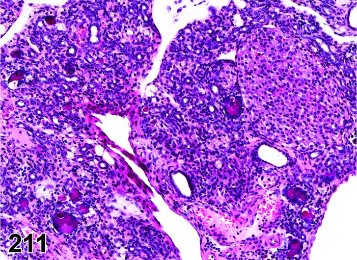
Mouse panceas. Atrophy, acinar cell.
Figure 212.
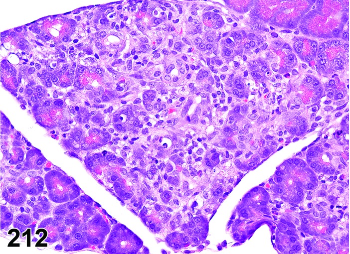
Rat pancreas. Atrophy, acinar cell, associated with infltrate mononuclear cell.
Pathogenesis: Decrease in number and/or size of acinar cells may be due to spontaneous or experimentally induced degenerative changes, apoptosis, or a sequel of chronic inflammation.
Diagnostic features
Focal, lobular, or diffuse.
Reduction in the number and/or size of acini.
Loss of acinar cell basophilia and decreased zymogen granules resulting in small acini lined by small columnar cells almost completely devoid of cytoplasm and with a small and inactive nucleus.
Relatively prominent intra and inter-lobular ducts.
Pyknotic and karyorrhectic nuclei, apoptotic bodies, and mitotic figures may be present.
Scattered to coalescing adipocytes within the interstitium.
Variable degree of interstitial fibrosis.
Variably dilated cyst-like or duct-like acini and/or ducts lined by cuboidal or flattened epithelial cells.
Transitional structures containing mixtures of normal acinar cells, atrophic acinar cells, and cuboidal ductal cells.
May be accompanied by minimal to mild infiltration of lymphocytes or macrophages.
Islets of Langerhans are unaffected.
Differential diagnoses
Degranulation, Acinar cell: Diffuse reduction of zymogen granules but maintenance of the basophilic basal cytoplasm; no fibrosis or adipocyte infiltration.
Peri-insular halos: Tele-insular acinar cells have relatively less zymogen granules and more RER when compared to peri-insular acinar cells. In certain planes of section, areas with more tele-insular regions may give a false impression of atrophy.
Inflammation, chronic: Infiltration of mononuclear inflammatory cells and interstitial fibrosis are frequently accompanied by acinar cell atrophy of variable degree.
Comment: Exocrine acinar atrophy is the most common spontaneous degenerative change in pancreas of both rats and mice. Acinar atrophy may range from focal atrophic exocrine acini with no inflammation or fibrosis to diffuse atrophy of exocrine acini with replacement by adipose tissue and residual ducts, vasculature and islets of Langerhans. Acinar atrophy frequently represents the sequel of chronic inflammation and, as such, is often accompanied by infiltrates of mononuclear cells and fibrosis. It is recommended to record in standard toxicity studies for such lesions either atrophy or chronic inflammation, but not both. Inflammation, chronic, should be recorded only, if the inflammatory changes are the predominant component. In rats, the acinar atrophy increases with age and the incidence varies with sex (males > females) and strain (BN/Bi/WAG/Rij(F1) > Crl:CD(SD)BR = BN/Bi > Slc:Wistar > Hap:(SD) > F344 > Osborne-Mendel). In B6C3F1 mice, the incidence of spontaneous exocrine pancreatic atrophy is 1–2% in 2-year studies (Boorman and Sills 1999). In rats, chronic protein or essential amino acid deficiency, copper deficiency, zinc deficiency, ethionine, hypophysectomy or high doses of glucagon induce loss of zymogen granules and pancreatic atrophy (Svoboda et al. 1966; Kitagawa and Ono 1986; Rao et al. 1987; Koo and Turk 1977). Malonaldehyde caused atrophy of exocrine pancreas in both male and female mice (NTP, 1988). Acinar atrophy has also been induced by pancreatic duct ligation (Watanabe et al. 1995; Eustis et al. 1990).
Mineralization
Figure 213.
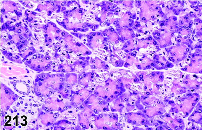
Rat panceas. Mineralization (dystrophic) of acinar cells.
Synonym: Calcification
Pathogenesis: Deposition of mineral either secondary to cellular necrosis (dystrophic mineralization) or secondary to hypercalemia (metastatic mineralization).
Diagnostic features
Focal, lobular, or diffuse.
Deposits of dark basophilic granular material (on H&E) within exocrine acini, ducts or interstitium.
Differential diagnoses
Artifacts: Hematoxylin stain deposits (distribution unrelated to tissue structures), acid hematin (use of unbuffered formalin; dark brown to black, tropism for erythrocytes).
Pigment: Lipofuscin and porphyrin appear brown to golden brown in H&E slides and may further be differentiated from mineralization by special stains.
Comment: Dystrophic calcification occurs in necrotic/injured tissue even in animals with reference range serum calcium levels, whereas metastatic calcification occurs in hypercalcemic animals throughout the body, especially within interstitial tissues and blood vessel walls. The two commonly used stains for detecting calcium are Alizarin red and von Kossa.
Amyloid
Figure 214.
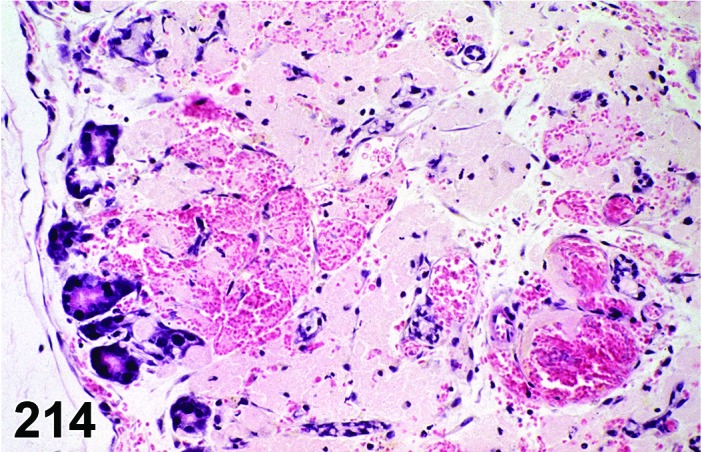
Mouse panceas. Amyloid.
Synonyms: Amyloidosis; amyloid deposition
Pathogenesis: Extracellular deposits of chemically diverse complex insoluble polypeptides.
Diagnostic features
Light eosinophilic homogenous amorphous extracellular material within the tunica media of blood vessels, within the interstitium and along basement membranes.
Variable degree of atrophy and loss of intervening acini and ducts may occur, if amyloid deposition is prominent.
Green birefringence using polarized light with Congo red stain.
Differential diagnoses
Necrosis: Loss of cellular detail and no extracellular material deposits, Congo red negative.
Fibrinoid change (necrosis) in vessel walls: Deep eosinophilic homogenization of vascular media.
Comment: Amyloid and amyloid-like material have been observed in various tissues of aged rodents, especially mice. Rats are much more resistant to develop amyloidosis than mice.
The characteristic morphologic appearance and location of amyloid in H&E sections that is usually adequate to make this diagnosis. Confirmation of the deposits as amyloid can be accomplished with light microscopy using special stains such as Congo red. Amyloid appears apple green under polarized light with this stain.
The term “hyaline” should be used in case of uncertainties about the nature of extracellular deposits hyaline material in H&E sections and the unavailability of a Congo red stain. Hyaline is also the appropriate term for homogenously eosinophilic extracellular deposits that react negative with Congo red.
Pigment
Figure 215.
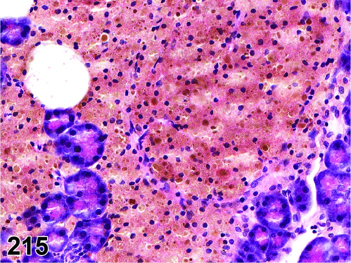
Rat pancreas. Pigment .
Synonyms: Pigment deposition; pigmentation; endogenous pigmentation; exogenous pigment deposition
Pathogenesis: Diverse, dependent on the nature of pigment.
Diagnostic features
Focal, lobular, diffuse.
Yellow to brown pigmentation within the interstitium, acini and ducts.
Differential diagnoses
Mineralization: Dark basophilic appearance in H&E stained sections; special stains like Alizarin red S and von Kossa can help identify calcium salts.
Artifacts: Hematoxylin stain deposits (distribution unrelated to tissue structures), acid hematin (use of unbuffered formalin; dark brown to black, tropism for erythrocytes).
Comment: In the rat, hemosiderin may be seen in neighboring islets of Langerhans where hemorrhage may have occurred previously (Imaoka et al. 2007).
The type of pigment can be identified by special stains, such as Schmorl’s (mature lipofuscin, melanin, bile), Fontana-Masson’s (melanin), Perl’s (Ferric Iron-hemosiderin) and Hall’s (bilirubin) stains. Formalin-heme pigment (acid-hematin) is observed in tissues fixed in unbuffered formalin. Polarization can help differentiate some pigments like lipofuscin (strong orange autofluorescence on unstained FFPE sections), porphyrin (bright red with a centrally located dark Maltese cross) and acid-hematin (birefringence on H&E sections).
c. Inflammatory Lesions
Infiltrate
Figure 216.
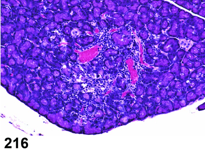
Rat panceas. Infiltrate, mononuclear cell.
Synonyms: Infiltrate inflammatory; infiltrate inflammatory cell; infiltration (plus modifier); infiltration, inflammatory; infiltration, inflammatory cell
Modifier: Type of inflammatory cell that represents the predominant cell type in the infiltrate
Pathogenesis: Infiltration with neutrophils (Infiltrate, neutrophil), eosinophils (Infiltrate, eosinophil), mononuclear cells (Infiltrate, mononuclear cell) or a combination of more than one type (Infiltrate, mixed) without other histological features of inflammation, e.g. hemorrhage, edema, fibroplasia.
Diagnostic features
Focal, multifocal or diffuse.
Presence of mononuclear or polymorphonuclear leukocytes but no other histological criteria of inflammation like edema, congestion or necrosis.
Usually no acinar cell degranulation.
Differential diagnoses
Inflammation: The inflammatory cell infiltrate is associated with additional morphological features of inflammation such as edema, hemorrhage, necrosis, and/or fibroplasia.
Granulocytic leukemia: Infiltration of neutrophil precursors and/or abnormal neutrophils will probably be present along with mature neutrophils; infiltration of similar neoplastic cells is likely present in other organs.
Lymphoma: Infiltration of monomorphic population of lymphocytes (often with atypical, increased and/or abnormal mitoses); infiltration of similar neoplastic cells is likely present in other organs.
Comment: In toxicity studies, inflammatory cell infiltrates are encountered more commonly than inflammation (pancreatitis). The term pancreatitis (or inflammation) should not be confused with or used in lieu of infiltrate (plus cell type). The term infiltrate (plus cell type) should be used instead of inflammation (or pancreatitis) when there is no tissue injury/reaction associated with the inflammatory cell infiltrate. Also, the inflammatory cell infiltrate should be qualified with the type (name) of the cells that are predominant, such as lymphocytic, neutrophilic, etc. The use of terminology summarizing the nature of the infiltrates as suppurative (in lieu of neutrophilic), granulomatous (in lieu of macrophage), and chronic (in lieu of lymphocytic, plasmacytic, mononuclear) is not recommended since these terms imply inflammation. The term inflammation (pancreatitis) should be reserved for lesions where the inflammatory cell infiltrate is associated with tissue injury/reaction such as edema, congestion, acinar cell degranulation, necrosis, vascular fibrinoid necrosis, hemorrhage, fat necrosis or saponification (Mann et al. 2012).
Inflammation
(Figures 217 and 218)
Figure 217.
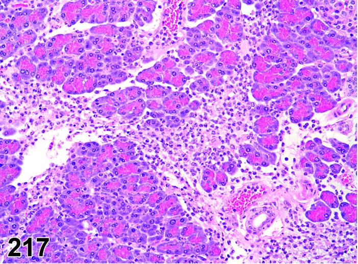
Mouse pancreas. Inflammation, neutrophil.
Figure 218.
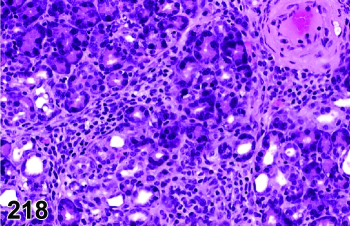
Rat pancreas. Inflammation, mononuclear cell. Note atrophic acini aquiring ductular morphology.
Synonym: Pancreatitis
Modifier: Type of inflammatory cell that represents the predominant cell type in the inflammation
Pathogenesis: Infiltration with neutrophils (Infiltrate, neutrophil), eosinophils (Infiltrate, eosinophil), mononuclear cells (Infiltrate, mononuclear cell) or a combination of more than one type (Infiltrate, mixed) with additional histological features of inflammation, e.g. hemorrhage, edema, fibroplasia.
In control rats, the most common cause of inflammation is spontaneous arteritis involving the pancreatoduodenal artery (Coleman et al. 1977). For pancreatitis induced by xenobiotics, the pathogenesis usually involves release of inactive proenzymes from exocrine pancreas and activation of these digestive enzymes locally and systemically.
Diagnostic features
Focally extensive or diffuse, involving predominantly the interstitium, but also acinar cells and intra- or inter-lobular ducts.
Infiltrate of mononuclear or polymorphonuclear leukocytes into the gland parenchyma.
Presence of other histological criteria of inflammation, e.g. hemorrhage, edema, fibroplasias.
Islets of Langerhans may be entrapped within the inflammation.
Inflammation, neutrophil
Infiltrate predominantly neutrophilic; frequently accompanied by edema and congestion; acinar cell degranulation and necrosis, fibrinoid necrosis of small vessels and hemorrhage as well as fat necrosis and saponification may be present.
Inflammation, mononuclear cell
Infiltrate predominantly mononuclear cell; frequently accompanied by acinar cell atrophy and mild acinar cell degeneration, ductal proliferation, interstitial fibrosis; mineralization of acini may be present.
Differential diagnoses
Infiltrate, inflammatory cell: Other histologic features of inflammation such as edema, hemorrhage, necrosis, and/or fibroplasias will be absent.
Granulocytic leukemia: Infiltration of neutrophil precursors and/or abnormal neutrophils will probably be present along with mature neutrophils; infiltration of similar neoplastic cells is likely present in other organs.
Lymphoma: Infiltration of monomorphic population of lymphocytes (often with atypical, increased and/or abnormal mitoses); infiltration of similar neoplastic cells is likely present in other organs.
Postmortem autolysis: uniform and often diffuse change in acinar cells starting with loss of basophilia, gradual loss of definition and final lysis; no inflammatory cell infiltration, congestion or edema.
Atrophic, acinar cell: Minimal to mild infiltrates of mononuclear cells and fibrosis, but atrophy is the predominant component.
Comment: When a mononuclear cell or mixed infiltrate is considered to represent an inflammatory process (inflammation, mononuclear cell or inflammation, mixed) and other morphologic features (acinar cell atrophy and mild acinar cell degeneration, ductal proliferation, interstitial fibrosis; mineralization of acini) associated with the infiltrate suggest an ongoing/continuing inflammatory process, the modifier ‘chronic’ may be included in the diagnosis (i.e. inflammation, mononuclear cell, chronic) to better characterize the finding.
Spontaneous inflammation, neutrophilic or mixed of the pancreas is very uncommon in control mice and rats. The severity of inflammation within pancreas can range from mild edematous inflammation to severe necrotizing inflammation. Acute inflammation is observed in humans and some rodents treated with several classes of drugs belonging to antimicrobials, estrogens, corticosteroids, diuretics, antibiotics, analgesics/anti-inflammatory agents, statins, ACE inhibitors, and Highly Active Antiretroviral Therapy (HAART) drugs (Badalov et al. 2007). Several animal models are used for studying pancreatitis such as choline-deficient ethionine supplemented diet; intraperitoneal injection of arginine; intraperitoneal or intravenous administration of secretogogues such as cholecystokinin analogue (caerulein), muscarinic receptor agonist (carbachol), anticholinesterases (organophosphate); sepsis (LPS); vascular compromise (hypovolemic shock, occlusion of pancreatoduodenal artery, occlusion of splenic and/or gastroduodenal vein occlusion); closed duodenal loop procedure; and obstruction of pancreatic duct or common biliopancreatic duct (Chan and Leung 2007). Acute pancreatitis and necrosis are also seen in cases of zinc toxicity, copper deficiency and extension of inflammation from other lesions such as gastritis secondary to oral gavage of irritating chemicals (Boorman and Sills 1999; Greaves 2012).
Spontaneous chronic inflammation is sporadically found in aged rats and mice. Chronic inflammation occurs more commonly in diabetic (BB) Wistar rats than their non-diabetic counterparts (Wright et al. 1983). Chronic exposure to manganese causes chronic pancreatitis, fibrosis and acinar atrophy in rats (Scheuhammer 1983). In many cases, chronic inflammation progresses into pancreatic atrophy and subsequent multifocal to diffuse fatty infiltration. Acinar atrophy frequently represents the sequel of chronic inflammation and, as such, is often accompanied by infiltrates of mononuclear cells and fibrosis. It is recommended to record in standard toxicity studies for such lesions the predominant morphology, either atrophy or chronic inflammation, not both. Inflammation, chronic, should be recorded only, if the inflammatory changes are the predominant component.
d. Vascular Lesions
Refer also to the INHAND monograph on the cardiovascular system.
Inflammation, vessel
Figure 219.
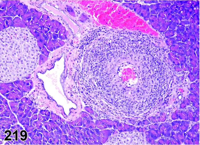
Mouse pancreas. Inflammation vessel.
Synonyms: Arteritis; vasculitis; polyarteritis
Pathogenesis: Spontaneous or induced inflammation of vascular wall in the pancreas.
Diagnostic features
Typically affects muscular wall of small to medium sized arteries or veins.
Variable mixed inflammatory cell infiltrate within and around vessel.
Possible presence of fibrinoid necrosis of vessel wall.
Proliferation of endothelium and thickening (fibrosis) of vessel wall.
Differential diagnoses
Inflammation, acute: Inflammatory processes predominantly within the parenchyma; vascular involvement only secondary and representing a minor proportion of the inflammatory process.
Infiltrate, inflammatory cell: Lack of vascular orientation, no other features of inflammation such as hemorrhage or fibrinoid necrosis.
Comment: Inflammation of the vasculature, if present, is frequently noted in histological sections of pancreas due to the prominent pancreato-duodenal artery and other small caliber interlobular pancreatic arteries, and mesenteric vessels in the vicinity. Arteritis lesions may be due to immune-mediated etiology as seen in (NZBXNZW)F1 hybrid, and MRL/Mp mice or due to hypertension as seen in spontaneously hypertensive rat (SHR) strains. Arteritis due to drug/chemical treatment is occasionally observed and may depend on the species, strain and sex. Examples include vasoactive agents like angiotensin, norepinephrine, dopamine agonists (like fenoldopam mesylate) and xanthine compounds which cause focal to diffuse medial necrosis and hemorrhage especially within the small and medium-caliber mesenteric, pancreatic and renal arteries; and non-vasoactive compounds like 2-amino-5-nitrophenol, nitrofurantoin, or phenacetin.
Edema
Figure 220.
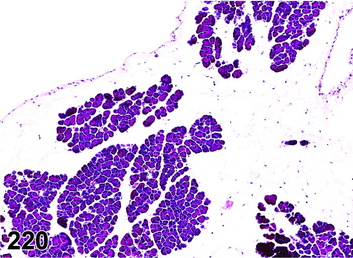
Mouse pancreas. Edema.
Pathogenesis: Accumulation of tissue fluid in the interstitium resulting from increased vascular permeability due to release of pancreatic enzymes, increased hydrostatic pressure in vasculature, decreased oncotic pressure, impaired coagulation system, or impaired lymphatic drainage.
Diagnostic features
Focally extensive or diffuse.
Distension of the interstitial tissue forming clear spaces or spaces filled with lightly eosinophilic material.
Minimal inflammatory cell infiltrate may be present in some cases.
Increase in total pancreatic volume and weight.
Differential diagnosis
Inflammation, acute: Usually associated with tissue and vascular damage resulting in exudative edema and inflammatory cell infiltrate.
Comment: Edema within the exocrine pancreas may be exudative (inflammatory edema due to capillary injury secondary to leakage of pancreatic enzymes) or transudative (hydrodynamic derangement secondary to increased hydrostatic osmotic pressure and decreased oncotic pressure). The exudative edema is usually accompanied by inflammatory cell infiltrate and tissue injury. Examples of chemical induced pancreatic edema have been documented in mice treated with p-nitrophenol and rats treated with D & C Yellow No. 11 and PCB mixtures in US-NTP studies.
Hemorrhage
Pathogenesis: Hemorrhage can occur from acinar necrosis, inflammation, vascular injury, or tumors.
Diagnostic features
Presence of erythrocytes in interstitial tissue, acini, ductules, or ducts.
Differential diagnoses
Angiectasis: Blood present within dilated vascular lumina.
Artifact: Erythrocytes on the surface of the pancreas sample or infiltrating from the surface into interstitial tissue; a result of necropsy procedure.
Comment: Islet hemorrhage has been described as a spontaneous finding in Sprague-Dawley rats (Imaoka et. al. 2007). If prominent, this may involve peri-insular exocrine tissue.
e. Miscellaneous Lesions
Hypertrophy, acinar cell
(Figures 221 and 222)
Figure 221.
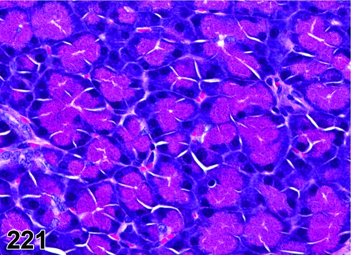
Rat pancreas. Normal acini – for comparison with Figure 222.
Figure 222.
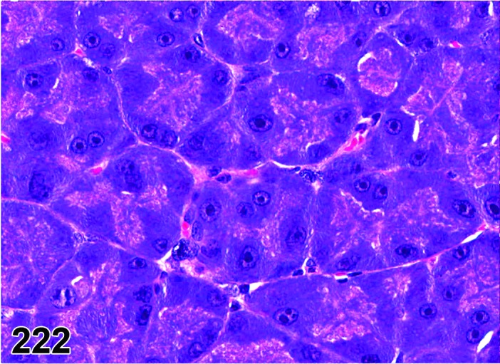
Rat pancreas. Hypertrophy acinar cell diffuse (same magnification as Figure 221).
Synonym: Cytomegaly
Pathogenesis: Increased size of pancreatic exocrine acinar cells due to increased trophic factors.
Diagnostic features
Usually diffuse distribution.
Enlarged acinar cells with more abundant cytoplasmic volume and more numerous zymogen granules.
No increase in the number of cells per acinus.
Larger nuclei with prominent nucleoli.
Increase in pancreas to body weight ratios.
Differential diagnoses
Halos, peri-insular, increased: characteristic distribution pattern; exocrine acini (peri-insular) surrounding the islets of Langerhans have more zymogen granules and slightly larger nuclei than their distal (tele-insular) counterparts.
Hyperplasia, acinar cell: Increased number of cells per acinus.
Comment: Exocrine pancreatic acinar hypertrophy results from dietary and trophic factors. Feeding rats with raw but not heat-treated soy flour (lack trypsin inhibitors) caused acinar hypertrophy initially. However, with continued feeding, the hypertrophic change progressed to hyperplasia with marked increase in DNA content. This hypertrophic change is attributed to the presence of trypsin inhibitors within the raw soy flour and its ability to stimulate the secretion of cholecystokinin by feed back mechanism (Crass and Morgan 1982; Folsch et al. 1978). Similarly, administration of gastrin (but not secretin analogues) like pentagastrin, pancreozymin, and peptavlon also caused pancreatic acinar cell hypertrophy (Rothman, and Wells 1967). Administration of isoprenaline (isoproterenol) also cased an increase in pancreatic weight due to acinar cell hypertrophy and increase in zymogen granules (Sturgess and Reid 1973). However, pancreas weight and pancreas to body weight ratios are not commonly recorded or calculated in toxicity studies.
Halos, peri-insular, increased
Figure 223.
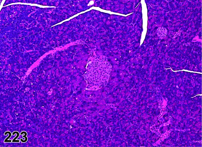
Mouse pancreas. Halo, peri-insular.
Synonyms: Eosinophilic change; focal eosinophilic hypertrophic cells; hypertrophy, peri-insular
Pathogenesis: Hypertrophy of pancreatic exocrine acinar cells surrounding the Islets of Langerhans.
Diagnostic features
Hypertrophy of exocrine pancreatic acini surrounding the islets of Langerhans.
Acinar cells with more abundant cytoplasmic volume with larger zymogen granules than tele-insular acinar cells (located distantly from the islets).
Larger nuclei with more nucleoli than tele-insular acinar cells.
Differential diagnosis
Hypertrophy, acinar cell: No local association with islets, usually multifocal to diffuse distribution.
Comment: The difference in the size of peri- and tele-insular acinar cells is greater in mice than in rats, resulting in more prominent peri-insular halos in mice. The peri-insular acinar cells are more resistant (3 hours) to pilocarpine-induced degranulation than the tele-insular acinar cells (1 hour). The size of peri-insular halos is markedly reduced in alloxan-induced diabetic pancreata compared to control non-diabetic rats. These features of the peri-insular halos are due to the hormones (mainly ghrelin and insulin) secreted by the beta-cells in islets of Langerhans that are locally enriched due to the insulo-acinar capillary anastomoses or due to diffusion. The peri-insular halos are not recorded in routine toxicity studies but recording any alterations within these halos may provide important information about chemicals affecting islet cells such as alloxan or about chemicals directly affecting the exocrine pancreas like pilocarpine.
Halos, peri-insular, decreased
Pathogenesis: Degranulation of the peri-insular acinar cells following loss or decrease in trophic factors released from the surrounding the islets of Langerhans.
Diagnostic features
Decreased zymogen granules within exocrine pancreatic acini surrounding the islets of Langerhans.
Loss of size distinction between the peri-insular acini located adjacent to the islets of Langerhans (usually larger) and the tele-insular acini located distantly from the islets (usually smaller than peri-insular acini)
Differential diagnosis
none
Comment: The difference in the size of peri- and tele-insular acinar cells is greater in mice than in rats, resulting in more prominent peri-insular halos in mice. The peri-insular acinar cells are more resistant (3 hours) to pilocarpine-induced degranulation than the tele-insular acinar cells (1 hour). The size of peri-insular halos is markedly reduced in alloxan-induced diabetic pancreata compared to control non-diabetic rats. These features of the peri-insular halos are due to the hormones (mainly insulin) secreted by the β-cells in islets of Langerhans that are locally enriched due to the insulo-acinar capillary anastomoses or due to diffusion. The peri-insular halos are not recorded in routine toxicity studies but recording any alterations within these halos may provide important information about chemicals affecting islet cells such as alloxan or about chemicals directly affecting the exocrine pancreas like pilocarpine.
Focus, basophilic
Figure 224.
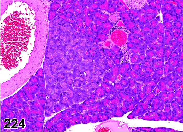
Rat pancreas. Focus, basophilic.
Synonyms: Basophilic-atypical acinar cell focus; focal cellular change; focal basophilic cellular change; cytological alteration
Pathogenesis: Focal formation of morphologically atypical pancreatic exocrine acinar cells.
Diagnostic features
Tinctorially distinct focus with decreased zymogen granules and increased basophilia due to abundance of RER.
Affects single or multiple contiguous acini with oval to irregular shape without encapsulation and with retained angular shape of the pancreatic lobe.
No compression or displacement of adjacent pancreatic acini
Cellular hypertrophy is commonly seen.
Slightly enlarged basal to parabasal nuclei with prominent nucleoli.
Differential diagnosis
Hyperplasia, acinar cell, focal: Increased number of cells per acinus, usually no increase of cytoplasmic basophilia.
Zymogen granules decreased, acinar cell: Reduced cell size due to loss of zymogen granules, nuclei inactive,
Comment: The incidence of basophilic foci is 6.6% in control and 5.4% in corn oil gavage historical control male F344 rats in the NTP database (Eustis et al. 1990). Basophilic foci occur also in mice, but at a much lower incidence than in rats (Roebuck et al. 1980). There is an increase in the incidence of basophilic foci in rats treated with 4-hydroxyaminoquinoline-1-oxide (4HAQO), 7,12-dimethyl-benzanthracene (DMBA), N5-(N-methyl-N-nitrosocarbamoyl)-L-ornithine (MNCO), raw soy bean flour, plant trypsin inhibitors, and azaserine. The proliferative ability of exocrine acinar cytological alterations as measured by tritiated thymidine autoradiography and mitotic index indicated that basophilic foci have similar proliferative ability as the surrounding normal exocrine acini (Rao et al. 1989). Thus, unlike focal hyperplasia, the basophilic foci should not be considered as preneoplastic (Scarpelli et al. 1984). The cytologic appearance of acinar cells within the basophilic foci may indicate an altered capacity for, or a defect in, the synthesis of secretory proteins. Basophilic foci stained positively with gamma-glutamyltranspetidase (GGT) while eosinophilic foci stained negatively. A microscopic morphological entity described as spontaneous pancreatic acinar hypertrophic foci in rats by Chiu (Chiu 1983) may indeed be basophilic foci because they share several features such as increased incidence with age, and they are non-hyperplastic and non-neoplastic.
Metaplasia, hepatocytic
Figure 225.
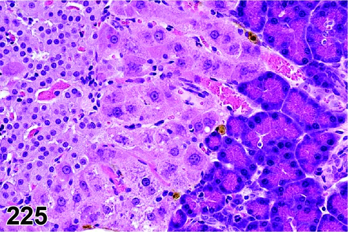
Rat pancreas. Metaplasia,hepatocytic at the interface between endocrine and exocrine tissue.
Pathogenesis: Periductal oval cells or acinar/endocrine intermediate transitional cells transdifferentiate into foci morphologically identical to hepatocytes within the liver.
Diagnostic features
Polygonal cells with central nuclei and abundant finely granular eosinophilic cytoplasm (morphologically identical to hepatocytes).
Located adjacent to ducts and islets of Langerhans.
Usually well integrated into surrounding tissue.
Hepatocytes have well developed bile canaliculi
Differential diagnoses
Hypertrophy, acinar cell: Pancreatic exocrine acini; focal to lobar distribution.
Halos, peri-insular: pancreatic exocrine acini; almost always surrounding islets.
Ectopic liver tissue: Usually circumscribed and not wholly integrated into the ‘host’ tissue; no evidence of dysplasia or history of tissue injury.
Comment: The incidence of metaplastic hepatic foci is less than 1% in control rats and mice. These lesions are occasionally observed in the rat pancreas and less frequently in the mouse pancreas. The pathogenesis of these sporadic lesions is not known. Experimentally, these metaplastic hepatic foci within the rat exocrine pancreas may be induced by several protocols such as copper depletion followed by copper repletion (s), ciprofibrate feed model (Reddy et al. 1984), and multiple subcutaneous injections of cadmium chloride (Konishi et al. 1990). Metaplastic hepatic foci in the pancreas were also induced in a transgenic mouse model where KGF (FGF7) was over-expressed in the pancreas under the control of the rat insulin promoter (Krakowski et al. 1999). An increase in metaplastic hepatic foci in toxicological studies may also be due to the result of test article-induced pancreatic atrophy and subsequent hepatic transdifferentiation.
Metaplasia, ductular
Synonym: Tubular complexes
Pathogenesis: Acinar cells transdifferentiate or take the appearance of ducts especially in areas of chronic pancreatitis.
Diagnostic features
Focal, circumscribed lesions surrounded by normal exocrine acini.
Variable caliber ductules lined by flat cuboidal epithelial cells.
Located in the acinar compartment rather than in the native ducts.
Intermixed with pale atrophic exocrine acini.
Islets not usually involved.
Differential diagnoses
Hyperplasia, Ductular: do not have intermixed pale exocrine acini.
Atrophy, acinar cell: ductular metaplasia may a component in some cases of atrophy and is considered a reparative process.
Comment: Exocrine acinar ductular metaplasia is usually secondary to chronic pancreatitis and atrophy and is considered a reparative process. The diagnosis of ductular metaplasia is not commonly used in toxicologic studies since it is one of the features of atrophy. In DMBA implantation studies in rats that resulted in pancreatic ductular adenocarcinoma, these lesions were considered preneoplastic; however, in the majority of cases these lesions do not progress to neoplasia and are of reparative or adaptive nature. The diagnosis of ductular metaplasia may be considered in those studies where a progression to ductular hyperplasia and neoplasia is noted.
Ectasia, duct
(Figures 226 and 227)
Figure 226.
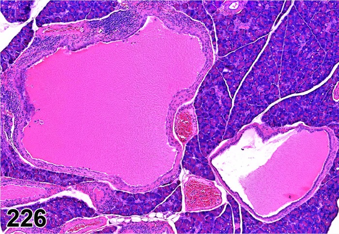
Mouse pancreas. Ectasia, duct.
Figure 227.
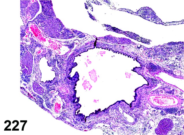
Mouse pancreas. Ectasia, duct.
Synonyms: Dilatation; luminal distension; cystic duct; ductal cyst
Pathogenesis: Luminal dilatation due blockage of the intralobular or interlobular ducts secondary to fibrosis or due to calculi.
Diagnostic features
Solitary or multiple tortuous irregular enlarged lumina.
Lined by flattened cuboidal epithelial cells.
May be accompanied by mild interstitial infiltration of lymphocytes and plasma cells.
Inspissated secretions may be present.
Differential diagnosis
None.
Comment: Duct ectasia is the most common age-associated ductal change in Albino and Gray Norway, August hooded, Fisher 344, Hap:(SD), and Crl:COBS CD (SD) rats (Denda et al. 1994). The lesions of duct ectasia or interlobular cystic ducts are uncommon and the incidence is usually less than 1% in rats and mice. These lesions usually result from blockage of inter- and intra-lobular ducts either proximally or distally due to chronic pancreatitis or rare inspissated secretory material.
Fibrosis
Synonym: Interstitial fibrosis
Pathogenesis: Pancreatic stellate cells reaction to acute or chronic inflammation or toxicity.
Diagnostic features
Focal, lobular, or diffuse.
Increase in the interstitial connective tissue due to pancreatic stellate cell proliferation and collagen deposition.
May be accompanied by inflammatory cell infiltrates.
Differential diagnosis
Amyloid: Abundant extracellular eosinophilic amorphous ground substance that exhibits apple green birefringence in Congo red stained sections under polarized light and absence of fibroblast cell proliferation.
Comment: Collagen may be identified by its birefringence under polarized light and can be stained histochemically using Masson’s trichrome, and van Gieson protocols. In some adult control rats, there may be some peri-insular interstitial fibrosis that extends for short distance into exocrine pancreas. This should be distinguished from fibrosis resulting from tissue insults based on the severity, distribution and the accompanying chronic inflammatory cell infiltrates.
f. Proliferative lesions of the exocrine pancreas
The lesions described here cover the currently known spectrum of proliferative findings in conventional strains of (wildtype) rats and mice. The majority of proliferative lesions in transgenic mouse models of human pancreatic cancer resemble the lesions of wildtype animals. Lesions that occur only in specific transgenic mouse models, like mucinous cystadenomas with ovarian-like stroma, are not described here (Hruban et al. 2006).
Hyperplasias and tumors of the exocrine pancreas are rare and age-related conditions in control rats and mice (Longnecker and Millar 1990; Boorman and Sills 1999). The separation of acinar hyperplasia and adenoma can be difficult and in the end may be arbitrary as these lesions seem to represent a continuum. Hyperplasia of ducts in circumscribed fibrous areas is often found in aged rats. Duct-like structures do not indicate an origin from pancreatic ducts as is the case in humans. Although hyperplasia of ducts may be seen in aged rats, it is still questionable if true ductular neoplasia occurs in rats such as humans (cystadenoma, cystadenocarcinoma) (Greaves and Faccini 1992). In mice, no spontaneous ductal cell hyperplasia or neoplasia has been described. Animal models with carcinogens revealed that rats develop acinar cell carcinomas whereas hamsters develop ductal carcinomas with the same carcinogen. This suggests that the species is a major determinant of the phenotype in carcinomas of the pancreas (Longnecker 1986; Longnecker 1994). Mice are relatively insensitive to the induction of pancreatic tumors with chemicals that are known to be tumorigens in rats (Boorman and Sills 1999).
g. Non-neoplastic Proliferative Lesions
Hyperplasia, acinar cell
(Figures 228 and 229)
Figure 228.
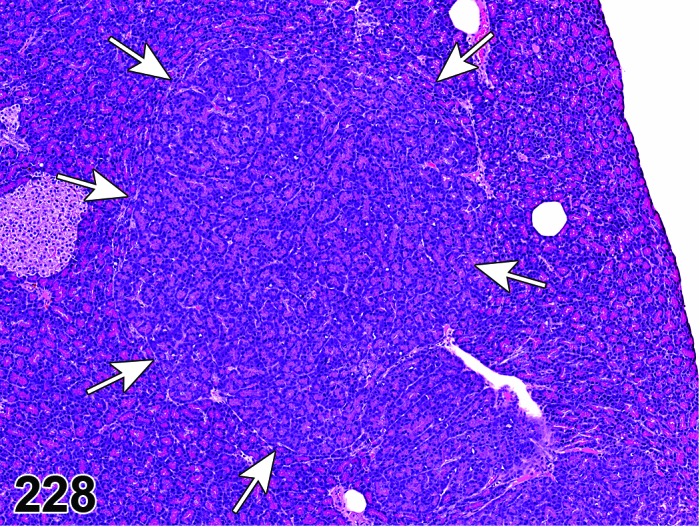
Rat pancreas. Hyperplasia, acinar cell.
Figure 229.
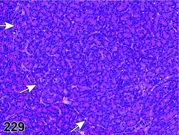
Rat pancreas. Hyperplasia, acinar cell. Higher magnification of Figure 228.
Synonym: Focus, eosinophilic
Modifier: Focal, diffuse
Histogenesis: Acinar cells.
Diagnostic features
Focal lesions usually larger than an average, unchanged islet.
Increased number of cells per acinus.
May exhibit tinctorial variation from surrounding tissue.
Merge usually imperceptibly with adjacent tissue; no or only minimal compression or displacement of adjacent acinar tissue.
Usually non-encapsulated.
Acinar architecture usually maintained or slightly altered at the most.
Low to high mitotic index, nuclear polymorphism and nuclear crowding possible.
Differential diagnoses
Adenoma, acinar cell: Distinct demarcation from the surrounding tissue with compression and sometimes encapsulation; altered architecture.
Focus, basophilic: Enlarged acinar structures with increased basophilia due to abundance of RER; number of cells per acinus not increased; no compression, acinar structure not altered.
Hypertrophy, acinar cell: Enlarged acinar cells with increased zymogen granules; number of cells per acinus not increased; usually diffuse distribution.
Comment: The separation of acinar hyperplasia and adenoma can be difficult and in the end may be arbitrary as these seem to be a continuum. Some foci of hyperplasia may be just at the edge of an adenoma and thus categorized as such due to plane of section. The grade of alteration of acinar structure and compression of adjacent tissue are the most important differential diagnostic criteria.
A sub-category of focal acinar hyperplasia with cell enlargement due to an increase of zymogen granules has been termed “focus, eosinophilic”. Eosinophilic foci are thought to be part of a continuum of lesions that subsequently progress into adenomas and adenocarcinoma (Rao et al. 1989; Scarpelli et al. 1984). In contrast to basophilic foci, these foci are positive for ATPase and glutathione-S-transferase µ histochemical stains. The incidence of eosinophilic foci is 2.6% in male F344 rats (Eustis and Boorman 1985). Eosinophilic foci occur also in mice, but at a much lower incidence than in rats (Roebuck et al. 1980).
Due to the difficulties to differentiate eosinophilic foci from focal hyperplasia in H&E stained sections, it is recommended to diagnose eosinophilic foci not separately but to collect them under focal hyperplasia in this nomenclature system.
Acinar hyperplasias have a low spontaneous incidence in rats and mice that increase with age (Longnecker and Millar 1990; Boorman and Sills 1999).
Hyperplasia, ductal cell
(Figures 230, 231 and Figure 232)
Figure 230.
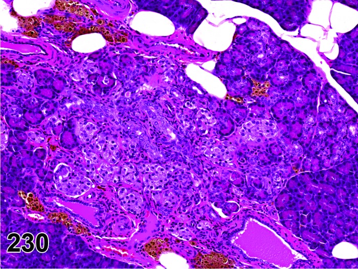
Rat pancreas. Hyperplasia, ductal cell, associated with pigment and minimal islet cell fibrosis.
Figure 231.
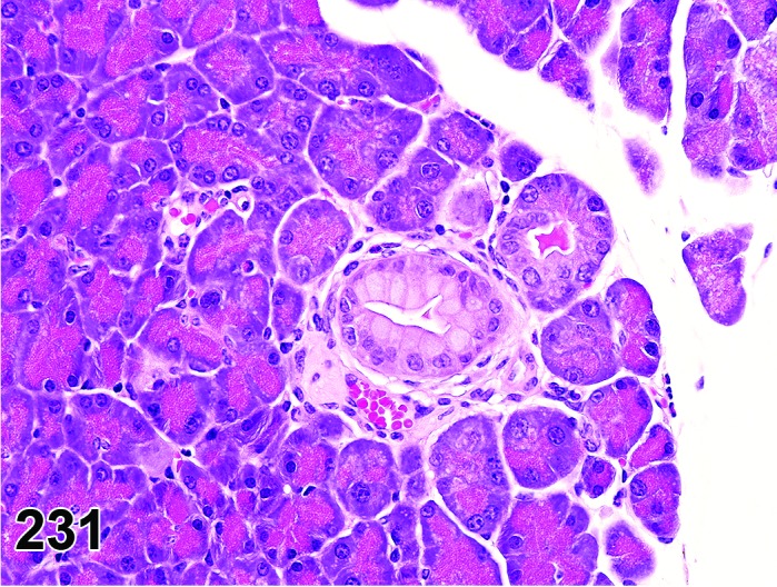
Mouse pancreas. Hyperplasia ductal cell. Note atypical character with mucin accumulation in tall columnar ductal cells in a transgenic mouse model of human pancreatic cancer; lesion resembles low-grade mPanIN.
Figure 232.
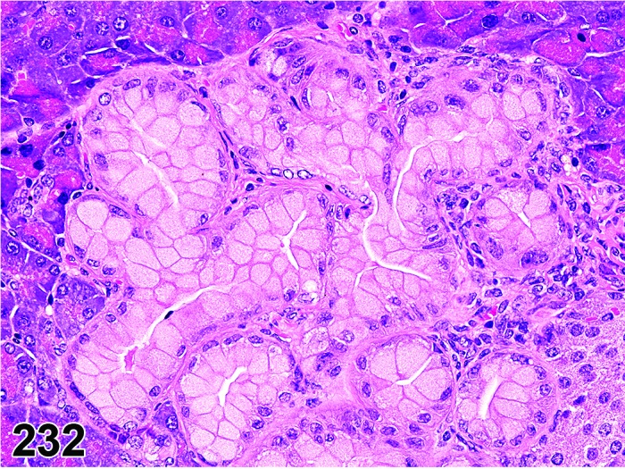
Mouse pancreas. Hyperplasia ductal cell. Note atypical character with cribriform growth pattern and mucin accumulation in hypertrophic ductal cells in a transgenic mouse model of human pancreatic cancer. Lesion resembles higher-grade mPanIN.
Histogenesis: Intra- and inter-lobular pancreatic ducts.
Diagnostic features
Multiple cross section profiles of tortuous hyperchromatic ductular cells within the acinar parenchyma.
Ducts with increased number of lining epithelial cells often forming papillae.
Protruding papillae may cause partial obliteration of the lumen.
Varying levels of cellular pleomorphism and atypia may be present.
Dilated tubules lined by flattened epithelium may occur.
Mucus metaplasia in hyperplastic duct epithelium may occur in transgenic mouse models of pancreatic neoplasia.
Differential diagnosis
Adenoma, ductal cell: Complex of ductular structures; focal acinar atrophy leading to increased numbers of ducts/unit area.
Comment: Hyperplasia ductal cell is more frequently noted within the intrapancreatic ducts than in the extrapancreatic ducts. Hyperplasia of ducts in circumscribed fibrous areas is often found in aged rats.
In mice, no spontaneous ductal cell hyperplasia or neoplasia has been described.
Ductal phenotype may occur under experimental conditions after administration of DMBA (Wendt et al. 2007), infection with reovirus 3 (Greaves 2012), feeding a Western-style diet (Xue et al. 1996), and in transgenic mice (Kras oncogene) (Grippo et al. 2003).
Transgenic mouse models for human pancreatic cancer may develop atypical ductal hyperplasia with mucin accumulation within tall columnar cells lining the ducts (Figure 231) (Hingorani et al. 2003; Aguirre et al. 2003). Following human pathology, these preneoplastic lesions are termed murine pancreatic intraepithelial neoplasia (mPanINs) (Hruban et al. 2006). Morphologically they range from ducts lined with an increased number of monomorphic tall columnar mucinous cells to multilayered lesions with cribriform growth and loss of cell polarity (Figure 232). A grading scheme of PanIN has been introduced (Hruban et al. 2006).
h. Neoplasms
Adenoma, acinar cell
(Figures 233 and 234)
Figure 233.
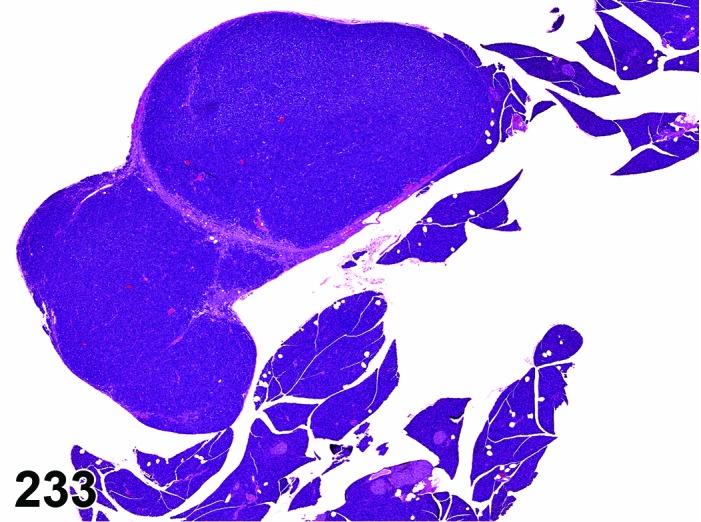
Rat pancreas. Adenoma, acinar cell.
Figure 234.
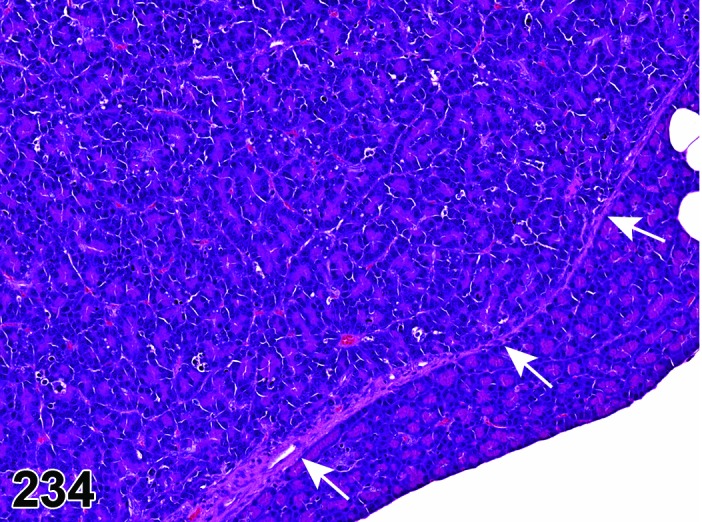
Rat pancreas. Adenoma, acinar cell. Higher magnification of Figure 233. Note sharp demarcation from surrounding tissue by delicate fibrous capsule (arrows).
Histogenesis: Acinar cells
Diagnostic features
Compression of adjacent tissue often present.
Encapsulation may occur but is often not present.
Sharp demarcation from surrounding normal pancreatic tissue.
Architecture is increasingly altered with increasing size.
Cells usually well-differentiated.
Exhibit variable degree of mitotic index, and nuclear polymorphism.
Differential diagnoses
Hyperplasia, acinar cell: No encapsulation or sharp demarcation, no or only slight alteration of acinar structure.
Adenocarcinoma, acinar cell: Local invasion or metastases.
Comment The separation of acinar hyperplasia and adenoma can be difficult and in the end may be arbitrary as these seem to be a continuum. Some foci of hyperplasia may be just at the edge of an adenoma and thus categorized as such due to plane of section.
Adenoma, ductal cell
(Figures 235 and 236)
Figure 235.
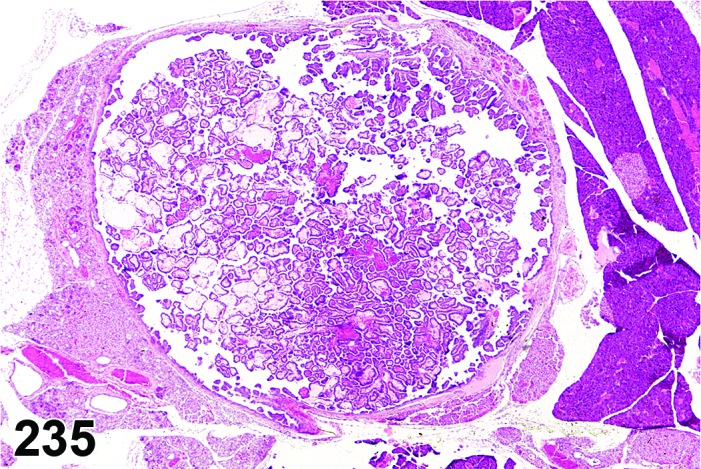
Mouse pancreas. Adenoma, ductal cell.
Figure 236.
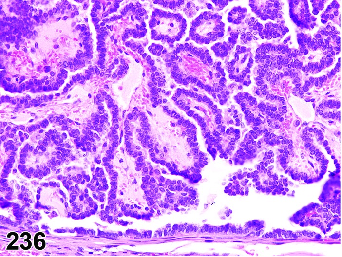
Mouse pancreas. Adenoma, ductal cell. Higher magnification of Figure 235.
Histogenesis: Intrapancreatic ducts.
Diagnostic features
Complex of ductular structures lined by a high cuboidal epithelium resembling that of normal ductules.
Differential diagnosis
Hyperplasia, ductal cell: Ducts with hyperplastic cells, only partly obliterating the lumen, found within focal fibrous areas.
Comment: Duct-like structures do not indicate an origin from pancreatic ducts such as occurs in humans. Although hyperplasia of ducts may be seen in aged rats, it is still questionable if true ductular neoplasia occurs in rats as it does in humans (cystadenoma, cystadenocarcinoma) (Greaves and Faccini 1992). In mice, no spontaneous ductal cell hyperplasia or neoplasia have been described. Pancreatic neoplasm with ductal phenotype may occur under experimental conditions after administration with DMBA (Wendt et al. 2007), infection with reo-virus 3 (Greaves 2012), feeding a Western-style diet (Xue et al. 1996), and in transgenic mice (Kras oncogene) (Grippo et al. 2003).
Adenocarcinoma, acinar cell
(Figures 237, 238 and 239)
Figure 237.
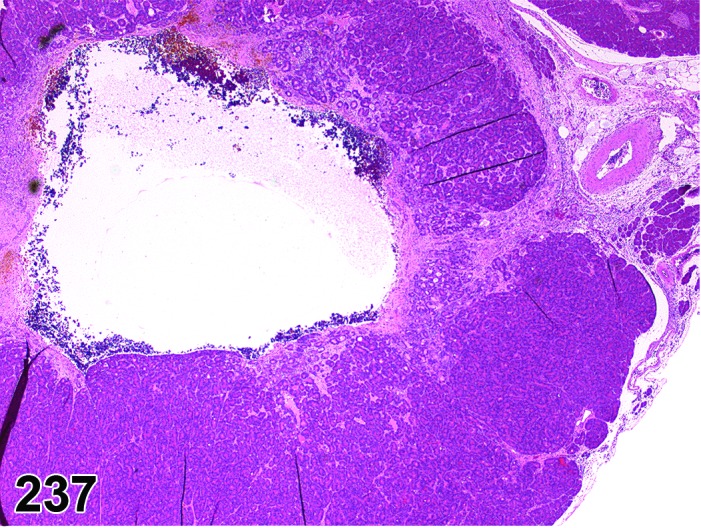
Rat pancreas. Adenocarcinoma, acinar cell.
Figure 238.
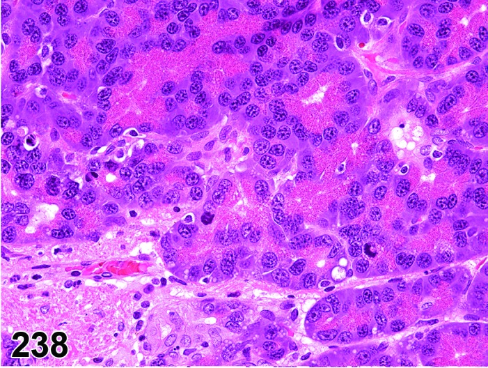
Rat pancreas. Adenocarcinoma, acinar cell. Higher magnification of Figure 237.
Figure 239.
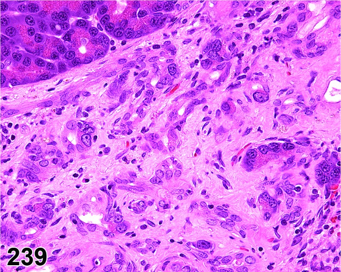
Rat pancreas. Adenocarcinoma, acinar cell. Higher magnification of Figure 237.
Histogenesis: Acinar cells.
Diagnostic features
Glandular, trabecular or solid growth pattern.
Loss of acinar architecture.
Cellular pleomorphism and anaplasia.
Local invasion or distant metastases.
May be associated with scirrhous reactions.
Differential diagnosis
Adenoma, acinar cell: No local invasion or metastases.
Comment: Animal models of chemical carcinogenesis revealed that rats develop acinar cell carcinomas whereas hamsters develop ductal carcinomas with the same carcinogen. This suggests that the species is a major determinant of the phenotype in carcinomas of the pancreas (Longnecker 1986; Longnecker 1994).
Azaserine is an effective pancreatic carcinogen in rats which initiates a sequence of focal proliferative changes in acinar cells that develop into carcinomas. The acinar phenotype is mostly retained but focal development of duct-like structures has been noted in a few carcinomas (Longnecker et al. 1992).
Mice are relatively insensitive to the induction of pancreatic tumors with chemicals that are known to be tumorigenic in rats (Boorman and Sills 1999). Squamous cell carcinoma of the exocrine pancreas has been experimentally induced in mice and has morphologic features of squamous cell carcinoma that occurs at other sites (Dixon and Maronpot 1994).
The transgenic mouse model bearing the elastase promoter-SV40-early antigen construct (Ela-1-SV40 T) develop focal acinar cell proliferation that progresses into acinar cell carcinomas while mostly retaining acinar differentiation, although in some cases they can develop into undifferentiated tumors (Glasner et al. 1992). A consensus report and recommendations for a standardized nomenclature with definitions and associated images was developed for the pathology of genetically-engineered mouse models of pancreatic exocrine neoplasia (Hruban et al. 2006).
Adenocarcinoma, ductal cell
(Figures 240 and 241)
Figure 240.
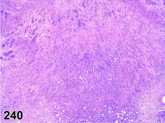
Rat pancreas. Adenocarcinoma, ductal cell. Note prominent scirrhous response.
Figure 241.
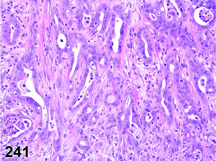
Rat pancreas. Adenocarcinoma, ductal cell. Higher magnification of Figure 240.
Histogenesis: Intrapancreatic ducts.
Diagnostic features
Malignant features, invasive growth.
Cell atypia.
Duct-like structures often accompanied by a dense fibrous stroma.
Differential diagnosis
Adenocarcinoma, acinar cell: Normally no ductular structure.
Comment: Duct-like structures do not indicate an origin from pancreatic ducts such as occurs in humans. Although hyperplasia of ducts may be seen in aged rats, it is still questionable if true ductular neoplasia occurs in rats as it does in humans (cystadenoma, cystadenocarcinoma) (Greaves and Faccini 1992).
Pancreatic adenocarcinomas induced by DMBA implantation in rats showed strong keratin, cytokeratin 19 and cytokeratin 20 expression by immunohistochemistry, consistent with a ductal phenotype (Jimenez et al. 1999). Implantation of DMBA in rats induced transdifferentiation of acinar cells to ductal cells and provided precursor lesions (tubular complexes) for the development of ductal adenocarcinoma (Bockman et al. 2003).
In mice, no spontaneous ductal cell hyperplasia or neoplasia has been described.
Pancreatic intraepithelial precursor lesions in small caliber pancreatic ducts and ductal adenocarcinoma were induced by DMBA implantation in mice (Wendt et al. 2007). Aggressive pancreatic ductal adenocarcinomas may occur in transgenic mice (Kras oncogene) after blockage of TGF-β signalling (Ijichi et al. 2006). A consensus report and recommendations for a standardized nomenclature with definitions and associated images was developed for the pathology of genetically-engineered mouse models of pancreatic exocrine neoplasia (Hruban et al. 2006). In genetically-engineered mice, PanIN has been described as a preneoplastic lesion for adenocarcinomas, but conclusive histopathogenesis has not been shown.
Acknowledgments
We thank Dr. Rupert Kellner for manuscript review and Ms. Beth Mahler, EPL Inc., for image editing. In addition, we acknowledge the tremendous help provided by Ms. Emily Singletary, EPL Inc., in providing several images from the National Toxicology Program image database. We also thank Dr. Kyathanahalli Janardhan, ILS Inc., for providing immunohistochemistry and Hematoxylin & esoin images of gastrointestinal stromal tumors and gastric neuroendocrine tumors.
VII. References
- Abe K, and Watanabe S. Apoptosis of mouse pancreatic acinar cells after duct ligation. Arch Histol Cytol. 58: 221–229. 1995. [DOI] [PubMed] [Google Scholar]
- Aguirre AJ, Bardeesy N, Sinha M, Lopez L, Tuveson DA, Horner J, Redston MS, and DePinho RA. Activated Kras and Ink4a/Arf deficiency cooperate to produce metastatic pancreatic ductal adenocarcinoma. Genes Dev. 17: 3112–3126. 2003. [DOI] [PMC free article] [PubMed] [Google Scholar]
- Aguirre SA, Liu L, Hosea NA, Scott W, May JR, Burns-Naas LA, Randolph S, Denlinger RH, and Han B. Intermittent oral coadministration of a gamma secretase inhibitor with dexamethasone mitigates intestinal goblet cell hyperplasia in rats. Toxicol Pathol. 42: 422–434. 2014. [DOI] [PubMed] [Google Scholar]
- Anderson LC. Salivary gland structure and function in experimental diabetes mellitus. Biomedical Reviews. 9: 107–119. 1998. [Google Scholar]
- Andersson P, Rubio C, Poellinger L, and Hanberg A. Gastric hamartomatous tumours in a transgenic mouse model expressing an activated dioxin/Ah receptor. Anticancer Res. 25(2A): 903–911. 2005. [PubMed] [Google Scholar]
- Andrew W. Age changes in the salivary glands of Wistar Institute rats with particular reference to the submandibular glands. J Gerontol. 4: 95–103. 1949. [DOI] [PubMed] [Google Scholar]
- Badalov N, Baradarian R, Iswara K, Li J, Steinberg W, and Tenner S. Drug-induced acute pancreatitis: an evidence-based review. Clin Gastroenterol Hepatol. 5: 648–661, quiz 644. 2007. [DOI] [PubMed] [Google Scholar]
- Baker DG. In Flynn’s Parasites of Laboratory Animals 2nd ed. American College of Laboratory Animal Medicine. Blackwell, Ames, Iowa. 2007. [Google Scholar]
- Barney GH, Orgebin-Crist MC, and Macapinalac MP. Genesis of esophageal parakeratosis and histologic changes in the testes of the zinc-deficient rat and their reversal by zinc repletion. J Nutr. 95: 526–534. 1968. [DOI] [PubMed] [Google Scholar]
- Becelli R, Perugini M, Gasparini G, Cassoni A, and Fabiani F. Abrikossoff’s tumor. J Craniofac Surg. 12: 78–81. 2001. [DOI] [PubMed] [Google Scholar]
- Bérard J, Gaboury L, Landers M, De Repentigny Y, Houle B, Kothary R, and Bradley WE. Hyperplasia and tumours in lung, breast and other tissues in mice carrying a RAR beta 4-like transgene. EMBO J. 13: 5570–5580. 1994. [DOI] [PMC free article] [PubMed] [Google Scholar]
- Berridge B, Betton GR, Boyle M, Clements P, Le Net JL, Mowat V, Nagai H, Nyska A, Okazaki Y, Rinke M, Snyder P, Wells MY. Proliferative and nonproliferative lesions of the rat and mouse cardiovascular system. In preparation.
- Bertram TA, Markovits JE, and Juliana MM. Non-proliferative lesions of the alimentary canal in rats. In Guides for Toxicologic Pathology. STP/ARP/AFIP Washington DC. 1996. [Google Scholar]
- Bertram TA, Ludlow JW, Basu J, and Muthupalani S. Gastrointestinal Tract. In: Handbook of Toxicologic Pathology, WM Haschek, CG Rousseaux, and MA Wallig (eds.). Academic Press, San Diego, CA. 2013. [Google Scholar]
- Betton GR, Dormer CS, Wells T, Pert P, Price CA, and Buckley P. Gastric ECL-cell hyperplasia and carcinoids in rodents following chronic administration of H2-antagonists SK&F 93479 and oxmetidine and omeprazole. Toxicol Pathol. 16: 288–298. 1988. [DOI] [PubMed] [Google Scholar]
- Betton GR, Whiteley LO, Anver MR, Brown R, Deschl U, Elwell M, Germann PG, Hartig F, Kuttler K, Mori H, Nolte T, Puschner H, and Tuch K. Gastrointestinal Tract. In: International Classification of Rodent Tumors: The Mouse. U Mohr (ed). Springer, Berlin. 23-58. 2001. [Google Scholar]
- Bhaskar SN, Bolden TE, and Weinmann JP. Experimental obstructive adenitis in the mouse. J Dent Res. 35: 852–862. 1956. [DOI] [PubMed] [Google Scholar]
- Bockman DE, Guo J, Büchler P, Müller MW, Bergmann F, and Friess H. Origin and development of the precursor lesions in experimental pancreatic cancer in rats. Lab Invest. 83: 853–859. 2003. [DOI] [PubMed] [Google Scholar]
- Bodner L, Dayan D, Chimovitz N, and Hammel I. Lipid pigment (lipofuscin) accumulation in rat tongue muscle with aging. Exp Gerontol. 26: 89–95. 1991. [DOI] [PubMed] [Google Scholar]
- Blazsek J, and Varga G. Secretion from minor salivary glands following ablation of the major salivary glands in rats. Arch Oral Biol. 44(Suppl 1): S45–S48. 1999. [DOI] [PubMed] [Google Scholar]
- Bogart BI. The effect of aging on the rat submandibular gland: an ultrastructural, cytochemical and biochemical study. J Morphol. 130: 337–351. 1970. [DOI] [PubMed] [Google Scholar]
- Boivin GP, Molina JR, Ormsby I, Stemmermann G, and Doetschman T. Gastric lesions in transforming growth factor beta-1 heterozygous mice. Lab Invest. 74: 513–518. 1996. [PubMed] [Google Scholar]
- Boivin GP, Washington K, Yang K, Ward JM, Pretlow TP, Russell R, Besselsen DG, Godfrey VL, Doetschman T, Dove WF, Pitot HC, Halberg RB, Itzkowitz SH, Groden J, and Coffey RJ. Pathology of mouse models of intestinal cancer: consensus report and recommendations. Gastroenterology. 124: 762–777. 2003. [DOI] [PubMed] [Google Scholar]
- Boorman GA, and Sills RC. Exocrine and endocrine pancreas. In: Pathology of the mouse: Reference and atlas, RR Maronpot, GA Boorman, and BW Gaul (eds). Cache Valley press, St. Louis. 185-206. 1999. [Google Scholar]
- Bosman FT, Carneiro F, Hruban RH, and Theise ND. IARC WHO Classification of Tumours of the Digestive System, 4th ed. Lyon. 417. 2010. [Google Scholar]
- Botts S, Jokinen M, Gaillard ET, Elwell MR, and Mann PC. Salivary, Harderian and Lacrimal glands. In: Maronpot RR, Boorman GA and Gaul BW (eds). Pathology of the mouse: Reference and atlas, Cache Valley press, St. Louis. 185-206. 1999. [Google Scholar]
- Boyd EM, Chen CP, and Muis LF. Resistance to starvation in albino rats fed from weaning on diets containing from 0 to 81 per cent of protein as casein. Growth. 34: 99–112. 1970. [PubMed] [Google Scholar]
- Boyle MC, Crabbs TA, Wyde ME, Painter JT, Hill GD, Malarkey DE, Lieuallen WG, and Nyska A. Intestinal lymphangiectasis and lipidosis in rats following subchronic exposure to indole-3-carbinol via oral gavage. Toxicol Pathol. 40: 561–576. 2012. [DOI] [PubMed] [Google Scholar]
- Brenner GM, and Stanton HC. Adrenergic mechanism responsible for submandibular salivary glandular hypertrophy in the rat. J Pharmacol Exp Ther. 173: 166–175. 1970. [PubMed] [Google Scholar]
- Brown HR, and Hardisty JF. Oral Cavity, Esophagus, and Stomach. In: Pathology of the Fischer Rat: Reference and Atlas. Academic Press, Inc. San Diego. 9-30. 1990. [Google Scholar]
- Brown HR, and Leininger JR. Changes in the Oral Cavity. In: Pathobiology of the Aging Rat, U Mohr, DL Dungworth, and CC Capen (eds). ILSI Press, Washington, DC. 309-322. 1994. [Google Scholar]
- Buchner A, and David R. Lipofuscin in salivary glands in health and disease. Oral Surg Oral Med Oral Pathol. 46: 79–86. 1978. [DOI] [PubMed] [Google Scholar]
- Burkhardt JE, Biehl ML, Kowsz KP, Kadyszewski E, Fisher DO, and Ochoa R. Effects of cholestyramine and diet on small intestinal histomorphometry in rats. Toxicol Pathol. 26: 271–275. 1998. [DOI] [PubMed] [Google Scholar]
- Cadwell K, Patel KK, Maloney NS, Liu TC, Ng AC, Storer CE, Head RD, Xavier R, Stappenbeck TS, and Virgin HW. Virus-plus-susceptibility gene interaction determines Crohn’s disease gene Atg16L1 phenotypes in intestine. Cell. 141: 1135–1145. 2010. [DOI] [PMC free article] [PubMed] [Google Scholar]
- Canpolat L, Sağiroğlu O, Kükner A, and Ozan E. Case report: a rare congenital esophageal malformation on double esophagus in the rat. Kaibogaku Zasshi. 73: 13–17. 1998. [PubMed] [Google Scholar]
- Cao X, Liu X, He W, Liu X, Hao Z, and Zhao Q. [Rat submandibular gland tumor induced by 3-methylcholanthrene]. Hua Xi Kou Qiang Yi Xue Za Zhi. 17: 128–130. 1999. [PubMed] [Google Scholar]
- Cao X, Liu X, Hao Z, Ma C, and Mao Z. [Establishment of rat submandibular gland squamous cell carcinoma induced by DMBA]. Hua Xi Kou Qiang Yi Xue Za Zhi. 18: 156–158. 2000. [PubMed] [Google Scholar]
- Cardiff RD, Leder A, Kuo A, Pattengale PK, and Leder P. Multiple tumor types appear in a transgenic mouse with the ras oncogene. Am J Pathol. 142: 1199–1207. 1993. [PMC free article] [PubMed] [Google Scholar]
- Chan YC, and Leung PS. Acute pancreatitis: animal models and recent advances in basic research. Pancreas. 34: 1–14. 2007. [DOI] [PubMed] [Google Scholar]
- Chandra SA, Nolan MW, and Malarkey DE. Chemical carcinogenesis of the gastrointestinal tract in rodents: an overview with emphasis on NTP carcinogenesis bioassays. Toxicol Pathol. 38: 188–197. 2010. [DOI] [PMC free article] [PubMed] [Google Scholar]
- Chiu T, and Chen HC. Spontaneous basophilic hypertrophic foci of the parotid glands in rats and mice. Vet Pathol. 23: 606–609. 1986. [DOI] [PubMed] [Google Scholar]
- Chiu T. Spontaneous hypertrophic foci of pancreatic acinar cells in CD rats. Toxicol Pathol. 11: 115–119. 1983. [DOI] [PubMed] [Google Scholar]
- Cohen RE, Noble B, Neiders ME, Bedi GS, and Comeau RL. Phenotypic characterization of resident macrophages in submandibular salivary glands of normal and isoproterenol-treated rats. Arch Oral Biol. 37: 503–509. 1992. [DOI] [PubMed] [Google Scholar]
- Coleman GL, Barthold W, Osbaldiston GW, Foster SJ, and Jonas AM. Pathological changes during aging in barrier-reared Fischer 344 male rats. J Gerontol. 32: 258–278. 1977. [DOI] [PMC free article] [PubMed] [Google Scholar]
- Crass RA, and Morgan RG. The effect of long-term feeding of soya-bean flour diets on pancreatic growth in the rat. Br J Nutr. 47: 119–129. 1982. [DOI] [PubMed] [Google Scholar]
- Damsch S, Eichenbaum G, Tonelli A, Lammens L, Van den Bulck K, Feyen B, Vandenberghe J, Megens A, Knight E, and Kelley M. Gavage-related reflux in rats: identification, pathogenesis, and toxicological implications (review) . Toxicol Pathol. 39: 348–360. 2011. [DOI] [PubMed] [Google Scholar]
- Dardick I, Jeans MT, Sinnott NM, Wittkuhn JF, Kahn HJ, and Baumal R. Salivary gland components involved in the formation of squamous metaplasia. Am J Pathol. 119: 33–43. 1985. [PMC free article] [PubMed] [Google Scholar]
- Dardick I, Ho J, Paulus M, Mellon PL, and Mirels L. Submandibular gland adenocarcinoma of intercalated duct origin in Smgb-Tag mice. Lab Invest. 80: 1657–1670. 2000. [DOI] [PubMed] [Google Scholar]
- Dawe CJ. Tumours of the salivary and lachrymal glands, nasal fossa and maxillary sinuses. In: Pathology of tumors in laboratory animals, Vol 2: Tumors of the mouse. Turusov VS (ed). IARC Scientific Publications No. 23. Lyon. 91-134. 1979. [PubMed] [Google Scholar]
- Dawrant MJ, Giles S, Bannigan J, and Puri P. Adriamycin produces a reproducible teratogenic model of vertebral, anal, cardiovascular, tracheal, esophageal, renal, and limb anomalies in the mouse. J Pediatr Surg. 42: 1652–1658. 2007. [DOI] [PubMed] [Google Scholar]
- Declercq J, Van Dyck F, Braem CV, Van Valckenborgh IC, Voz M, Wassef M, Schoonjans L, Van Damme B, Fiette L, and Van de Ven WJ. Salivary gland tumors in transgenic mice with targeted PLAG1 proto-oncogene overexpression. Cancer Res. 65: 4544–4553. 2005. [DOI] [PubMed] [Google Scholar]
- Denda A, Tsutsumi M, and Konishi Y. Exocrine pancreas. In: Pathobiology of the aging rat. U Mohr, DL Dungworth, CC Capen (eds). ILSI press. Washington, D.C. 351-360. 1994. [Google Scholar]
- de Rijk EP, Ravesloot WT, Hafmans TG, and van Esch E. Multifocal ductal cell hyperplasia in the submandibular salivary glands of Wistar rats chronically treated with a novel steroidal compound. Toxicol Pathol. 31: 1–9. 2003. [DOI] [PubMed] [Google Scholar]
- Deschl U, Ernst H, Frantz JD, Goodman DG, Hartig F, Konishi Y, Küttler K, Popp JA, Püschner H, Squire RA, Tatematsu M, Tuch K, Ward JM, and Whiteley LO. Digestive System. In: International Classification of Rodent Tumours. Part I: The rat. U Mohr, CC Capen, DL Dungworth, RA Griesemer, N Ito, VS Turusov (eds). IARC Scientific Publications No. 122. Lyon. 16-27. 1997. [Google Scholar]
- Deschl U, Cattley RC, Harada T, Küttler K, Haily JR, Hartig F, Leblanc B, Marsman DS, and Shirai T. Liver, Gallbladder, and Exocrine Pancreas. In: International Classification of Rodent Tumors: The mouse. U Mohr (ed). Springer, Berlin. 82-86. 2001. [Google Scholar]
- Dethloff LA, Robertson DG, Tierney BM, Breider MA, and Bestervelt LL. Gastric gland degeneration induced in monkeys by the CCK-B/gastrin receptor antagonist CI-988. Toxicol Pathol. 25: 441–448. 1997. [DOI] [PubMed] [Google Scholar]
- Dixon D, and Maronpot RR. Tumours of the pancreas. Pathology of tumours in laboratory animals, Vol 2. Tumours of the mouse, 2nd ed. IARC Scientific Publications No. 111. Lyon. 281–303. 1994. [PubMed] [Google Scholar]
- Dixon D, Alison R, Bach U, Colman K, Foley GL, Harleman JH, Haworth R, Herbert R, Heuser A, Long G, Mirsky M, Regan K, Van Esch E, Westwood FR, Vidal J, and Yoshida M. Nonproliferative and proliferative lesions of the rat and mouse female reproductive system. J Toxicol Pathol. 27(3-4 Suppl): 1S-107S. 2014. [DOI] [PMC free article] [PubMed]
- Doi R, Wada M, Hosotani R, Lee JU, Koshiba T, Fujimoto K, Mori C, Nakamura N, Shiota K, and Imamura M. Role of apoptosis in duct obstruction-induced pancreatic involution in rats. Pancreas. 14: 39–46. 1997. [DOI] [PubMed] [Google Scholar]
- Dunnick JK, Haseman JK, Lilja HS, and Wyand S. Toxicity and carcinogenicity of chlorodibromomethane in Fischer 344/N rats and B6C3F1 mice. Fundam Appl Toxicol. 5: 1128–1136. 1985. [DOI] [PubMed] [Google Scholar]
- Eaton KA, Opp JS, Gray BM, Bergin IL, and Young VB. Ulcerative typhlocolitis associated with Helicobacter mastomyrinus in telomerase-deficient mice. Vet Pathol. 48: 713–725. 2011. [DOI] [PMC free article] [PubMed] [Google Scholar]
- Elangbam CS, Clevidence KJ, Jackson CB, and Holson JF. A communicating intestinal duplication in a Sprague-Dawley rat. Toxicol Pathol. 26: 290–293. 1998. [DOI] [PubMed] [Google Scholar]
- Elmore S. Apoptosis: a review of programmed cell death. Toxicol Pathol. 35: 495–516. 2007. [DOI] [PMC free article] [PubMed] [Google Scholar]
- Elwell MR, and Leininger JR. Tumours of the salivary and lacrimal glands. In: Pathology of tumours in laboratory animals, Vol 1. Tumours of the rat, 2nd ed. VS Turusov, U Mohr (eds). IARC Scientific Publications No. 99. Lyon. 89-107. 1990. [PubMed] [Google Scholar]
- Elwell MR, and McConnell EE. Small and large intestine. In: Pathology of the Fischer Rat. Reference and atlas. GA Boorman, SL Eustis, MR Elwell, CA Montgomery, WF MacKenzie (eds). Academic Press, San Diego. 1990. [Google Scholar]
- Era S, Harada KH, Toyoshima M, Inoue K, Minata M, Saito N, Takigawa T, Shiota K, and Koizumi A. Cleft Palate Caused by Perfluorooctane Sulfonate Is Caused Mainly by Extrinsic Factors. Toxicology. 4:256(1-2): 42-47. 2009. [DOI] [PubMed]
- Erdman SE, and Poutahidis T. Roles for inflammation and regulatory T cells in colon cancer. Toxicol Pathol. 38: 76–87. 2010. [DOI] [PMC free article] [PubMed] [Google Scholar]
- Eustis SL, and Boorman GA. Proliferative lesions of the exocrine pancreas: relationship to corn oil gavage in the National Toxicology Program. J Natl Cancer Inst. 75: 1067–1073. 1985. [PubMed] [Google Scholar]
- Eustis SL, Boorman GA, and Hayashi Y. Exocrine Pancreas. In: Pathology of the fisher rat: Reference and atlas. GA Boorman, SL Eustis, MR Elwell, CA Montgomery Jr., and WF MacKenzie (eds). Academic Press, Inc, New York. 95-108. 1990. [Google Scholar]
- Ewald D, Li M, Efrat S, Auer G, Wall RJ, Furth PA, and Hennighausen L. Time-sensitive reversal of hyperplasia in transgenic mice expressing SV40 T antigen. Science. 273: 1384–1386. 1996. [DOI] [PubMed] [Google Scholar]
- Facinni JM, Abbott DP, and Paulus GJJ. Mouse histopathology. Amsterdam: Elsevier. 1990. [Google Scholar]
- Ferguson MW. Developmental mechanisms in normal and abnormal palate formation with particular reference to the aetiology, pathogenesis and prevention of cleft palate. Br J Orthod. 8: 115–137. 1981. [DOI] [PubMed] [Google Scholar]
- Fernandez-Salguero PM, Ward JM, Sundberg JP, and Gonzalez FJ. Lesions of aryl-hydrocarbon receptor-deficient mice. Vet Pathol. 34: 605–614. 1997. [DOI] [PubMed] [Google Scholar]
- Fitzgerald PJ, and Alvizouri M. Rapid restitution of the rat pancreas following acinar cell necrosis subsequent to ethionine. Nature. 170: 929–930. 1952. [DOI] [PubMed] [Google Scholar]
- Fölsch UR, Winckler K, and Wormsley KG. Influence of repeated administration of cholecystokinin and secretin on the pancreas of the rat. Scand J Gastroenterol. 13: 663–671. 1978. [DOI] [PubMed] [Google Scholar]
- Fox JG, Rogers AB, Whary MT, Ge Z, Ohtani M, Jones EK, and Wang TC. Accelerated progression of gastritis to dysplasia in the pyloric antrum of TFF2 -/- C57BL6 x Sv129 Helicobacter pylori-infected mice. Am J Pathol. 171: 1520–1528. 2007. [DOI] [PMC free article] [PubMed] [Google Scholar]
- Frantz JD, Betton G, Cartwright ME, Crissman JW, Macklin AW, and Maronpot RR. Proliferative Lesions of the non-glandular and glandular stomach in rats. In: Guides for Toxicologic Pathology. STP/ARP/AFIP. Washington DC. 1-20. 1991. [Google Scholar]
- Frith CH, and Heath JE. Adenoma, adenocarcinoma, salivary gland, mouse. In: Monographs on pathology of laboratory animals. Digestive system. TC Jones, U Mohr, RD Hunt (eds). Springer, Berlin. 190-192. 1985. [Google Scholar]
- Frith CH, and Heath JE. Tumours of the salivary gland. In: Pathology of tumours in laboratory animals, Vol. 2. Tumours of the mouse. VS Turusov and U Mohr (eds). IARC Scientific Publications No. 111. Lyon. 115-126. 1994. [PubMed] [Google Scholar]
- Frith CH, and Ward JM. Color atlas of neoplastic and non-neoplastic lesions in aging mice. Elsevier, Amsterdam. 109. 1988. [Google Scholar]
- Fujimoto H, Shibutani M, Kuroiwa K, Inoue K, Woo GH, U M, and Hirose M. A case report of a spontaneous gastrointestinal stromal tumor (GIST) occurring in a F344 rat. Toxicol Pathol. 34: 164–167. 2006. [DOI] [PubMed] [Google Scholar]
- Fukuda M. The influence of isoprenaline and propranolol on the submaxillary gland of the rat. Jpn J Pharmacol. 18: 185–199. 1968. [DOI] [PubMed] [Google Scholar]
- Fukuda A, Kawaguchi Y, Furuyama K, Kodama S, Horiguchi M, Kuhara T, Koizumi M, Boyer DF, Fujimoto K, Doi R, Kageyama R, Wright CVE, and Chiba T. Ectopic pancreas formation in Hes1 -knockout mice reveals plasticity of endodermal progenitors of the gut, bile duct, and pancreas. J Clin Invest. 116: 1484–1493. 2006. [DOI] [PMC free article] [PubMed] [Google Scholar]
- Fukushima S, and Ito N. Papilloma, squamous cell carcinoma, forestomach, rat. In: Monographs on pathology of laboratory animals. Digestive system. TC Jones, U Mohr, and Hunt RD (eds). Springer, Berlin. 289–295. 1985. [Google Scholar]
- Galluzzi L, Vitale I, Abrams JM, Alnemri ES, Baehrecke EH, Blagosklonny MV, Dawson TM, Dawson VL, El-Deiry WS, Fulda S, Gottlieb E, Green DR, Hengartner MO, Kepp O, Knight RA, Kumar S, Lipton SA, Lu X, Madeo F, Malorni W, Mehlen P, Nuñez G, Peter ME, Piacentini M, Rubinsztein DC, Shi Y, Simon HU, Vandenabeele P, White E, Yuan J, Zhivotovsky B, Melino G, and Kroemer G. Molecular definitions of cell death subroutines: recommendations of the Nomenclature Committee on Cell Death 2012. Cell Death Differ. 19: 107–120. 2012. [DOI] [PMC free article] [PubMed] [Google Scholar]
- Garrett JR. The proper role of nerves in salivary secretion: a review. J Dent Res. 66: 387–397. 1987. [DOI] [PubMed] [Google Scholar]
- Garrett JR, Suleiman AM, Anderson LC, and Proctor GB. Secretory responses in granular ducts and acini of submandibular glands in vivo to parasympathetic or sympathetic nerve stimulation in rats. Cell Tissue Res. 264: 117–126. 1991. [DOI] [PubMed] [Google Scholar]
- Ghanayem BI, Maronpot RR, and Matthews HB. Ethyl acrylate-induced gastric toxicity. I. Effect of single and repetitive dosing. Toxicol Appl Pharmacol. 80: 323–335. 1985. [DOI] [PubMed] [Google Scholar]
- Ghaisas S, Saranath D, and Deo MG. ICRC mouse with congenital mega-esophagus as a model to study esophageal tumorigenesis. Carcinogenesis. 10: 1847–1854. 1989. [DOI] [PubMed] [Google Scholar]
- Glasner S, Memoli V, and Longnecker DS. Characterization of the ELSV transgenic mouse model of pancreatic carcinoma. Histologic type of large and small tumors. Am J Pathol. 140: 1237–1245. 1992. [PMC free article] [PubMed] [Google Scholar]
- Glucksmann A, and Cherry CP. Tumours of the salivary glands. In: Pathology of tumours in laboratory animals, Vol. 1. Tumours of the rat, Part 1. VS Turusov (ed). IARC Scientific Publications No. 5. Lyon. 75-86. 1973. [PubMed] [Google Scholar]
- Gopinath C, Prentice DE, and Lewis DJ. Atlas of Experimental Toxicological Pathology. MTP Press. Norwell. 1987. [Google Scholar]
- Greaves P, and Faccini JM. Rat histopathology. A glossary for use in toxicity and carcinogenicity studies. Elsevier, Amsterdam. 118–126. 1992. [Google Scholar]
- Greaves P. Histopathology of preclinical toxicity studies. Interpretation and relevance in drug safety evaluation. Elsevier, Amsterdam. 2012. [Google Scholar]
- Greaves P, Chouinard L, Ernst H, Mecklenburg L, Pruimboom-Brees IM, Rinke M, Rittinghausen S, Thibault S, Von Erichsen J, and Yoshida T. Proliferative and non-proliferative lesions of the rat and mouse soft tissue, skeletal muscle and mesothelium. J Toxicol Pathol. 26(Suppl): 1S–26S. 2013. [DOI] [PMC free article] [PubMed] [Google Scholar]
- Greenman DL, Sheldon W, Schieferstein G, Allen R, and Allaben WT. Triprolidine: 104-week feeding study in rats. Fundam Appl Toxicol. 27: 223–231. 1995. [DOI] [PMC free article] [PubMed] [Google Scholar]
- Gresik EW. The granular convoluted tubule (GCT) cell of rodent submandibular glands. Microsc Res Tech. 27: 1–24. 1994. [DOI] [PubMed] [Google Scholar]
- Grippo PJ, Nowlin PS, Demeure MJ, Longnecker DS, and Sandgren EP. Preinvasive pancreatic neoplasia of ductal phenotype induced by acinar cell targeting of mutant Kras in transgenic mice. Cancer Res. 63: 2016–2019. 2003. [PubMed] [Google Scholar]
- Gukovskaya AS, and Gukovsky I. Autophagy and pancreatitis. Am J Physiol Gastrointest Liver Physiol. 303: G993–G1003. 2012. [DOI] [PMC free article] [PubMed] [Google Scholar]
- Haines DC, Gorelick PL, Battles JK, Pike KM, Anderson RJ, Fox JG, Taylor NS, Shen Z, Dewhirst FE, Anver MR, and Ward JM. Inflammatory large bowel disease in immunodeficient rats naturally and experimentally infected with Helicobacter bilis. Vet Pathol. 35: 202–208. 1998. [DOI] [PubMed] [Google Scholar]
- Haines DC, Chattopadhyay S, and Ward JM. Pathology of aging B6;129 mice. Toxicol Pathol. 29: 653–661. 2001. [DOI] [PubMed] [Google Scholar]
- Hall HD, and Schneyer CA. Salivary Gland Atrophy in Rat Induced by Liquid Diet. Proc Soc Exp Biol Med. 117: 789–793. 1964. [DOI] [PubMed] [Google Scholar]
- Hare WV, and Stewart HL. Chronic gastritis of the glandular stomach, adenomatous polyps of the duodenum, and calcareous pericarditis in strain DBA mice. J Natl Cancer Inst. 16: 889–911. 1956. [PubMed] [Google Scholar]
- Harkness JE, and Ferguson FG. Idiopathic megaesophagus in a rat (Rattus norvegicus). Lab Anim Sci. 29: 495–498. 1979. [PubMed] [Google Scholar]
- Harvey H. Sexual dimorphism of submaxillary glands in mice in relation to reproductive maturity and sex hormones. Physiol Zool. 25: 205–222. 1952. [Google Scholar]
- Haschek WM, Rousseaux CG, and Wallig MA. Gastrointestinal tract. In: Fundamentals of Toxicologic Pathology. Academic Press, San Diego. 2010. [Google Scholar]
- Hebel R, and Stromberg MW. Digestive System. In: Anatomy of the Laboratory Rat. The Williams & Wilkins Company, Baltimore, Maryland. 43-54. 1976. [Google Scholar]
- Helin H, Mero M, Markkula H, and Helin M. Pancreatic acinar ultrastructure in human acute pancreatitis. Virchows Arch A Pathol Anat Histol. 387: 259–270. 1980. [DOI] [PubMed] [Google Scholar]
- Hingorani SR, Petricoin EF, Maitra A, Rajapakse V, King C, Jacobetz MA, Ross S, Conrads TP, Veenstra TD, Hitt BA, Kawaguchi Y, Johann D, Liotta LA, Crawford HC, Putt ME, Jacks T, Wright CVE, Hruban RH, Lowy AM, and Tuveson DA. Preinvasive and invasive ductal pancreatic cancer and its early detection in the mouse. Cancer Cell. 4: 437–450. 2003. [DOI] [PubMed] [Google Scholar]
- Horn VJ, Redman RS, and Ambudkar IS. Response of rat salivary glands to mastication of pelleted vitamin A-deficient diet. Arch Oral Biol. 41: 769–777. 1996. [DOI] [PubMed] [Google Scholar]
- Hosokawa S, Imai T, Hayakawa K, Fukuta T, and Sagami F. Parotid gland papillary cystadenocarcinoma in a Fischer 344 rat. Contemp Top Lab Anim Sci. 39: 31–33. 2000. [PubMed] [Google Scholar]
- Hosoi K, Kobayashi S, and Ueha T. Sex difference in L-glutamine D-fructose-6-phosphate aminotransferase activity of mouse submandibular gland. Biochim Biophys Acta. 543: 283–292. 1978. [DOI] [PubMed] [Google Scholar]
- Hotz J, Goberna R, and Clodi PH. Reserve capacity of the exocrine pancreas. Enzyme secretion and fecal fat assimilation in the 95 percent pancreatectomized rat. Digestion. 9: 212–223. 1973. [DOI] [PubMed] [Google Scholar]
- Hruban RH, Adsay NV, Albores-Saavedra J, Anver MR, Biankin AV, Boivin GP, Furth EE, Furukawa T, Klein A, Klimstra DS, Klöppel G, Lauwers GY, Longnecker DS, Lüttges J, Maitra A, Offerhaus GJA, Pérez-Gallego L, Redston M, and Tuveson DA. Pathology of genetically engineered mouse models of pancreatic exocrine cancer: consensus report and recommendations. Cancer Res. 66: 95–106. 2006. [DOI] [PubMed] [Google Scholar]
- Ijichi H, Chytil A, Gorska AE, Aakre ME, Fujitani Y, Fujitani S, Wright CVE, and Moses HL. Aggressive pancreatic ductal adenocarcinoma in mice caused by pancreas-specific blockade of transforming growth factor-β signaling in cooperation with active Kras expression. Genes Dev. 20: 3147–3160. 2006. [DOI] [PMC free article] [PubMed] [Google Scholar]
- Imai T, Fukuta K, Hasumura M, Cho YM, Ota Y, Takami S, Nakagama H, and Hirose M. Significance of inflammation-associated regenerative mucosa characterized by Paneth cell metaplasia and beta-catenin accumulation for the onset of colorectal carcinogenesis in rats initiated with 1,2-dimethylhydrazine. Carcinogenesis. 28: 2199–2206. 2007. [DOI] [PubMed] [Google Scholar]
- Imaoka M, Satoh H, and Furuhama K. Age- and sex-related differences in spontaneous hemorrhage and fibrosis of the pancreatic islets in Sprague-Dawley rats. Toxicol Pathol. 35: 388–394. 2007. [DOI] [PubMed] [Google Scholar]
- Inomata A, Nakano-Ito K, Fujikawa Y, Sonoda J, Hayakawa K, Ohta E, Taketa Y, Van Gessel Y, Akare S, Hutto D, Hosokawa S, and Tsukidate K. Brunner’s gland lesions in rats induced by a vascular endothelial growth factor receptor inhibitor. Toxicol Pathol. 42: 1267–1274. 2014. [DOI] [PubMed] [Google Scholar]
- Imura H, Shimada A, Naota M, Morita T, Togawa M, Hasegawa T, and Seko Y. Vanadium toxicity in mice: possible impairment of lipid metabolism and mucosal epithelial cell necrosis in the small intestine. Toxicol Pathol. 41: 842–856. 2013. [DOI] [PubMed] [Google Scholar]
- Inanaga A, Habu T, Tanaka E, Taniguchi T, Nishiura T, Ishibashi K, Naruse S, and Abe K. Age changes in secretory function of male and female rat parotid glands in response to methoxamine and pilocarpine. J Dent Res. 67: 565–573. 1988. [DOI] [PubMed] [Google Scholar]
- Iovanna J, and Vaccaro MI. Quantitation of autophagy in the pancreas at the tissue and cell levels. The Pancreapedia. Version 1.0, Sept. 9, 2010.
- Ishihara K, Ohara S, Azuumi Y, Goso K, and Hotta K. Changes of gastric mucus glycoproteins with aspirin administration in rats. Digestion. 29: 98–102. 1984. [DOI] [PubMed] [Google Scholar]
- Ishikawa Y, Nishimori K, Tanaka K, and Kadota K. Naturally occurring mucoepidermoid carcinoma in the submandibular salivary gland of two mice. J Comp Pathol. 118: 145–149. 1998. [DOI] [PubMed] [Google Scholar]
- Ito K, Ishida K, Shishido T, Tabata H, Miura H, Okamiya H, and Hanada T. Acute parietal and chief cell changes induced by a lethal dose of lipopolysaccharide in mouse stomach before thrombus formation. Toxicol Pathol. 28: 304–309. 2000. [DOI] [PubMed] [Google Scholar]
- Jackson CD, and Blackwell BN. Subchronic studies of doxylamine in Fischer 344 rats. Fundam Appl Toxicol. 10: 243–253. 1988. [DOI] [PubMed] [Google Scholar]
- Jackson CD, and Blackwell BN. 2-year toxicity study of doxylamine succinate in the Fischer 344 rat. J Am Coll Toxicol. 12: 1–11. 1993. [Google Scholar]
- Jacoby RO. Sialodacryoadenitis (SDA) infection, rat. In: Monographs on pathology of laboratory animals. Digestive system. TC Jones, U Mohr, and RD Hunt (eds). Springer, Berlin. 201-206. 1985. [Google Scholar]
- Jacoby RO, Bhatt PN, and Jonas AM. Pathogenesis of sialodacryoadenitis in gnotobiotic rats. Vet Pathol. 12: 196–209. 1975. [DOI] [PubMed] [Google Scholar]
- Jimenez RE, Z’graggen K, Hartwig W, Graeme-Cook F, Warshaw AL, and Fernandez-del Castillo C. Immunohistochemical characterization of pancreatic tumors induced by dimethylbenzanthracene in rats. Am J Pathol. 154: 1223–1229. 1999. [DOI] [PMC free article] [PubMed] [Google Scholar]
- Jones TC, Hunt RD, and King NW. Veterinary Pathology. Baltimore MD: Williams and Wilkins. 1997. [Google Scholar]
- Kalter H, and Warkany J. Experimental production of congenital malformations in strains of inbred mice by maternal treatment with hypervitaminosis A. Am J Pathol. 38: 1–21. 1961. [PMC free article] [PubMed] [Google Scholar]
- Karam SM. Lineage commitment and maturation of epithelial cells in the gut. Front Biosci. 4: D286–D298. 1999. [DOI] [PubMed] [Google Scholar]
- Karam SM, and Alexander G. Blocking of histamine H2 receptors enhances parietal cell degeneration in the mouse stomach. Histol Histopathol. 16: 469–480. 2001. [DOI] [PubMed] [Google Scholar]
- Kaufmann W, Bolon B, Bradley A, Butt M, Czasch S, Garman RH, George C, Gröters S, Krinke G, Little P, McKay J, Narama I, Rao D, Shibutani M, and Sills R. Proliferative and nonproliferative lesions of the rat and mouse central and peripheral nervous systems. Toxicol Pathol. 40(Suppl): 87S–157S. 2012. [DOI] [PubMed] [Google Scholar]
- Kazacos EA, and Van Vleet JF. Sequential ultrastructural changes of the pancreas in zinc toxicosis in ducklings. Am J Pathol. 134: 581–595. 1989. [PMC free article] [PubMed] [Google Scholar]
- Keren DF, Elliott HL, Brown GD, and Yardley JH. Atrophy of villi with hypertrophy and hyperplasia of Paneth cells in isolated (thiry-Vella) ileal loops in rabbits. Light-microscopic studies. Gastroenterology. 68: 83–93. 1975. [PubMed] [Google Scholar]
- King WW, and Russell SP. Metabolic, traumatic, and miscellaneous diseases. In: The Laboratory Rat. MA Suchow, SH Weisbroth, CL Franklin (eds). Elsevier, Amsterdam. 2006. [Google Scholar]
- Kitagawa T, and Ono K. Ultrastructure of pancreatic exocrine cells of the rat during starvation. Histol Histopathol. 1: 49–57. 1986. [PubMed] [Google Scholar]
- Klein-Szanto AJP, Martin D, and Sega M. Hyperkeratinization and hyperplasia of the forestomach epithelium in vitamin A deficient rats. Virchows Arch B Cell Pathol Incl Mol Pathol. 40: 387–394. 1982; . [DOI] [PubMed] [Google Scholar]
- Koerker RM. The effects of hypophysectomy on the digestive glands of the mouse. Am J Anat. 121: 571–599. 1967. [DOI] [PubMed] [Google Scholar]
- Konishi N, Ward JM, and Waalkes MP. Pancreatic hepatocytes in Fischer and Wistar rats induced by repeated injections of cadmium chloride. Toxicol Appl Pharmacol. 104: 149–156. 1990. [DOI] [PubMed] [Google Scholar]
- Koo SI, and Turk DE. Effect of zinc deficiency on the ultrastructure of the pancreatic acinar cell and intestinal epithelium in the rat. J Nutr. 107: 896–908. 1977. [DOI] [PubMed] [Google Scholar]
- Korenaga T, Fu X, Xing Y, Matsusita T, Kuramoto K, Syumiya S, Hasegawa K, Naiki H, Ueno M, Ishihara T, Hosokawa M, Mori M, and Higuchi K. Tissue distribution, biochemical properties, and transmission of mouse type A AApoAII amyloid fibrils. Am J Pathol. 164: 1597–1606. 2004. [DOI] [PMC free article] [PubMed] [Google Scholar]
- Kosa P, Szabo R, Molinolo AA, and Bugge TH. Suppression of Tumorigenicity-14, encoding matriptase, is a critical suppressor of colitis and colitis-associated colon carcinogenesis. Oncogene. 31: 3679–3695. 2012. [DOI] [PMC free article] [PubMed] [Google Scholar]
- Krakowski ML, Kritzik MR, Jones EM, Krahl T, Lee J, Arnush M, Gu D, and Sarvetnick N. Pancreatic expression of keratinocyte growth factor leads to differentiation of islet hepatocytes and proliferation of duct cells. Am J Pathol. 154: 683–691. 1999. [DOI] [PMC free article] [PubMed] [Google Scholar]
- Kramer JA, O’Neill E, Phillips ME, Bruce D, Smith T, Albright MM, Bellum S, Gopinathan S, Heydorn WE, Liu X, Nouraldeen A, Payne BJ, Read R, Vogel P, Yu X-Q, and Wilson AGE. Early toxicology signal generation in the mouse. Toxicol Pathol. 38: 452–471. 2010. [DOI] [PubMed] [Google Scholar]
- Kurtzman CP, Robnett CJ, Ward JM, Brayton C, Gorelick P, and Walsh TJ. Multigene phylogenetic analysis of pathogenic candida species in the Kazachstania (Arxiozyma) telluris complex and description of their ascosporic states as Kazachstania bovina sp. nov., K. heterogenica sp. nov., K. pintolopesii sp. nov., and K. slooffiae sp. nov. J Clin Microbiol. 43: 101–111. 2005. [DOI] [PMC free article] [PubMed] [Google Scholar]
- Kuwamura M, Okajima R, Yamate J, Kotani T, Kuramoto T, and Serikawa T. Pancreatic metaplasia in the gastro-achlorhydria in WTC-dfk rat, a potassium channel Kcnq1 mutant. Vet Pathol. 45: 586–591. 2008. [DOI] [PubMed] [Google Scholar]
- Laine VJO, Nyman KM, Peuravuori HJ, Henriksen K, Parvinen M, and Nevalainen TJ. Lipopolysaccharide induced apoptosis of rat pancreatic acinar cells. Gut. 38: 747–752. 1996. [DOI] [PMC free article] [PubMed] [Google Scholar]
- Langberg CW, and Hauer-Jensen M. Influence of fraction size on the development of late radiation enteropathy. An experimental study in the rat. Acta Oncol. 35: 89–94. 1996. [DOI] [PubMed] [Google Scholar]
- Leininger JR, McDonald MM, and Abbott DP. Hepatocytes in the mouse stomach. Toxicol Pathol. 18: 678–686. 1990. [DOI] [PubMed] [Google Scholar]
- Leininger JR, Jokinen MP, Dangler CA, and Whiteley LO. Oral Cavity, esophagus and stomach. In: Pathology of the Mouse. RR Maronpot (ed). Cache River Press, Vienna. 1999. [Google Scholar]
- Leininger JR, and Jokinen MP. Tumors of the Stomach. In: Pathology of tumours in laboratory animals, Vol 2. Tumours of the mouse, 2nd ed. VS Turusov, U Mohr (eds). IARC Scientific Publications No. 111. Lyon. 169–172. 1994. [Google Scholar]
- Levin S, Bucci TJ, Cohen SM, Fix AS, Hardisty JF, LeGrand EK, Maronpot RR, and Trump BF. The nomenclature of cell death: recommendations of an ad hoc Committee of the Society of Toxicologic Pathologists. Toxicol Pathol. 27: 484–490. 1999. [DOI] [PubMed] [Google Scholar]
- Lipman RD, Gaillard ET, Harrison DE, and Bronson RT. Husbandry factors and the prevalence of age-related amyloidosis in mice. Lab Anim Sci. 43: 439–444. 1993. [PubMed] [Google Scholar]
- Loftus SK, Cannons JL, Incao A, Pak E, Chen A, Zerfas PM, Bryant MA, Biesecker LG, Schwartzberg PL, and Pavan WJ. Acinar cell apoptosis in Serpini2-deficient mice models pancreatic insufficiency. PLoS Genet. 1: e38 2005. [DOI] [PMC free article] [PubMed] [Google Scholar]
- Longnecker DS. Experimental models of exocrine pancreatic tumours. In: The exocrine pancreas: Biology, pathobiology and diseases. VLW Go, JD Gardener, FP Brooks, E Lebenthal, EP Diamagno, and GA Scheele (eds). Raven Press, New York. 443–458. 1986. [Google Scholar]
- Longnecker DS. Preneoplastic and neoplastic lesions of the pancreas in hamsters, mouse and rats. In: Pathology of neoplasia and preneoplasia in rodents. P Bannasch, and W Gössner (eds). Schattauer, Stuttgart. 64-81. 1994. [Google Scholar]
- Longnecker DS, and Wilson GL. Pancreas. In: Handbook of Toxicologic pathology, 2nd ed. WM Haschek, CG Rousseaux, MA Wallig. Academic Press, Amsterdam. 227-254. 2002. [Google Scholar]
- Longnecker DS, Memoli V, and Pettengill OS. Recent results in animal models of pancreatic carcinoma: histogenesis of tumors. Yale J Biol Med. 65: 457–464, discussion 465–469. 1992. [PMC free article] [PubMed] [Google Scholar]
- Longnecker DS, and Millar PM. Tumours of the pancreas. In: Pathology of tumours in laboratory animal, Vol 1. Tumours of the rat, 2nd ed. VS Turusov, U Mohr (eds). IARC Scientific Publications No. 99. Lyon. 241–257. 1990. [PubMed] [Google Scholar]
- MacKinnon JE. A yeast in the stomach of the mouse. Mycopathol Mycol Appl. 10: 207–208. 1959. [DOI] [PubMed] [Google Scholar]
- Maekawa A. Changes in the esophagus and stomach. In: Pathobiology of the aging rat, Vol. 2. U Mohr, DL Dungworht, CC Capen (eds). ILSI Press, Washington D.C. 1994. [Google Scholar]
- Maekawa A, Enomoto M, Hirouchi Y, and Yamakawa S. Changes in the upper digestive tract and stomach. In: Pathobiology of the Aging Mouse, Volume 2. U Mohr, DL Dungworth, CC Capen, WW Carlton, JP Sundberg, JM Ward (eds). ILSI Press, Washington D.C. 1996. [Google Scholar]
- Maggio-Price L, Bielefeldt-Ohmann H, Treuting P, Iritani BM, Zeng W, Nicks A, Tsang M, Shows D, Morrissey P, and Viney JL. Dual infection with Helicobacter bilis and Helicobacter hepaticus in p-glycoprotein-deficient mdr1a-/- mice results in colitis that progresses to dysplasia. Am J Pathol. 166: 1793–1806. 2005. [DOI] [PMC free article] [PubMed] [Google Scholar]
- Mahler M, Rozell B, Mahler JF, Merlino G, Devor-Henneman D, Ward JM, and Sundberg JP. Pathology of the Gastrointestinal Tract in Genetical Engineered Mice. In: Pathology of Genetically Engineered Mice. JM Ward, JF Mahler, RR Maronpot, and JP Sundberg (eds). Iowa State University Press, Ames. 269-297. 2000. [Google Scholar]
- Maiuri MC, Zalckvar E, Kimchi A, and Kroemer G. Self-eating and self-killing: crosstalk between autophagy and apoptosis. Nat Rev Mol Cell Biol. 8: 741–752. 2007. [DOI] [PubMed] [Google Scholar]
- Mann PC, Vahle J, Keenan CM, Baker JF, Bradley AE, Goodman DG, Harada T, Herbert R, Kaufmann W, Kellner R, Nolte T, Rittinghausen S, and Tanaka T. International harmonization of toxicologic pathology nomenclature: an overview and review of basic principles. Toxicol Pathol. 40(Suppl): 7S–13S. 2012. [DOI] [PubMed] [Google Scholar]
- Mazue G, Vic P, Gouy D, Remandet B, Lacheretz F, Berthe J, Barchewitz G, and Gagnol JP. Recovery from amiodarone-induced lipidosis in laboratory animals: a toxicological study. Fundam Appl Toxicol. 4: 992–999. 1984. [DOI] [PubMed] [Google Scholar]
- McInnes EF. Background lesions in laboratory animals. A color atlas, 1st ed. Elsevier, Edinburgh. 2012. [Google Scholar]
- Miller CL, Muthupalani S, Shen Z, and Fox JG. Isolation of Helicobacter spp. from mice with rectal prolapses. Comp Med. 64: 171–178. 2014. [PMC free article] [PubMed] [Google Scholar]
- Miyoshi H, Nakau M, Ishikawa TO, Seldin MF, Oshima M, and Taketo MM. Gastrointestinal hamartomatous polyposis in Lkb1 heterozygous knockout mice. Cancer Res. 62: 2261–2266. 2002. [PubMed] [Google Scholar]
- Morgan RW, Ward JM, and Hartman PE. Aroclor 1254-induced intestinal metaplasia and adenocarcinoma in the glandular stomach of F344 rats. Cancer Res. 41: 5052–5059. 1981. [PubMed] [Google Scholar]
- Mukaisho K, Miwa K, Totsuka Y, Shimomura A, Sugihara H, Wakabayashi K, and Hattori T. Induction of gastric GIST in rat and establishment of GIST cell line. Cancer Lett. 231: 295–303. 2006. [DOI] [PubMed] [Google Scholar]
- Murgatroyd LB. A morphological and histochemical study of a drug-induced enteropathy in the Alderley Park rat. Br J Exp Pathol. 61: 567–578. 1980. [PMC free article] [PubMed] [Google Scholar]
- Nakai N, Ishikawa T, Nishitani A, Liu NN, Shincho M, Hao H, Isozaki K, Kanda T, Nishida T, Fujimoto J, and Hirota S. A mouse model of a human multiple GIST family with KIT-Asp820Tyr mutation generated by a knock-in strategy. J Pathol. 214: 302–311. 2008. [DOI] [PubMed] [Google Scholar]
- National Toxicology Program NTP Toxicology and carcinogenesis studies of iodinated glycerol (Organidin(R).) (CAS No. 5634-39-9) in F344/N rats and B6C3F1 mice (gavage studies). Natl Toxicol Program Tech Rep Ser. 340: 1–171. 1990. [PubMed] [Google Scholar]
- National Toxicology Program NTP Toxicology and Carcinogenesis Studies of Malonaldehyde, Sodium Salt (3-Hydroxy-2-propenal, Sodium Salt) (CAS No. 24382-04-5) in F344/N Rats and B6C3F1 Mice (Gavage Studies). Natl Toxicol Program Tech Rep Ser. 331: 1–182. 1988. [PubMed] [Google Scholar]
- National Toxicology Program NTP technical report on the toxicity studies of Glyphosate (CAS No. 1071-83-6) Administered In Dosed Feed To F344/N Rats And B6C3F1 Mice. Toxic Rep Ser. 16: 1–D3. 1992. a. [PubMed] [Google Scholar]
- National Toxicology Program NTP technical report on the toxicity studies of Diethanolamine (CAS No. 111-42-2) Administered Topically and in Drinking Water to F344/N Rats and B6C3F1 Mice. Toxic Rep Ser. 20: 1–D10. 1992. b. [PubMed] [Google Scholar]
- National Toxicology Program NTP Toxicology and Carcinogenesis Studies of Methyleugenol (CAS NO. 93-15-2) in F344/N Rats and B6C3F1 Mice (Gavage Studies). Natl Toxicol Program Tech Rep Ser. 491: 1–412. 2000. [PubMed] [Google Scholar]
- National Toxicology Program NTP Toxicology and Carcinogenesis Studies of Scopolamine Hydrobromide Trihydrate (CAS No. 6533-68-2) in F344 Rats and B6C3F1 Mice (Gavage Studies). Natl Toxicol Program Tech Rep Ser. 445: 1–277. 1997a. [PubMed] [Google Scholar]
- National Toxicology Program NTP Toxicology and Carcinogenesis Studies of D&C Yellow No. 11 (CAS No. 8003-22-3) in F344/N Rats (Feed Studies). Natl Toxicol Program Tech Rep Ser. 463: 1–190. 1997b. [PubMed] [Google Scholar]
- National Toxicology Program Toxicology and carcinogenesis studies of sodium dichromate dihydrate (Cas No. 7789-12-0) in F344/N rats and B6C3F1 mice (drinking water studies). Natl Toxicol Program Tech Rep Ser. 546: 1–192. 2008. [PubMed] [Google Scholar]
- Neuenschwander SB, and Elwell MR. Salivary glands. In: Pathology of the fisher rat: Reference and atlas. GA Boorman, SL Eustis, MR Elwell, CA Montgomery Jr., and WF Mackenzie (eds). Academic Press, New York. 31-42. 1990. [Google Scholar]
- Newberne PM, Conner MW, and Estes P. The influence of food additives and related materials on lower bowel structure and function. Toxicol Pathol. 16: 184–197. 1988. [DOI] [PubMed] [Google Scholar]
- Nielsen LL, Discafani CM, Gurnani M, and Tyler RD. Histopathology of salivary and mammary gland tumors in transgenic mice expressing a human Ha-ras oncogene. Cancer Res. 51: 3762–3767. 1991. [PubMed] [Google Scholar]
- Nielsen LL, Gurnani M, Catino JJ, and Tyler RD. In wap-ras transgenic mice, tumor phenotype but not cyclophosphamide-sensitivity is affected by genetic background. Anticancer Res. 15: 385–392. 1995. [PubMed] [Google Scholar]
- Nishikawa S, Sano F, Takagi K, Okada M, Sugimoto J, and Takagi S. Spontaneous poorly differentiated carcinoma with cells positive for vimentin in a salivary gland of a young rat. Toxicol Pathol. 38: 315–318. 2010. [DOI] [PubMed] [Google Scholar]
- Nyska A, Waner T, Galiano A, and Fich A. Constipation and megacolon in rats related to treatment with oxodipine, a calcium antagonist. Toxicol Pathol. 22: 589–594. 1994. [DOI] [PubMed] [Google Scholar]
- Oates PS, Morgan RGH, and Light AM. Cell death (apoptosis) during pancreatic involution after raw soya flour feeding in the rat. Am J Physiol. 250: G9–G14. 1986. [DOI] [PubMed] [Google Scholar]
- Obert LA, Sobocinski GP, Bobrowski WF, Metz AL, Rolsma MD, Altrogge DM, and Dunstan RW. An immunohistochemical approach to differentiate hepatic lipidosis from hepatic phospholipidosis in rats. Toxicol Pathol. 35: 728–734. 2007. [DOI] [PubMed] [Google Scholar]
- Ohyama Y, Carroll VA, Deshmukh U, Gaskin F, Brown MG, and Fu SM. Severe focal sialadenitis and dacryoadenitis in NZM2328 mice induced by MCMV: a novel model for human Sjögren’s syndrome. J Immunol. 177: 7391–7397. 2006. [DOI] [PubMed] [Google Scholar]
- Olson WA, Habermann RT, Weisburger EK, Ward JM, and Weisburger JH. Induction of stomach cancer in rats and mice by halogenated aliphatic fumigants. J Natl Cancer Inst. 51: 1993–1995. 1973. [DOI] [PubMed] [Google Scholar]
- Ouellette AJ. Paneth cells and innate mucosal immunity. Curr Opin Gastroenterol. 26: 547–553. 2010. [DOI] [PubMed] [Google Scholar]
- Ozono S, Onozuka M, Sato K, and Ito Y. Immunohistochemical evidence for the presence of progesterone receptor in rat submandibular glands. Cell Struct Funct. 16: 511–513. 1991. [DOI] [PubMed] [Google Scholar]
- Pandiri AR, Sills RC, Hoenerhoff MJ, Peddada SD, Ton TV, Hong HH, Flake GP, Malarkey DE, Olson GR, Pogribny IP, Walker NJ, and Boudreau MD. Aloe vera non-decolorized whole leaf extract-induced large intestinal tumors in F344 rats share similar molecular pathways with human sporadic colorectal tumors. Toxicol Pathol. 39: 1065–1074. 2011. [DOI] [PMC free article] [PubMed] [Google Scholar]
- Patnaik AK. Histologic and immunohistochemical studies of granular cell tumors in seven dogs, three cats, one horse, and one bird. Vet Pathol. 30: 176–185. 1993. [DOI] [PubMed] [Google Scholar]
- Percy DH, and Barthold SW. Pathology of Laboratory Rodents and Rabbits, 3rd ed. Iowa State University Press, Ames. 2007. [Google Scholar]
- Pirmohamed M, James S, Meakin S, Green C, Scott AK, Walley TJ, Farrar K, Park BK, and Breckenridge AM. Adverse drug reactions as cause of admission to hospital: prospective analysis of 18 820 patients. BMJ. 329: 15–19. 2004. [DOI] [PMC free article] [PubMed] [Google Scholar]
- Proctor GB, and Carpenter GH. Regulation of salivary gland function by autonomic nerves. Auton Neurosci. 133: 3–18. 2007. [DOI] [PubMed] [Google Scholar]
- Puurunen J, Huttunen P, and Hirvonen J. Is ethanol-induced damage of the gastric mucosa a hyperosmotic effect? Comparative studies on the effects of ethanol, some other hyperosmotic solutions and acetylsalicylic acid on rat gastric mucosa. Acta Pharmacol Toxicol (Copenh). 47: 321–327. 1980. [DOI] [PubMed] [Google Scholar]
- Raju J. Azoxymethane-induced rat aberrant crypt foci: relevance in studying chemoprevention of colon cancer. World J Gastroenterol. 14: 6632–6635. 2008. [DOI] [PMC free article] [PubMed] [Google Scholar]
- Ramiro-Ibáñez F, Trajkovic D, and Jessen B. Gastric and pancreatic lesions in rats treated with a pan-CDK inhibitor. Toxicol Pathol. 33: 784–791. 2005. [DOI] [PubMed] [Google Scholar]
- Randelia HP, and Lalitha VS. Megaoesophagus in ICRC mice. Lab Anim. 22: 23–26. 1988. [DOI] [PubMed] [Google Scholar]
- Randelia HP, Panicker KN, and Lalitha VS. Megaoesophagus in the mouse: histochemical and ultrastructural studies. Lab Anim. 24: 78–86. 1990. [DOI] [PubMed] [Google Scholar]
- Rao MS, Subbarao V, Yeldandi AV, and Reddy JK. Pancreatic acinar cell regeneration following copper deficiency-induced pancreatic necrosis. Int J Pancreatol. 2: 71–85. 1987. [DOI] [PubMed] [Google Scholar]
- Rao MS, Upton MP, Subbarao V, and Scarpelli DG. Two populations of cells with differing proliferative capacities in atypical acinar cell foci induced by 4-hydroxyaminoquinoline-1-oxide in the rat pancreas. Lab Invest. 46: 527–534. 1982. [PubMed] [Google Scholar]
- Rao MS, Dwivedi RS, Yeldandi AV, Subbarao V, Tan X, Usman MI, Thangada S, Nemali MR, Kumar S, Scarpelli DG, and Reddy JK. Role of periductular and ductular epithelial cells of the adult rat pancreas in pancreatic hepatocyte lineage. Am J Pathol. 134: 1069–1086. 1989. [PMC free article] [PubMed] [Google Scholar]
- Rao MS, Yeldandi AV, Subbarao V, and Reddy JK. Role of periductal and ductular epithelial cells of the adult rat pancreas in pancreatic hepatocyte lineage. A change in the differentiation commitment. Am J Pathol. 142: 1952–1957. 1993. [PMC free article] [PubMed] [Google Scholar]
- Reasor MJ, Hastings KL, and Ulrich RG. Drug-induced phospholipidosis: issues and future directions. Expert Opin Drug Saf. 5: 567–583. 2006. [DOI] [PubMed] [Google Scholar]
- Reddy JK, Rao MS, Qureshi SA, Reddy MK, Scarpelli DG, and Lalwani ND. Induction and origin of hepatocytes in rat pancreas. J Cell Biol. 98: 2082–2090. 1984. [DOI] [PMC free article] [PubMed] [Google Scholar]
- Reid LE, and Walker NI. Acinar cell apoptosis and the origin of tubular complexes in caerulein-induced pancreatitis. Int J Exp Pathol. 80: 205–215. 1999. [DOI] [PMC free article] [PubMed] [Google Scholar]
- Redman RS. Morphologic diversity of the minor salivary glands of the rat: fertile ground for studies in gene function and proteomics. Biotech Histochem. 87: 273–287. 2012. [DOI] [PubMed] [Google Scholar]
- Rees J, Spencer A, Wilson S, Reid A, and Harpur E. Time course of stomach mineralization, plasma, and urinary changes after a single intravenous administration of gadolinium(III) chloride in the male rat. Toxicol Pathol. 25: 582–589. 1997. [DOI] [PubMed] [Google Scholar]
- Renne R, Brix A, Harkema J, Herbert R, Kittel B, Lewis D, March T, Nagano K, Pino M, Rittinghausen S, Rosenbruch M, Tellier P, and Wohrmann T. Proliferative and nonproliferative lesions of the rat and mouse respiratory tract. Toxicol Pathol. 37(Suppl): 5S–73S. 2009. [DOI] [PubMed] [Google Scholar]
- Rex DK, Ahnen DJ, Baron JA, Batts KP, Burke CA, Burt RW, Goldblum JR, Guillem JG, Kahi CJ, Kalady MF, O’Brien MJ, Odze RD, Ogino S, Parry S, Snover DC, Torlakovic EE, Wise PE, Young J, and Church J. Serrated lesions of the colorectum: review and recommendations from an expert panel. Am J Gastroenterol. 107: 1315–1329, quiz 1314, 1330. 2012. [DOI] [PMC free article] [PubMed] [Google Scholar]
- Roebuck BD, Lilja HS, Curphey TJ, and Longnecker DS. Pathologic and biochemical effects of azaserine in inbred Wistar/Lewis rats and noninbred CD-1 mice. J Natl Cancer Inst. 65: 383–389. 1980. [PubMed] [Google Scholar]
- Rothman SS, and Wells H. Enhancement of pancreatic enzyme synthesis by pancreozymin. Am J Physiol. 213: 215–218. 1967. [DOI] [PubMed] [Google Scholar]
- Rogers AB, and Fox JG. Inflammation and Cancer. I. Rodent models of infectious gastrointestinal and liver cancer. Am J Physiol Gastrointest Liver Physiol. 286: G361–G366. 2004. [DOI] [PubMed] [Google Scholar]
- Rogers AB, and Houghton J-M. Helicobater-based models of digestive system carcinogenesis. In: Inflammation and Cancer, Methods in Molecular Biology. SV Kozlov (ed). 511: 267-295. 2009. [DOI] [PubMed] [Google Scholar]
- Rogers AB. Histologic scoring of gastritis and gastric cancer in mouse models. Methods Mol Biol. 921: 189–203. 2012. [DOI] [PubMed] [Google Scholar]
- Ruben Z, Rohrbacher E, and Miller JE. Esophageal impaction in BHE rats. Lab Anim Sci. 33: 63–65. 1983. [PubMed] [Google Scholar]
- Ruehl-Fehlert C, Kittel B, Morawietz G, Deslex P, Keenan C, Mahrt CR, Nolte T, Robinson M, Stuart BP, Deschl U. RITA Group NACAD Group Revised guides for organ sampling and trimming in rats and mice—part 1. Exp Toxicol Pathol. 55: 91–106. 2003. [PubMed] [Google Scholar]
- Sagulin GB, and Roomans GM. Effects of thyroxine and dexamethasone on rat submandibular glands. J Dent Res. 68: 1247–1251. 1989. [DOI] [PubMed] [Google Scholar]
- Sagström S, Scarlett SM, Sagulin GB, and Roomans GM. Early effects of alloxan on rat submandibular gland. J Submicrosc Cytol. 19: 555–559. 1987. [PubMed] [Google Scholar]
- Sashima M. Age-related changes of rat submandibular gland: a morphometric and ultrastructural study. J Oral Pathol. 15: 507–512. 1986. [DOI] [PubMed] [Google Scholar]
- Savage DC, and Dubos RJ. Localization of indigenous yeast in the murine stomach. J Bacteriol. 94: 1811–1816. 1967. [DOI] [PMC free article] [PubMed] [Google Scholar]
- Savage NW, Barber MT, and Adkins KF. Pigmentary changes in rat oral mucosa following antimalarial therapy. J Oral Pathol. 15: 468–471. 1986. [DOI] [PubMed] [Google Scholar]
- Scarlett SM, Sagström S, Sagulin GB, and Roomans GM. Effects of chronic furosemide treatment on rat exocrine glands. Exp Mol Pathol. 48: 206–215. 1988. [DOI] [PubMed] [Google Scholar]
- Scarpelli DG, Rao MS, and Reddy JK. Studies of pancreatic carcinogenesis in different animal models. Environ Health Perspect. 56: 219–227. 1984. [PMC free article] [PubMed] [Google Scholar]
- Scheuhammer AM. Chronic manganese exposure in rats: histological changes in the pancreas. J Toxicol Environ Health. 12: 353–360. 1983. [DOI] [PubMed] [Google Scholar]
- Schuepbach PM, and Schroeder HE. Prenatal repair of experimentally induced clefts in the secondary palate of the rat. Teratology. 30: 131–142. 1984. [DOI] [PubMed] [Google Scholar]
- Scott J, and Gunn DL. A comparative quantitative histological investigation of atrophic changes in the major salivary glands of liquid-fed rats. Arch Oral Biol. 36: 855–857. 1991. [DOI] [PubMed] [Google Scholar]
- Seamons A, Treuting PM, Brabb T, and Maggio-Price L. Characterization of dextran sodium sulfate-induced inflammation and colonic tumorigenesis in Smad3(-/-) mice with dysregulated TGFβ. PLoS ONE. 8: e79182 2013. [DOI] [PMC free article] [PubMed] [Google Scholar]
- Sela J, Schechter D, Rosenmann E, and Boss JH. Acinar cell hydropic degeneration induced by intraductal instillation of solutions into the parotid glands of the rats Virchows Arch a Pathol Anat Histol 13. 374(1): 63-69. 1977. [DOI] [PubMed]
- Selye H, Veilleux R, and Cantin M. Excessive stimulation of salivary gland growth by isoproterenol. Science. 133: 44–45. 1961. [DOI] [PubMed] [Google Scholar]
- Shackelford CC, and Elwell MR. Small and large intestine, and mesentery. In: Pathology of the Mouse. RR Maronpot (ed). Cache River Press. Vienna. 1999. [Google Scholar]
- Sigler RE, Dominick MA, and McGuire EJ. Subacute toxicity of a halogenated pyrrole hydroxymethylglutaryl-coenzyme A reductase inhibitor in Wistar rats. Toxicol Pathol. 20: 595–602. 1992. [DOI] [PubMed] [Google Scholar]
- Simson JAV. Evidence of cell damage in rat salivary glands after isoproterenol. Anat Rec. 173: 437–451. 1972. [DOI] [PubMed] [Google Scholar]
- Sodicoff M, Sinesi MS, and Pratt NE. Terbutaline-induced enlargement of the rat parotid gland. Arch Oral Biol. 25: 781–783. 1980. [DOI] [PubMed] [Google Scholar]
- Spencer AJ, Barbolt TA, Henry DC, Eason CT, Sauerschell RJ, and Bonner FW. Gastric morphological changes including carcinoid tumors in animals treated with a potent hypolipidemic agent, ciprofibrate. Toxicol Pathol. 17: 7–15. 1989. [DOI] [PubMed] [Google Scholar]
- Standish SM, and Shafer WG. Serial histologic effects of rat submaxillary and sublingual salivary gland duct and blood vessel ligation. J Dent Res. 36: 866–879. 1957. [DOI] [PubMed] [Google Scholar]
- Stanger BZ, and Hebrok M. Control of cell identity in pancreas development and regeneration. Gastroenterology. 144: 1170–1179. 2013. [DOI] [PMC free article] [PubMed] [Google Scholar]
- Stottmann RW, Bjork BC, Doyle JB, and Beier DR. Identification of a Van der Woude syndrome mutation in the cleft palate 1 mutant mouse. Genesis. 48: 303–308. 2010. [DOI] [PMC free article] [PubMed] [Google Scholar]
- Sumitomo S, Hashimura K, and Mori M. Growth pattern of experimental squamous cell carcinoma in rat submandibular glands—an immunohistochemical evaluation. Eur J Cancer B Oral Oncol. 32B: 97–105. 1996. [DOI] [PubMed] [Google Scholar]
- Sundberg JP, Hanson CA, Roop DR, Brown KS, and Bedigian HG. Myoepitheliomas in inbred laboratory mice. Vet Pathol. 28: 313–323. 1991. [DOI] [PubMed] [Google Scholar]
- Sundberg JP, Kenty GA, Beamer WG, and Adkison DL. Forestomach papillomas in flaky skin and steel-Dickie mutant mice. J Vet Diagn Invest. 4: 312–317. 1992. [DOI] [PubMed] [Google Scholar]
- Sturgess J, and Reid L. The effect of isoprenaline and pilocarpine on (a) bronchial mucus-secreting tissue and (b) pancreas, salivary glands, heart, thymus, liver and spleen. Br J Exp Pathol. 54: 388–403. 1973. [PMC free article] [PubMed] [Google Scholar]
- Svoboda D, Grady H, and Higginson J. The effects of chronic protein deficiency in rats. II. Biochemical and ultrastructural changes. Lab Invest. 15: 731–749. 1966. [PubMed] [Google Scholar]
- Szabo S, and Cho CH. From cysteamine to MPTP: structure-activity studies with duodenal ulcerogens. Toxicol Pathol. 16: 205–212. 1988. [DOI] [PubMed] [Google Scholar]
- Takahashi M, and Hasegawa R. Tumors of the Stomach. In: Pathology of tumours in laboratory animals. Vol I. Tumours of the rat, 2nd ed. VS Turusov, and U Mohr (eds). IARC Scientific Publications No. 99. Lyon. 129–157. 1990. [PubMed] [Google Scholar]
- Takai Y, Murase N, Hosaka M, Kawamura K, and Mori M. Immunohistochemical localization of keratin in experimental carcinoma of the mouse submandibular gland. J Oral Pathol. 15: 5–10. 1986. [DOI] [PubMed] [Google Scholar]
- Takaku K, Miyoshi H, Matsunaga A, Oshima M, Sasaki N, and Taketo MM. Gastric and duodenal polyps in Smad4 (Dpc4) knockout mice. Cancer Res. 59: 6113–6117. 1999. [PubMed] [Google Scholar]
- Takegawa K, Mitsumori K, Onodera H, Yasuhara K, Kitaura K, Shimo T, and Takahashi M. Induction of squamous cell carcinomas in the salivary glands of rats by potassium iodide. Jpn J Cancer Res. 89: 105–109. 1998. [DOI] [PMC free article] [PubMed] [Google Scholar]
- Tamai Y, Nakajima R, Ishikawa T, Takaku K, Seldin MF, and Taketo MM. Colonic hamartoma development by anomalous duplication in Cdx2 knockout mice. Cancer Res. 59: 2965–2970. 1999. [PubMed] [Google Scholar]
- Thoolen B, Maronpot RR, Harada T, Nyska A, Rousseaux C, Nolte T, Malarkey DE, Kaufmann W, Küttler K, Deschl U, Nakae D, Gregson R, Vinlove MP, Brix AE, Singh B, Belpoggi F, and Ward JM. Proliferative and nonproliferative lesions of the rat and mouse hepatobiliary system. Toxicol Pathol. 38(Suppl): 5S–81S. 2010. [DOI] [PubMed] [Google Scholar]
- Til HP, Woutersen RA, Feron VJ, and Clary JJ. Evaluation of the oral toxicity of acetaldehyde and formaldehyde in a 4-week drinking-water study in rats. Food Chem Toxicol. 26: 447–452. 1988. [DOI] [PubMed] [Google Scholar]
- Treuting PM, and Dintzis SM. Lower gastrointestinal tract. In: Comparative Anatomy and Histology. PM Treuting, and SM Dintzis. Academic Press, San Diego. 177-192. 2012. [Google Scholar]
- Tsukamoto T, Mizoshita T, and Tatematsu M. Animal models of stomach carcinogenesis. Toxicol Pathol. 35: 636–648. 2007. [DOI] [PubMed] [Google Scholar]
- Tsunenari I, Yamate J, and Sakuma S. Poorly differentiated carcinoma of the parotid gland in a six-week-old Sprague-Dawley rat. Toxicol Pathol. 25: 225–228. 1997. [DOI] [PubMed] [Google Scholar]
- Tucker AS. Salivary gland development. Semin Cell Dev Biol. 18: 237–244. 2007. [DOI] [PubMed] [Google Scholar]
- van de Loo S, Thunnissen E, Postmus P, and van der Waal I. Granular cell tumor of the oral cavity; a case series including a case of metachronous occurrence in the tongue and the lung. Med Oral Patol Oral Cir Bucal. 20: e30–e33. 2015. [DOI] [PMC free article] [PubMed] [Google Scholar]
- Van Esch E, Dreef-van der Meulen HC, and Feron VJ. Spontaneous hyperplastic and metaplastic duct epithelium in the sublingual salivary glands of Wistar rats. Lab Anim. 20: 127–131. 1986. [DOI] [PubMed] [Google Scholar]
- Visscher GE, Robison RL, and Hartman HA. Chemically induced lipidosis of the small intestinal villi in the rat. Toxicol Appl Pharmacol. 55: 535–544. 1980. [DOI] [PubMed] [Google Scholar]
- Walker NI, Winterford CM, Williamson RM, and Kerr JFR. Ethionine-induced atrophy of rat pancreas involves apoptosis of acinar cells. Pancreas. 8: 443–449. 1993. [DOI] [PubMed] [Google Scholar]
- Wallig MA, and Sullivan JM. Exocrine pancreas. In: Haschek and Rousseaux’s handbook of toxicologic pathology. 3rd ed. WM Haschek, CG Rousseaux, and MA Wallig (eds). Academic Press, Amsterdam. 2361-2391. 2013. [Google Scholar]
- Ward JM, Yamamoto RS, Weisburger JH, and Benjamin T. Transplantation of chemically induced metastatic mucinous adenocarcinomas of the jejunum and colon in rats. J Natl Cancer Inst. 51: 1997–1999. 1973. [DOI] [PubMed] [Google Scholar]
- Ward JM. Morphogenesis of chemically induced neoplasms of the colon and small intestine in rats. Lab Invest. 30: 505–513. 1974. [PubMed] [Google Scholar]
- Ward JM, Anver MR, Haines DC, Melhorn JM, Gorelick P, Yan L, and Fox JG. Inflammatory large bowel disease in immunodeficient mice naturally infected with Helicobacter hepaticus. Lab Anim Sci. 46: 15–20. 1996. [PubMed] [Google Scholar]
- Ward JM, Yoon M, Anver MR, Haines DC, Kudo G, Gonzalez FJ, and Kimura S. Hyalinosis and Ym1/Ym2 gene expression in the stomach and respiratory tract of 129S4/SvJae and wild-type and CYP1A2-null B6, 129 mice. Am J Pathol. 158: 323–332. 2001. [DOI] [PMC free article] [PubMed] [Google Scholar]
- Watanabe S, Abe K, Anbo Y, and Katoh H. Changes in the mouse exocrine pancreas after pancreatic duct ligation: a qualitative and quantitative histological study. Arch Histol Cytol. 58: 365–374. 1995. [DOI] [PubMed] [Google Scholar]
- Wells H. The neural regulation of salivary gland growth. Ann N Y Acad Sci. 106: 654–667. 1963. [DOI] [PubMed] [Google Scholar]
- Wendt LR, Osvaldt AB, Bersch VP, Schumacher RC, Edelweiss MI, and Rohde L. Pancreatic intraepithelial neoplasia and ductal adenocarcinoma induced by DMBA in mice: effects of alcohol and caffeine. Acta Cir Bras. 22: 202–209. 2007. [DOI] [PubMed] [Google Scholar]
- Whiteley LO, Anver MR, Botts S, and Jokinen MP. Proliferative lesions of the intestine, salivary glands, oral cavity and esophagus in rats. In: Guides for Toxicologic Pathology. STP/ARP/AFIP. Washington, DC. 18. 1996. [Google Scholar]
- Wolff MS, Mirels L, Lagner J, and Hand AR. Development of the rat sublingual gland: a light and electron microscopic immunocytochemical study. Anat Rec. 266: 30–42. 2002. [DOI] [PubMed] [Google Scholar]
- Appel M, Visser C, and Woutersen R. Cell proliferation and apoptosis in the exocrine pancreas of azaserine-treated rats and N-nitrosobis(2-oxopropyl)amine-treated hamsters. Int J Oncol. 8: 1171–1178. 1996. [DOI] [PubMed] [Google Scholar]
- Wright JR, Jr , Yates AJ, Sharma HM, and Thibert P. Pathological lesions in the spontaneously diabetic BB Wistar rat: a comprehensive autopsy study. Metabolism. 32(Suppl 1): 101–105. 1983. [DOI] [PubMed] [Google Scholar]
- Xue L, Yang K, Newmark H, Leung D, and Lipkin M. Epithelial cell hyperproliferation induced in the exocrine pancreas of mice by a western-style diet. J Natl Cancer Inst. 88: 1586–1590. 1996. [DOI] [PubMed] [Google Scholar]
- Yoshitomi K, Brown HR, and Eustis S. Fordyce’s granules of the incisor and molar gingiva in F344 rats. Vet Pathol. 27: 432–438. 1990. [DOI] [PubMed] [Google Scholar]
- Yoshimura K, Delbarre SG, Kraus E, and Boland CR. The effects of omeprazole and famotidine on mucin and PGE2 release in the rat stomach. Aliment Pharmacol Ther. 10: 111–117. 1996. [DOI] [PubMed] [Google Scholar]
- Yoshizawa K, Marsh T, Foley JF, Cai B, Peddada S, Walker NJ, and Nyska A. Mechanisms of exocrine pancreatic toxicity induced by oral treatment with 2,3,7,8-tetrachlorodibenzo-p-dioxin in female Harlan Sprague-Dawley Rats. Toxicol Sci. 85: 594–606. 2005. [DOI] [PubMed] [Google Scholar]
- Yura Y, Tsujimoto H, Kusaka J, Yoshida H, and Sato M. Effects of testosterone on tumor induction and epidermal growth factor production in the mouse submandibular gland. J Oral Pathol Med. 24: 303–308. 1995. [DOI] [PubMed] [Google Scholar]
- Zaman A, Kohgo T, Shindoh M, Iizuka T, and Amemiya A. Induction of adenocarcinomas in the submandibular salivary glands of female Wistar rats treated with 7,12-dimethylbenz(a)anthracene. Arch Oral Biol. 41: 221–224. 1996. [DOI] [PubMed] [Google Scholar]
- Zeiss CJ. The apoptosis-necrosis continuum: insights from genetically altered mice. Vet Pathol. 40: 481–495. 2003. [DOI] [PubMed] [Google Scholar]
- Zhang L, Zhang J, Shea K, Xu L, Tobin G, Knapton A, Sharron S, and Rouse R. Autophagy in pancreatic acinar cells in caerulein-treated mice: immunolocalization of related proteins and their potential as markers of pancreatitis. Toxicol Pathol. 42: 435–457. 2014. [DOI] [PubMed] [Google Scholar]


