

Abstract
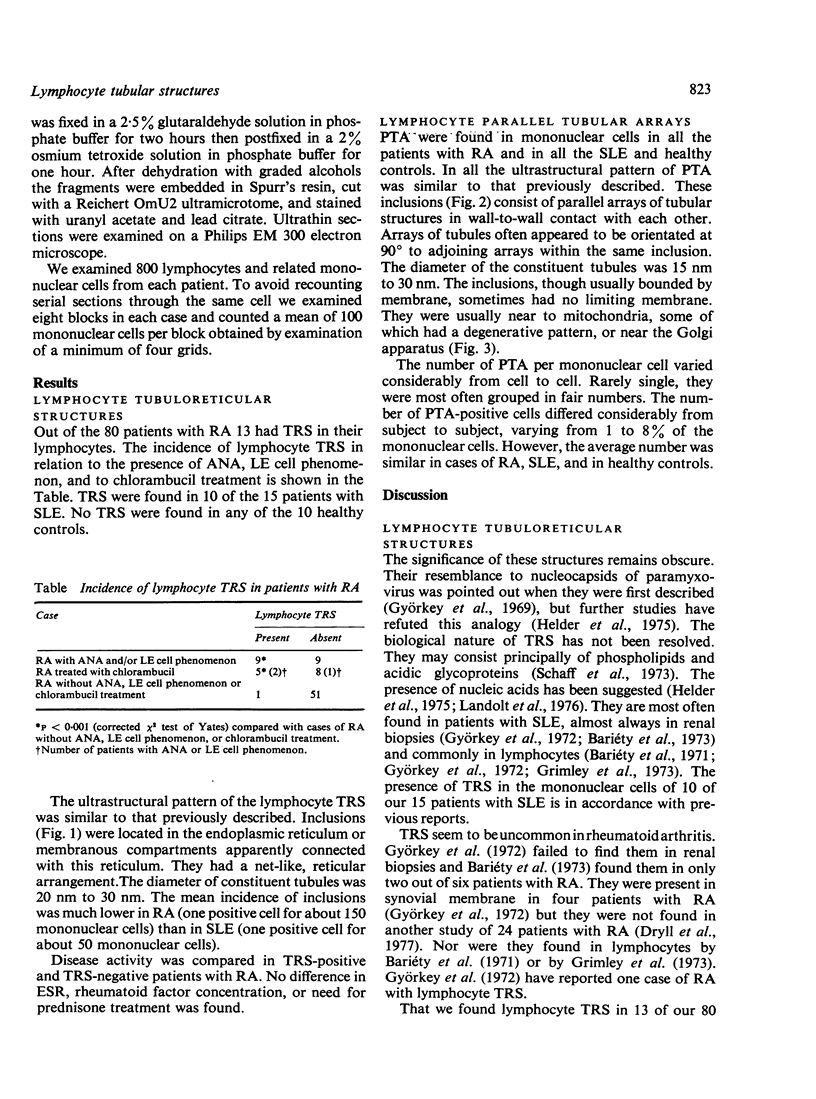
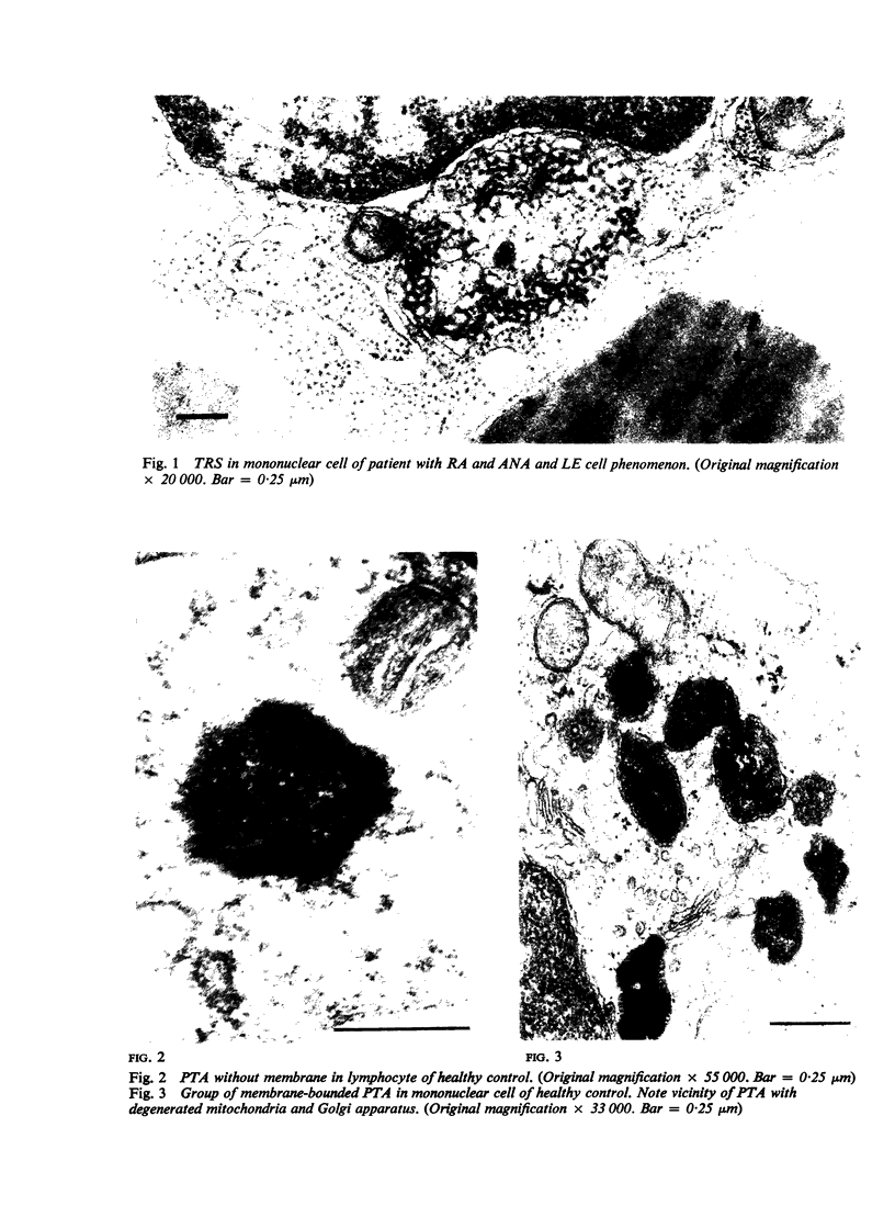
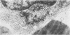
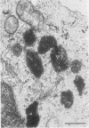
Two types of lymphocyte tubular structures were studied by electron microscopy in 80 patients with classic or definite rheumatoid arthritis (RA). Fifteen patients with unequivocal systemic lupus erythematosus (SLE) and 10 healthy persons were studied as controls. Lymphocyte tubulo-reticular structures were found in 13 of the 80 patients with RA and in 10 of the 15 patients with SLE. No tubuloreticular structures were found in any of the healthy subjects. In the RA patients antinuclear antibodies, LE cell phenomenon, and chlorambucil treatment were associated significantly with these inclusions but disease activity was not related to their presence. In cases in which tubuloreticular structures were present the number of cells containing inclusions was on average much lower in patients with RA than in those with SLE. Lymphocyte tubular parallel arrays were found in all the patients with RA and SLE and in all the healthy persons. In all three groups the average number of cells containing parallel tubular arrays was similar.
Full text
PDF
Images in this article
Selected References
These references are in PubMed. This may not be the complete list of references from this article.
- Bariety J., Amor B., Kahan A., Balafrej J. L., Delbarre F. Ultrastructural anomalies in mononuclear cells of peripheral blood in S.L.E.: Presence of virus-like inclusions. Rev Eur Etud Clin Biol. 1971 Aug-Sep;16(7):715–720. [PubMed] [Google Scholar]
- Bariéty J., Idatte J. M., Bedrossian J., Callard P., Appay M. D. Frequency of renal intraendothelial microtubular inclusions in kidney transplants. An electron microscopy study. Transplantation. 1974 Jan 1;17(1):140–142. [PubMed] [Google Scholar]
- Bariéty J., Richer D., Appay M. D., Grossetete J., Callard P. Frequency of intraendothelial 'virus-like' particles: an electron microscopy study of 376 human renal biopsies. J Clin Pathol. 1973 Jan;26(1):21–24. doi: 10.1136/jcp.26.1.21. [DOI] [PMC free article] [PubMed] [Google Scholar]
- Belcher R. W., Czarnetzki B. M., Campbell P. B. Ultrastructure of inclusions in peripheral blood mononuclear cells in sarcoidosis. Am J Pathol. 1975 Mar;78(3):461–468. [PMC free article] [PubMed] [Google Scholar]
- Brunning R. D., Parkin J. Ultrastructural studies of parallel tubular arrays in human lymphocytes. Am J Pathol. 1975 Jan;78(1):59–70. [PMC free article] [PubMed] [Google Scholar]
- Dryll A., Lansaman J., Cazalis P., Peltier A. P., De Seze S. Light and electron microscopy study of capillaries in normal and inflammatory human synovial membrane. J Clin Pathol. 1977 Jun;30(6):556–562. doi: 10.1136/jcp.30.6.556. [DOI] [PMC free article] [PubMed] [Google Scholar]
- Grrimley P. M., Decker J. L., Michelitch H. J., Frantz M. M. Abnormal structures in circulating lymphocytes from patients with systemic lupus erythematosus and related diseases. Arthritis Rheum. 1973 May-Jun;16(3):313–323. doi: 10.1002/art.1780160305. [DOI] [PubMed] [Google Scholar]
- Györkey F., Min K. W., Sincovics J. G., Györkey P. Systemic lupus erythematosus and myxovirus. N Engl J Med. 1969 Feb 6;280(6):333–333. doi: 10.1056/nejm196902062800620. [DOI] [PubMed] [Google Scholar]
- Györkey F., Sinkovics J. G., Min K. W., Györkey P. A morphologic study on the occurrence and distribution of structures resembling viral nucleocapsids in collagen diseases. Am J Med. 1972 Aug;53(2):148–158. doi: 10.1016/0002-9343(72)90125-8. [DOI] [PubMed] [Google Scholar]
- Hovig T., Jeremic M., Stavem P. A new type of inclusion bodies in lymphocytes. Scand J Haematol. 1968;5(2):81–96. doi: 10.1111/j.1600-0609.1968.tb01723.x. [DOI] [PubMed] [Google Scholar]
- Landolt A. M., Ryffel U., Hosbach H. U., Wyler R. Ultrastructure of tubular inclusions in endothelial cells of pituitary tumors associated with acromegaly. Virchows Arch A Pathol Anat Histol. 1976 May 3;370(2):129–140. doi: 10.1007/BF00430809. [DOI] [PubMed] [Google Scholar]
- Lymphocyte tubuloreticular structures in lupus erythematosus. Correlation with disease activity. Ann Intern Med. 1974 Sep;81(3):355–357. doi: 10.7326/0003-4819-81-3-355. [DOI] [PubMed] [Google Scholar]
- ROPES M. W., BENNETT G. A., COBB S., JACOX R., JESSAR R. A. 1958 Revision of diagnostic criteria for rheumatoid arthritis. Bull Rheum Dis. 1958 Dec;9(4):175–176. [PubMed] [Google Scholar]
- Schaff Z., Barry D. W., Grimley P. M. Cytochemistry of tubuloreticular structures in lymphocytes from patients with systemic lupus erythematosus and in cultured human lymphoid cells: comparison to a paramyxovirus. Lab Invest. 1973 Dec;29(6):577–586. [PubMed] [Google Scholar]
- White J. G. Giant organelles containing tubules in Chediak-Higashi lymphocytes. Am J Pathol. 1972 Nov;69(2):225–238. [PMC free article] [PubMed] [Google Scholar]