Abstract
Sphingomonas paucimobilis strain LCT-SP1 is a glucose-nonfermenting Gram-negative, chemoheterotrophic, strictly aerobic bacterium. The major feature of strain LCT-SP1, isolated from the Chinese spacecraft Shenzhou X, together with the genome draft and annotation are described in this paper. The total size of strain LCT-SP1 is 4,302,226 bp with 3,864 protein-coding and 50 RNA genes. The information gained from its sequence is potentially relevant to the elucidation of microbially mediated corrosion of various materials.
Keywords: genome sequence, Sphingomonas paucimobilis, corrosion
Introduction
Sphingomonas paucimobilis strain LCT-SP1 is a glucose-nonfermenting Gram-negative, chemoheterotrophic, strictly aerobic bacterium [1]. LCT-SP1, based on 16S rRNA gene sequences, is most closely related to Sphingomonas haloaromaticamans, which is isolated from water and soil. Several studies suggest that S. paucimobilis can degrade many compounds or materials, such as ferulic acid [2], lignin [3], and biphenyl [4]. LCT-SP1 was isolated from the condensate water in the Chinese spacecraft Shenzhou X.
LCT-SP1 can corrode numerous materials including epoxy resin, ester polyurethane, and ethers polyurethane. Therefore, the strain may be a suitable model for examining the properties of genes involved in microbial corrosion of materials used in aerospace applications. This study mainly aims to describe the draft genome of S. paucimobilis strain LCT-SP1 together with the genomic sequencing and annotation, which may be helpful in investigating the possible mechanisms in the microbial corrosion of materials.
Organism information
Classification and features
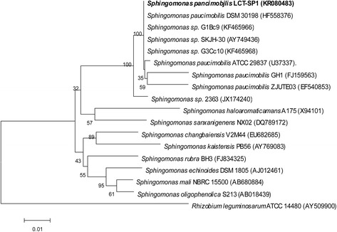
A phylogenetic tree was constructed with MEGA 5 [5] along with the sequences of representative members of the genus Sphingomonas using the maximum likelihood method based on 16S rRNA gene phylogeny (Fig. 1). Figure 1 shows that LCT-SP1 is most closely related to Sphingomonas sp.DSM 30198 (HF558376), G1Bc9 (KF465966), SKJH-30 (AY749436), and G3Cc10 (KF465968), with a sequence similarity of 100 % based on BLAST analysis. In addition, considering that the ANI is an important index in terms of phylogenetic analysis [6], the ANIs between LCT-SP1 and Sphingomonas paucimobilisNBRC 13935 were also calculated. The ANI result was 99.68 %, which is greater than 95 % (the species ANI cutoff value). Therefore, LCT-SP1 is asssumed to belongs to the species of Sphingomonas paucimobilis.
Fig. 1.

Phylogenetic tree highlighting the position of the Sphingomonas paucimobilis strain LCT-SP1 relative to selected Sphingomonas species using the Rhizobium leguminosarum ATCC 14480 as the outgroup. The strains and their corresponding GenBank accession numbers of 16S rRNA genes are indicated. Bar: 0.01 substitutions per nucleotide position
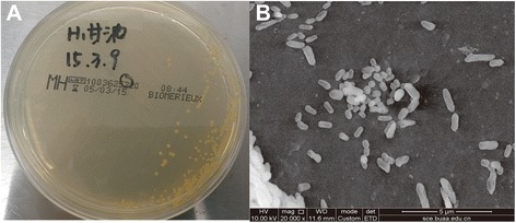
The general information of LCT-SP1 is shown in Table 1. LCT-SP1 is an aerobic, Gram-negative, rod-shaped, glucose-nonfermenting, slowly motile, and non-sporulating bacterium (Fig. 2-b). The strain grew optimally in the following conditions: pH 7.2, 35 °C, and at low salinity (NaCl range 0–1.0 %). On aerobic LB agar, LCT-SP1 formed several small, yellow-pigmented, round colonies (Fig. 2-a). LCT-SP1 was able to use a range of carbon substrates including D-glucose, maltose, lactose, sucrose, fucose, malic acid, acetic acid, and Tween-40.
Table 1.
Classification and general features of Sphingomonas paucimobilis strain LCT-SP1 according to the MIGS recommendations [22]
| MIGS ID | Property | Term | Evidence codea |
|---|---|---|---|
| Classification | Domain Bacteria | TAS [23] | |
| Phylum Proteobacteria | TAS [24] | ||
| Class Alphaproteobacteria | TAS [25, 26] | ||
| Order Sphingomonadales | TAS [25, 26] | ||
| Family Sphingomonadaceae | TAS [27, 28] | ||
| Genus Sphingomonas | TAS [27, 28] | ||
| Species Sphingomonas paucimobilis | TAS [1, 29] | ||
| (Type) strain: LCT-SP1 | IDA | ||
| Gram stain | Negative | TAS [1] | |
| Cell shape | Rod-shaped | TAS [1] | |
| Motility | Slow motility | TAS [1] | |
| Sporulation | Non-sporulating | TAS [1] | |
| Temperature range | 30-38 °C | NAS | |
| Optimum temperature | 35 °C | NAS | |
| pH range; Optimum | 6.0-7.5; 7.2 | IDA | |
| Carbon source | D-glucose, maltose, lactose, sucrose, fucose, malic acid, acetic acid, Tween-40 | IDA | |
| MIGS-6 | Habitat | Space cabin surface | IDA |
| MIGS-6.3 | Salinity | 0-1.0 % NaCl (w/v) | IDA |
| MIGS-22 | Oxygen requirement | Aerobic | TAS [1] |
| MIGS-15 | Biotic relationship | Free-living | NAS |
| MIGS-14 | Pathogenicity | Opportunistic pathogen | TAS [1, 30, 31] |
| MIGS-4 | Geographic location | Inner Mongolia, China | IDA |
| MIGS-5 | Sample collection | June 5, 2013 | NAS |
| MIGS-4.1 | Latitude | Not recorded | |
| MIGS-4.2 | Longitude | Not recorded | |
| MIGS-4.4 | Altitude | Not recorded |
aEvidence codes -IDA Inferred from Direct Assay, TAS Traceable Author Statement (i.e., a direct report exists in the literature), NAS Non-traceable Author Statement (i.e., not directly observed for the living, isolated sample, but based on a generally accepted property for the species, or anecdotal evidence). These evidence codes are from the Gene Ontology project [32]
Fig. 2.

Images of the Sphingomonas paucimobilis strain LCT-SP1: (a) colonies of the strains on Luria Bertani agar plates, and (b) scanning electron micrographs of the strain
Genome sequencing information
Genome project history
A summary of the main project information of the S. paucimobilis strain LCT-SP1 is shown in Table 2. This organism was isolated from the condensate water in the Shenzhou X spacecraft, and was selected for sequencing for its phylogenetic affiliation with a lineage of S. paucimobilis. The genome sequences of this organism were deposited in GenBank under accession number KR080483, which belongs to the 16s ribosomal RNA coding gene sequence of LCT-SP1.
Table 2.
Project information
| MIGS ID | Property | Term |
|---|---|---|
| MIGS-31 | Finishing quality | Improved high quality draft |
| MIGS-28 | Libraries used | One 300bp Illumina genomic library |
| MIGS-29 | Sequencing platforms | Illumina HiSeq2000 |
| MIGS-31.2 | Fold coverage | 50× |
| MIGS-30 | Assemblers | SOAPdenovo 1.05 |
| MIGS-32 | Gene calling method | Glimmer 3.0 |
| Locus Tag | ACJ66 | |
| Genbank ID | LDUA01000000 | |
| Genbank Date of Release | June 18, 2015 | |
| GOLD ID | Gs0115809 | |
| BIOPROJECT | PRJNA282437 | |
| MIGS-13 | Source Material Identifier | LCT-SP1 |
| Project relevance | Environment |
Growth conditions and genomic DNA preparation
S. paucimobilis strain LCT-SP1 was grown overnight on an aerobic LB agar plate at 35 °C. The total genomic DNA was extracted from 20 mL of cells using a CTAB bacterial genomic DNA isolation method [7] with kits provided by Illumina Inc. according to the manufacturer's instructions. DNA quality and quantity was determined by spectrophotometry.
Genome sequencing and assembly
The genome of LCT-SP1 was sequenced using paired-end sequencing technology [8] with Illumina HiSeq2000 (Illumina, SanDiego, CA, USA) at Majorbio Bio-pharm Technology Co., Ltd. (Shanghai, China). Draft assemblies were based on 6,986,766 readings, totaling 1,754 Mbp of 300 bp the PCR-free library, and 3,442,511 readings, totaling 1,556 Mbp of the 6,000 bp index library.
The assembly was performed using the SOAPdenovo software package version 1.05 [9]. The gaps among scaffolds were closed by custom primer walks or by PCR amplification, followed by DNA sequencing to achieve optimal assembly results. The genome contained 3,884 candidate protein-encoding genes (with an average size of 958 bp), giving a coding intensity of 87.7%. A total of 1,906 proteins were assigned to 25 COG families [10]. A total of 47 tRNA genes and 3 rRNA genes were identified.
Genome annotation
Protein-coding genes of the draft genome assemblies were established using Glimmer version 3.0 [11]. The predicted CDSs were translated and employed to search the KEGG, COG, String, NR, and GO databases. These data sources were brought together to assert a product description for each predicted protein. tRNAs and rRNAs were predicted using tRNAscan-SE [12] and RNAmmer [13], respectively. Automatic gene annotation was performed by the National Center for Biotechnology Information Prokaryotic Genomes Automatic Annotation Pipeline [14].
Genome properties
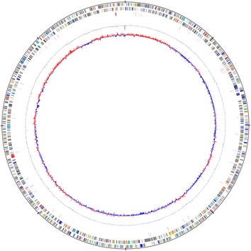
The LCT-SP1 genome consisted of 4,302,226 bp circular chromosomes with a GC content of 65.66 % (Table 3). Of the 3,934 predicted genes, 3,884 (98.73 %) were protein-coding genes, and 50 (1.27 %) were RNA genes (3 rRNA genes, and 47 tRNA genes). In addition, among the total predicted genes, 1,906 (48.45 %) represented COG functional categories. Of these, the most abundant COG category was “General function prediction only” (211 proteins) followed by “Amino acid transport and metabolism” (171 proteins), “Translation” (141 proteins), “Energy production and conversion” (140 proteins), “Replication, recombination and repair” (130 proteins), “Function unknown” (124 proteins), “Inorganic ion transport and metabolism” (210 proteins), and “Replication, recombination and repair” (201 proteins). The properties and statistics of the genome are summarized in Table 3. The draft genome map of S. paucimobilis strain LCT-SP1 is illustrated in Fig. 3, and the distribution of genes into COG functional categories is presented in Table 4.
Table 3.
Genome statistics
| Attribute | Value | % of total |
|---|---|---|
| Genome size (bp) | 4,302,226 | 100.00 |
| DNA coding (bp) | 3,772,440 | 87.69 |
| DNA G + C (bp) | 2,824,842 | 65.66 |
| DNA scaffolds | 91 | 100.00 |
| Total genes | 3,934 | 100.00 |
| Protein coding genes | 3,884 | 98.73 |
| RNA genes | 50 | 1.27 |
| Pseudo genes | 0 | 0.00 |
| Genes in internal clusters | 1,610 | 40.93 |
| Genes with function prediction | 3,911 | 99.42 |
| Genes assigned to COGs | 1,906 | 48.45 |
| Genes with Pfam domains | 2,571 | 65.35 |
| Genes with signal peptides | 367 | 9.33 |
| Genes with transmembrane helices | 846 | 21.50 |
| CRISPR repeats | 6 | - |
Fig. 3.

Circular map of the draft genome of the Sphingomonas paucimobilis strain LCT-SP1. From outside to the center: Genes on the forward strand (colored by the predicted coding sequences), genes on the reverse strand (colored by COG categories), RNA genes, GC content, and GC skew. The map was created using the DNAPlotter according to the method described by Carver et al. (2009) [33]. DNAPlotter reads the common sequence formats (EMBL, Genbank, GFF) using the Artemis file-reading library and displays the sequence as the circular plot. Additional feature files can be read in and overlaid on the sequence
Table 4.
Number of genes associated with general COG functional categories
| Code | Value | % age | Description |
|---|---|---|---|
| J | 141 | 3.58 | Translation, ribosomal structure and biogenesis |
| A | 0 | 0.00 | RNA processing and modification |
| K | 115 | 2.92 | Transcription |
| L | 130 | 3.30 | Replication, recombination and repair |
| B | 1 | 0.03 | Chromatin structure and dynamics |
| D | 14 | 0.36 | Cell cycle control, Cell division, chromosome partitioning |
| V | 27 | 0.69 | Defense mechanisms |
| T | 78 | 1.98 | Signal transduction mechanisms |
| M | 75 | 1.91 | Cell wall/membrane biogenesis |
| N | 27 | 0.69 | Cell motility |
| U | 57 | 1.45 | Intracellular trafficking and secretion |
| O | 89 | 2.26 | Posttranslational modification, protein turnover, chaperones |
| C | 140 | 3.56 | Energy production and conversion |
| G | 109 | 2.77 | Carbohydrate transport and metabolism |
| E | 171 | 4.35 | Amino acid transport and metabolism |
| F | 47 | 1.19 | Nucleotide transport and metabolism |
| H | 94 | 2.39 | Coenzyme transport and metabolism |
| I | 85 | 2.16 | Lipid transport and metabolism |
| P | 114 | 2.90 | Inorganic ion transport and metabolism |
| Q | 57 | 1.45 | Secondary metabolites biosynthesis, transport and catabolism |
| R | 211 | 5.36 | General function prediction only |
| S | 124 | 3.15 | Function unknown |
| - | 2,028 | 51.55 | Not in COGs |
Insights from the genome sequence
Several studies suggest that the genus S. paucimobilis can degrade many compounds or materials, such as ferulic acid [2], lignin [3], and biphenyl [4]. Arens et al. believed that the localized corrosion of copper cold-water pipes resulted from the genus Sphingomonas, leading to surface erosions, covered tubercles, and through-wall pinhole pits on the inner surface of the pipe [15]. S. paucimobilis strain LCT-SP1 can corrode several materials including epoxy resin, ester polyurethane, and ethers polyurethane (unpublished data). LCT-SP1 was isolated from the condensation water in the Chinese spacecraft Shenzhou X. Therefore, LCT-SP1 could be a suitable model for studying the properties of genes involved in microbial corrosion of aerospace related materials.
Additionally, EC 1.14.11.2, gloA, and arsC gene were present in LCT-SP1, which was identified with 100% similarity to Sphingomonas sp. S17 [16]. EC 1.14.11.2 is categorized as a procollagen-proline catalyzing enzyme [17]. The gloA gene encodes a glyoxalase that can reduce methylglyoxal toxicity in a cell [18]. Furthermore, arsC gene produces an arsenate reductase that can convert arsenate into arsenite, which is accordingly exported from cells by an energy-dependent efflux process [19]. Therefore, the genes mentioned above are likely responsible for the ability of LCT-SP1 to degrade various recalcitrant aromatic compounds and polysaccharides.
The LCT-SP1 genome also contained an NhaA-type CDS for the Na+/H+ antiporter and some subunits of the multisubunit cation antiporter (Na+/H+) [20], which suggested that this strain should be compatible with its alkaline and hypersaline environment, and could corrode metallic materials by changing the pH balance of their surface.
Also, biofilms from bacteria may be beneficial for corrosion control because of the removal of corrosive agents and the generation of a protective layer by biofilms [21]. LCT-SP1 included the gene encoding biofilm dispersion protein BdlA and biofilm growth-associated repressor that could inhibit the formation of biofilm, which may explain the microbial corrosion of materials. Further studies are needed to investigate these corrosion-based gene-coding sequences to reveal the role of LCT-SP1 in the microbial corrosion of materials.
Conclusions
The genome of S. paucimobilis strain LCT-SP1 isolated from the condensate water in the Chinese spacecraft Shenzhou X was sequenced. The strain LCT-SP1 genome included numerous genes that are likely responsible for their ability to degrade various recalcitrant aromatic compounds and polysaccharides. Further study of these corrosion-based gene-coding sequences may reveal the role of S. paucimobilis LCT-SP1 in microbial corrosion of materials, especially in aerospace applications. The genome sequence has been deposited at DDBJ/EMBL/GenBank under accession number LDUA00000000.
Acknowledgements
This work was performed at the Chinese PLA General Hospital. We gratefully acknowledge the China Astronaut Research and Training Centre for providing strains. This work was financially supported by the National Basic Research Program of China (973 program, no. 2014CB744400), the Key Program of Medical Research in the Military “12th 5-year Plan” (no. BWS12J046), and the Program of Manned Spaceflight (no. 040203). This work was also partially supported by the National Natural Science Foundation of China (no. 81350020) and the National Significant Science Foundation of China (no. 2015ZX09J15102-003).
Abbreviations
- ANI
Average nucleotide identity
- LB
Luria–Bertani
- CTAB
Cetyl Trimethyl Ammonium Bromide
- COG
Clusters of orthologous group
- CDS
Coding sequences
Footnotes
Competing interests
The authors declare that they have no competing interests.
Authors’ contributions
CTL initiated and supervised the study. LP drafted the first manuscript. LCG performed electron microscopy. LP, HZ, JL, JG and CX annotated the genome. LP, HZ and JL worked on genome sequencing and assembly. LP, HZ, JL, BH, XLZ and CTL discussed, analyzed the data and revised the manuscript. LP, HZ and JL contributed equally to this work. All authors read and approved the final manuscript.
References
- 1.Yabuuchi E, Yano I, Oyaizu H, Hashimoto Y, Ezaki T, Yamamoto H. Proposals of Sphingomonaspaucimobilis gen. nov. and comb. nov., Sphingomonas parapaucimobilis sp. nov., Sphingomonasyanoikuyae sp. nov., Sphingomonas adhaesiva sp. nov., Sphingomonas capsulata comb. nov., and two genospecies of the genus Sphingomonas. Microbiol Immunol. 1990;34(2):99–119. doi: 10.1111/j.1348-0421.1990.tb00996.x. [DOI] [PubMed] [Google Scholar]
- 2.Masai E, Harada K, Peng X, Kitayama H, Katayama Y, Fukuda M. Cloning and characterization of the ferulic acid catabolic genes of Sphingomonas paucimobilis SYK-6. Appl Environ Microb. 2002;68(9):4416–24. doi: 10.1128/AEM.68.9.4416-4424.2002. [DOI] [PMC free article] [PubMed] [Google Scholar]
- 3.Nishikawa S, Sonoki T, Kasahara T, Obi T, Kubota S, Kawai S, et al. Cloning and sequencing of the Sphingomonas (Pseudomonas) paucimobilis gene essential for the O demethylation of vanillate and syringate. Appl Environ Microb. 1998;64(3):836–42. doi: 10.1128/aem.64.3.836-842.1998. [DOI] [PMC free article] [PubMed] [Google Scholar]
- 4.Peng X, Masai E, Kitayama H, Harada K, Katayama Y, Fukuda M. Characterization of the 5-carboxyvanillate decarboxylase gene and its role in lignin-related biphenyl catabolism in Sphingomonas paucimobilis SYK-6. Appl Environ Microb. 2002;68(9):4407–15. doi: 10.1128/AEM.68.9.4407-4415.2002. [DOI] [PMC free article] [PubMed] [Google Scholar]
- 5.Tamura K, Peterson D, Peterson N, Stecher G, Nei M, Kumar S. MEGA5: molecular evolutionary genetics analysis using maximum likelihood, evolutionary distance, and maximum parsimony methods. Mol Biol Evol. 2011;28(10):2731–9. doi: 10.1093/molbev/msr121. [DOI] [PMC free article] [PubMed] [Google Scholar]
- 6.Figueras MJ, Beaz-Hidalgo R, Hossain MJ, Liles MR. Taxonomic affiliation of new genomes should be verified using average nucleotide identity and multilocus phylogenetic analysis. Genome Announc. 2014;2(6). doi:10.1128/genomeA.00927-14. [DOI] [PMC free article] [PubMed]
- 7.van Embden JD, Cave MD, Crawford JT, Dale JW, Eisenach KD, Gicquel B, et al. Strain identification of Mycobacterium tuberculosis by DNA fingerprinting: recommendations for a standardized methodology. J Clin Microbiol. 1993;31(2):406–9. doi: 10.1128/jcm.31.2.406-409.1993. [DOI] [PMC free article] [PubMed] [Google Scholar]
- 8.Bentley DR, Balasubramanian S, Swerdlow HP, Smith GP, Milton J, Brown CG, et al. Accurate whole human genome sequencing using reversible terminator chemistry. Nature. 2008;456(7218):53–9. doi: 10.1038/nature07517. [DOI] [PMC free article] [PubMed] [Google Scholar]
- 9.Li R, Zhu H, Ruan J, Qian W, Fang X, Shi Z, et al. De novo assembly of human genomes with massively parallel short read sequencing. Genome Res. 2010;20(2):265–72. doi: 10.1101/gr.097261.109. [DOI] [PMC free article] [PubMed] [Google Scholar]
- 10.Tatusov RL, Galperin MY, Natale DA, Koonin EV. The COG database: a tool for genome-scale analysis of protein functions and evolution. Nucleic Acids Res. 2000;28(1):33–6. doi: 10.1093/nar/28.1.33. [DOI] [PMC free article] [PubMed] [Google Scholar]
- 11.Delcher AL, Harmon D, Kasif S, White O, Salzberg SL. Improved microbial gene identification with GLIMMER. Nucleic Acids Res. 1999;27(23):4636–41. doi: 10.1093/nar/27.23.4636. [DOI] [PMC free article] [PubMed] [Google Scholar]
- 12.Lowe TM, Eddy SR. tRNAscan-SE: a program for improved detection of transfer RNA genes in genomic sequence. Nucleic Acids Res. 1997;25(5):955–64. doi: 10.1093/nar/25.5.0955. [DOI] [PMC free article] [PubMed] [Google Scholar]
- 13.Lagesen K, Hallin P, Rodland EA, Staerfeldt HH, Rognes T, Ussery DW. RNAmmer: consistent and rapid annotation of ribosomal RNA genes. Nucleic Acids Res. 2007;35(9):3100–8. doi: 10.1093/nar/gkm160. [DOI] [PMC free article] [PubMed] [Google Scholar]
- 14.Angiuoli SV, Gussman A, Klimke W, Cochrane G, Field D, Garrity G, et al. Toward an online repository of Standard Operating Procedures (SOPs) for (meta)genomic annotation. OMICS. 2008;12(2):137–41. doi: 10.1089/omi.2008.0017. [DOI] [PMC free article] [PubMed] [Google Scholar]
- 15.Arens P, Tuschewitzki GJ, Wollmann M, Follner H, Jacobi H. Indicators for microbiologically induced corrosion of copper pipes in a cold-water plumbing system. Zentralbl Hyg Umweltmed. 1995;196(5):444–54. [PubMed] [Google Scholar]
- 16.Farias ME, Revale S, Mancini E, Ordonez O, Turjanski A, Cortez N, et al. Genome sequence of Sphingomonas sp. S17, isolated from an alkaline, hyperarsenic, and hypersaline volcano-associated lake at high altitude in the Argentinean Puna. J Bacteriol. 2011;193(14):3686–7. doi: 10.1128/JB.05225-11. [DOI] [PMC free article] [PubMed] [Google Scholar]
- 17.Berg RA, Prockop DJ. Affinity column purification of protocollagen proline hydroxylase from chick embryos and further characterization of the enzyme. J Biol Chem. 1973;248(4):1175–82. [PubMed] [Google Scholar]
- 18.Ng J, Kidd SP. The concentration of intracellular nickel in Haemophilus influenzae is linked to its surface properties and cell-cell aggregation and biofilm formation. Int J Med Microbiol. 2013;303(3):150–7. doi: 10.1016/j.ijmm.2013.02.012. [DOI] [PubMed] [Google Scholar]
- 19.Ji G, Silver S. Reduction of arsenate to arsenite by the ArsC protein of the arsenic resistance operon of Staphylococcus aureus plasmid pI258. Proc Natl Acad Sci U S A. 1992;89(20):9474–8. doi: 10.1073/pnas.89.20.9474. [DOI] [PMC free article] [PubMed] [Google Scholar]
- 20.Padan E, Tzubery T, Herz K, Kozachkov L, Rimon A, Galili L. NhaA of Escherichia coli, as a model of a pH-regulated Na+/H + antiporter. Biochim Biophys Acta. 2004;1658(1-2):2–13. doi: 10.1016/j.bbabio.2004.04.018. [DOI] [PubMed] [Google Scholar]
- 21.Zuo R. Biofilms: strategies for metal corrosion inhibition employing microorganisms. Appl Microbiol Biotechnol. 2007;76(6):1245–53. doi: 10.1007/s00253-007-1130-6. [DOI] [PubMed] [Google Scholar]
- 22.Field D, Garrity G, Gray T, Morrison N, Selengut J, Sterk P, et al. The minimum information about a genome sequence (MIGS) specification. Nat Biotechnol. 2008;26(5):541–7. doi: 10.1038/nbt1360. [DOI] [PMC free article] [PubMed] [Google Scholar]
- 23.Woese CR, Kandler O, Wheelis ML. Towards a natural system of organisms: proposal for the domains Archaea, Bacteria, and Eucarya. Proc Natl Acad Sci U S A. 1990;87(12):4576–9. doi: 10.1073/pnas.87.12.4576. [DOI] [PMC free article] [PubMed] [Google Scholar]
- 24.Garrity GM, Bell JA, Lilburn T. Phylum XIV. Proteobacteria phyl nov. In: Garrity GM, Brenner DJ, Krieg NR, Staley JT, editors. Bergey’s Manual of Systematic Bacteriology. Volume 2, Part B. 2. New York: Springer; 2005. p. 1. [Google Scholar]
- 25.Yabuuchi E, Kosako Y. Order IV. Sphingomonadales ord. nov. In: Garrity GM, Brenner DJ, Krieg NR, Staley JT, editors. Bergey's Manual of Systematic Bacteriology, Second Edition, Volume 2, Part C. New York: Springer; 2005. pp. 230–233. [Google Scholar]
- 26.List Editor. Validation List No. 107. List of new names and new combinations previously effectively, but not validly, published. Int J Syst Evol Microbiol 2006; 56:1-6. [DOI] [PubMed]
- 27.Kosako Y, Yabuuchi E, Naka T, Fujiwara N, Kobayashi K. Proposal of Sphingomonadaceae fam. nov., consisting of Sphingomonas Yabuuchi et al. 1990, Erythrobacter Shiba and Shimidu 1982, Erythromicrobium Yurkov et al. 1994, Porphyrobacter Fuerst et al. 1993, Zymomonas Kluyver and van Niel 1936, and Sandaracinobacter Yurkov et al. 1997, with the type genus Sphingomonas Yabuuchi et al. 1990. Microbiol Immunol. 2000;44(7):563–75. doi: 10.1111/j.1348-0421.2000.tb02535.x. [DOI] [PubMed] [Google Scholar]
- 28.Validation of publication of new names and new combinations previously effectively published outside the IJSEM. Int J Syst Evol Micr. 2000;50 Pt 6:1953. [DOI] [PubMed]
- 29.Validation of the publication of new names and new combinations previously effectively published outside the IJSB. List no. 39. Int J Syst Bacteriol. 1991;41(4):580-1. [DOI] [PubMed]
- 30.Perola O, Nousiainen T, Suomalainen S, Aukee S, Karkkainen UM, Kauppinen J, et al. Recurrent Sphingomonas paucimobilis -bacteraemia associated with a multi-bacterial water-borne epidemic among neutropenic patients. J Hosp Infect. 2002;50(3):196–201. doi: 10.1053/jhin.2001.1163. [DOI] [PubMed] [Google Scholar]
- 31.Adams WE, Habib M, Berrington A, Koerner R, Steel DH. Postoperative endophthalmitis caused by Sphingomonas paucimobilis. J Cataract Refract Surg. 2006;32(7):1238–40. doi: 10.1016/j.jcrs.2006.01.094. [DOI] [PubMed] [Google Scholar]
- 32.Ashburner M, Ball CA, Blake JA, Botstein D, Butler H, Cherry JM, et al. Gene ontology: tool for the unification of biology. The Gene Ontology Consortium. Nat Genet. 2000;25(1):25–9. doi: 10.1038/75556. [DOI] [PMC free article] [PubMed] [Google Scholar]
- 33.Carver T, Thomson N, Bleasby A, Berriman M, Parkhill J. DNAPlotter: circular and linear interactive genome visualization. Bioinformatics. 2009;25(1):119–20. doi: 10.1093/bioinformatics/btn578. [DOI] [PMC free article] [PubMed] [Google Scholar]


