

Abstract
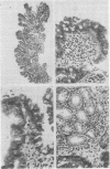
The results of histological and immunohistochemical examination of gastric and duodenal biopsy specimens from 50 volunteers without a clinical history of gastrointestinal disease are reported. Multiple specimens of tissue from standard sites in the stomach and duodenum were carefully orientated, and serially sectioned for examination by light microscopy and for immunohistochemical characterisation of plasma cells within the lamina propria. The antrum and fundus were normal in 32 of the 50 subjects but the other 18 showed histopathological evidence of gastritis in either the antrum or fundus. The latter appeared to be age-related. There was considerable variation in the appearance of the surface epithelium of the duodenum within as well as among individual subjects. Superficial gastric metaplasia in one or more biopsy specimens from the duodenal bulb was found in 64% of individuals. Histopathological examination of the duodenum revealed signs of chronic inflammation in 12% of the subjects. In two individuals there was active inflammation but in only one of these was the diagnosis made on endoscopic appearances. Histological criteria important for the diagnosis of duodenitis are discussed. The number of plasma cells in different biopsy specimens from subjects not showing histological signs of inflammation was variable. The ratio IgA:IgG:IgM producing plasma cells was remarkably constant from subject to subject as well as from specimen to specimen.
Full text
PDF


Images in this article

Selected References
These references are in PubMed. This may not be the complete list of references from this article.
- Avrameas S., Ternynck T. Peroxidase labelled antibody and Fab conjugates with enhanced intracellular penetration. Immunochemistry. 1971 Dec;8(12):1175–1179. doi: 10.1016/0019-2791(71)90395-8. [DOI] [PubMed] [Google Scholar]
- Beck I. T., Kahn D. S., Lacerte M., Solymar J., Callegarini U., Geokas M. C. 'Chronic duodenitis': a clinical pathological entity? Gut. 1965 Aug;6(4):376–383. doi: 10.1136/gut.6.4.376. [DOI] [PMC free article] [PubMed] [Google Scholar]
- Bosman F. T., Lindeman J., Kuiper G., van der Wal A., Kreunig J. The influence of fixation on immunoperoxidase staining of plasmacells in paraffin sections of intestinal biopsy specimens. Histochemistry. 1977 Jul 18;53(1):57–62. doi: 10.1007/BF00511210. [DOI] [PubMed] [Google Scholar]
- Chaput J. C., Petite J. P., Rain B., Buffet C., Camillieri J. P., Etienne J. P. Les duodénites non spécifiques. Etude de 80 cas. Arch Fr Mal App Dig. 1974 Dec;63(8):611–623. [PubMed] [Google Scholar]
- Graham R. C., Jr, Karnovsky M. J. The early stages of absorption of injected horseradish peroxidase in the proximal tubules of mouse kidney: ultrastructural cytochemistry by a new technique. J Histochem Cytochem. 1966 Apr;14(4):291–302. doi: 10.1177/14.4.291. [DOI] [PubMed] [Google Scholar]
- Hijmans W., Schuit H. R., Klein F. An immunofluorescence procedure for the detection of intracellular immunoglobulins. Clin Exp Immunol. 1969 Apr;4(4):457–472. [PMC free article] [PubMed] [Google Scholar]
- Hoedemaeker P. J. Heterotopic gastric mucosa in the duodenum. Digestion. 1970;3(3):165–173. doi: 10.1159/000197027. [DOI] [PubMed] [Google Scholar]
- Korn E. R., Foroozan P. Endoscopic biopsies of normal duodenal mucosa. Gastrointest Endosc. 1974 Nov;21(2):51–54. doi: 10.1016/s0016-5107(74)73790-7. [DOI] [PubMed] [Google Scholar]
- Meikle D. D., Taylor K. B., Truelove S. C., Whitehead R. Gastritis duodenitis, and circulating levels of gastrin in duodenal ulcer before and after vagotomy. Gut. 1976 Sep;17(9):719–728. doi: 10.1136/gut.17.9.719. [DOI] [PMC free article] [PubMed] [Google Scholar]
- Perera D. R., Weinstein W. M., Rubin C. E. Symposium on pathology of the gastrointestinal tract-Part II. Small intestinal biopsy. Hum Pathol. 1975 Mar;6(2):157–217. doi: 10.1016/s0046-8177(75)80176-6. [DOI] [PubMed] [Google Scholar]
- Skinner J. M., Whitehead R. The plasma cells in inflammatory disease of the colon: a quantitative study. J Clin Pathol. 1974 Aug;27(8):643–646. doi: 10.1136/jcp.27.8.643. [DOI] [PMC free article] [PubMed] [Google Scholar]
- Söltoft J. Immunoglobulin-containing cells in normal jejunal mucosa and in ulcerative colitis and regional enteritis. Scand J Gastroenterol. 1969;4(4):353–360. doi: 10.3109/00365526909180616. [DOI] [PubMed] [Google Scholar]
- Vermeer B. J., Lindeman J., van der Harst-Oostveen C. J., Peña A. S., van Vloten W. A. The immunoglobulin-bearing cells in the lamina propria and the clinical response to a gluten-free diet in dermatitis herpetiformis. Arch Dermatol Res. 1977 May 27;258(3):223–230. doi: 10.1007/BF00561123. [DOI] [PubMed] [Google Scholar]
- Whitehead R., Roca M., Meikle D. D., Skinner J., Truelove S. C. The histological classification of duodenitis in fibreoptic biopsy specimens. Digestion. 1975;13(3):129–136. doi: 10.1159/000197701. [DOI] [PubMed] [Google Scholar]