Abstract
Introduction
International resuscitation guidelines suggest to use positive end-expiratory pressure (PEEP) during manual ventilation of neonates. Aim of our study was to test the reliability of self-inflating bags (SIB) with single-use PEEP valves regarding PEEP delivery and the effect of different peak inflation pressures (PIP) and ventilation rates (VR) on the delivered PEEP.
Methods
Ten new single-use PEEP valves from 5 manufacturers were tested by ventilating an intubated 1kg neonatal manikin containing a lung model with a SIB that was actuated by an electromechanical plunger device. Standard settings: PIP 20cmH2O, VR 60/min, flow 8L/min. PEEP settings of 5 and 10cmH2O were studied. A second test was conducted with settings of PIP 40cmH2O and VR 40/min. The delivered PEEP was measured by a respiratory function monitor (CO2SMO+).
Results
Valves from one manufacturer delivered no relevant PEEP and were excluded. The remaining valves showed a continuous decay of the delivered pressure during expiration. The median (25th and 75th percentile) delivered PEEP with standard settings was 3.4(2.7–3.8)cmH2O when set to 5cmH2O and 6.1(4.9–7.1)cmH2O when set to 10cmH2O. Increasing the PIP from 20 to 40 cmH2O led to a median (25th and 75th percentile) decrease in PEEP to 2.3(1.8–2.7)cmH2O and 4.3(3.2–4.8)cmH2O; changing VR from 60 to 40/min led to a PEEP decrease to 2.8(2.1–3.3)cmH2O and 5.0(3.5–6.2)cmH2O for both PEEP settings.
Conclusion
Single-use PEEP valves do not reliably deliver the set PEEP. PIP and VR have an effect on the delivered PEEP. Operators should be aware of these limitations when manually ventilating neonates.
Introduction
Self-inflating bags (SIB) are widely used for neonatal resuscitation. They are the most commonly used devices for manual ventilation of neonates in several countries.[1, 2] Perceived advantages of SIBs are: 1) their comparatively low cost; 2) their ease of use; 3) they can be used independently of a gas source, i.e. outside the hospital or in resource-limited settings;[3, 4] 4) they can be used with a gas source attached to deliver higher oxygen concentrations; 5) they can provide positive end-expiratory pressure (PEEP) when used in conjunction with additional PEEP valves. Several animal studies demonstrated the benefits of applying PEEP during ventilation: PEEP helps establish and maintain functional residual capacity,[5, 6] which is essential during transition to extrauterine life.[7, 8] Therefore, recent resuscitation guidelines suggest the use of PEEP for resuscitation of preterm newborn infants.[9, 10] However, studies by our group as well as others have found that PEEP valves often do not reliably deliver the PEEP as intended.[11–13] In a recent study we tested 10 factory new multi-use PEEP valves throughout 30 cycles of thermo-sterilization and demonstrated that these procedures further decreased their reliability.[14]
Consequently, SIBs with single-use PEEP valves might represent a more reliable alternative than SIBs with multi-use PEEP valves. However, single-use PEEP valves have not been well studied and very little is known about their ability to generate the set PEEP. Therefore, the aim of our study was to test the reliability of single-use PEEP valves from different manufacturers during simulated resuscitation of preterm infants in the delivery room and to investigate the effect of peak inflation pressure (PIP) and ventilation rate (VR) settings on the delivered PEEP.
Material and Methods
We tested 10 factory new single-use PEEP valves from 5 different manufacturers: two valves each from Ambu® (0–20 cmH2O; Ballerup, Denmark), DROH® (0–10 cmH2O; Mainz, Germany), Medisize® (5–20 cmH2O; Hillegom, Netherlands), The Bag II® (5–20 cmH2O; Laerdal Medical, Stavanger, Norway) and Vital Signs Inc. ® (5–20 cmH2O; Totowa, NJ, USA). All had a 30 mm connection and a freely adjustable spring valve for setting the PEEP to the above given range.
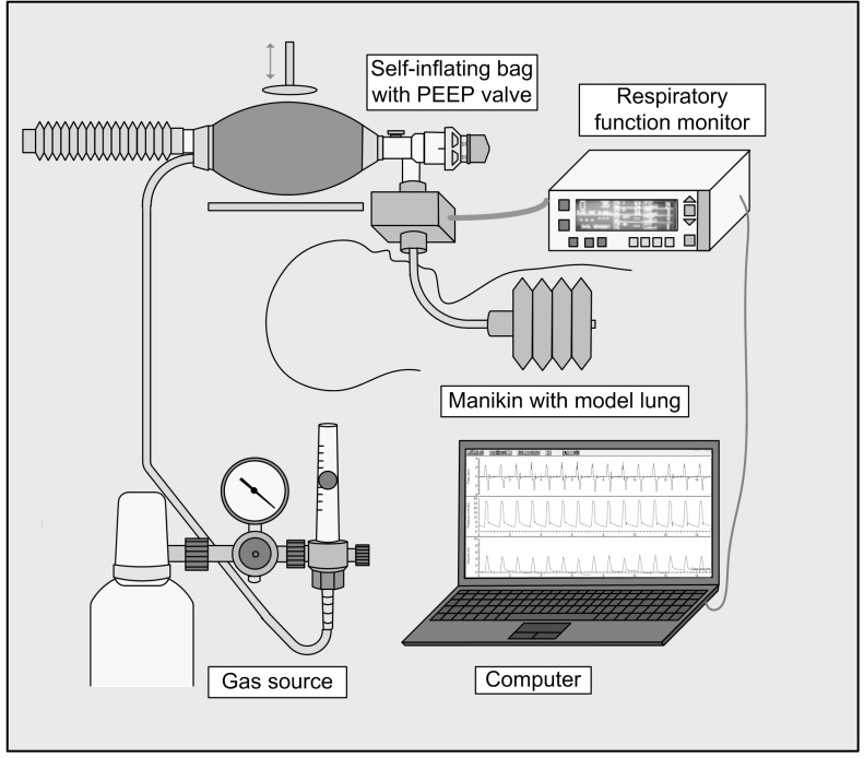
The valves were attached to a new disposable Ambu SPUR II Baby Resuscitator (Ambu®, Ballerup, Denmark) with a maximum stroke volume of 150 ml that was designed to ventilate neonates and infants of up to 10 kg bodyweight. The bag had a pressure-relief valve set at 40 cmH2O. An oxygen reservoir tube with a volume of approximately 100 ml was attached and the bag was connected to medical gas (Flow 8 l/min, FiO2 0.21). An intubated, leak free neonatal manikin (Fisher & Paykel Healthcare®, Auckland, New Zealand) simulating a 1kg neonate, that contained a neonatal lung model with a compliance of 2.0 ml*kPa-1 was ventilated. Airway pressure, gas flow and volume were detected using a CO2SMO+ PLUS! monitor (Novametrix Medical Systems®, Wallingford, CT, USA). The experimental setup is shown in Fig 1. Data were recorded by a laptop computer using the CO2SMO+ Software (for Windows, Novametrix, version 1.0).
Fig 1. Schematic diagram of the experimental setup: The single-use PEEP valves were consecutively attached to a mechanically driven self-inflating bag.
A gas source was connected. Subsequently a manikin simulating a 1kg neonate was ventilated. The delivered pressures and volume were measured using a respiratory function monitor and analyzed with a laptop computer. Standard settings were: Peak inspiratory pressure = 20 cmH2O, ventilation rate = 60/min, flow = 8 l/min.
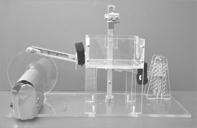
We constructed an electromechanical device that allowed compression of the SIB under standardized laboratory conditions (Fig 2). Positive inspiratory pressure (PIP) could be set by modifying the indentation depth of a plunger compressing the bag. The device was actuated by an electrical engine that was connected to an adjustable power supply. The ventilation rate (VR) could be set by altering the motor voltage.
Fig 2. Electromechanical device constructed to compress the SIB under standardized conditions.
Standard settings of PIP = 20 cmH2O, gas flow = 8 l/min, VR = 60 breaths per minute and PEEP = 5 and 10 cmH2O were used and extended by measurements with PIP = 40 cmH2O and VR = 40 /min to study the effect of PIP and VR on the delivered PEEP. PIP was adjusted mechanically without the PEEP valve attached (indentation depth of the plunger) and VR was adjusted by the motor voltage according to the measured and displayed PIP and VR values of the CO2SMO+. The device was set before the valves were attached to guarantee equal conditions for all the measurements. The adjustment of PIP = 20 cmH2O could be performed without any problems, however, after attachment of the PEEP valve there was a slight increase of the PIP: The median PIP (25th and 75th percentile) was 26.3. (25.0–27.6), 26.0 (24.5–27.5), 20.8 (20.0–22.2), 25.3 (23.8–26.9), 25.4 (23.7–27.1) cmH2O for valves of manufacturer 1–5. For a set PIP of 40 cmH2O the targeted value could not be reached during most measurements due to early opening of the pressure relief valve. The median PIP (25th and 75th percentile) for manufacturer 1–5 was 37.4 (37.2–37.6), 37.4 (37.3–37.5), 37.1 (37.0–37.2), 37.3 (37.3–37.4), 37.4 (37.3–37.5) cmH2O. VR during the measurements showed no relevant deviations from the set values. Eight 30-second-measurements with the different combinations of parameter settings were performed with each valve and repeated 5 times to test reproducibility of our data (adding up to 400 measurements in total). PEEP was defined as the pressure at the end of the expiratory phase, just before the start of the subsequent inflation and was calculated by the software of the CO2SMO+ respiratory function monitor. Ten consecutive inflation cycles of each measurement were evaluated and the median PEEP was calculated. Because there was no statistically significant difference in the delivered PEEP between both valves of the same manufacturer, the data were pooled.
All measured PEEP values are presented as medians with 25th and 75th percentile in brackets and were compared using the Mann-Whitney or Kruskal-Wallis test, as appropriate. For each parameter setting the reproducibility of serial PEEP values during the 30-second-measurements was described by coefficient of variation (CV [%] = 100*SD / mean) of 10 consecutive inflation cycles and compared using the Kruskal-Wallis test. The effect of changes in PIP and VR on the delivered PEEP was tested separately by the Wilcoxon test for paired samples. The relationship between the decrease of the delivered PEEP and the increase of the delivered volume was investigated by linear regression analysis. Statistical analysis was performed using Statgraphics Centurion® software (Version 16.0, Statpoint Inc., Herndon, Virginia, USA) and MedCalc (Version 9.2.0.2; MedCalc Software, Mariakerke, Belgium). A p-value less than 0.05 was considered statistically significant.
Results
Reproducibility
The coefficient of variation of 10 consecutive inflations of each PEEP valve was calculated. Table 1 shows median and 25th and 75th percentile of 80 series of measurements (two valves of each manufacturer measured with 8 different combinations of parameter settings, repeated 5 times) per valve. As valve 3 did not deliver any relevant PEEP, it was excluded from the analysis. There were no statistically significant differences in the CVs between valves 1, 2 and 5. Only valve 4 showed a weak but statistically significantly (p<0.001) higher variability in the delivered PEEP. However, median CV of all remaining valves (except valve 3) was <2% and negligible from the clinical point of view, indicating highly reproducible PEEP during the individual measurements.
Table 1. Coefficient of variation.
| Valve 1 | Valve 2 | Valve 3 | Valve 4 | Valve 5 | p-value | |
|---|---|---|---|---|---|---|
| CVPEEP (%) | 1.12 (0.66–2.04) | 1.36 (0.74–2.24) | - | 1.89 (1.11–3.07) | 1.08 (0–2.32) | <0.001 (0.158)1) |
1)without valve 4
Coefficient of variation (CV) of serial PEEP values of the PEEP valves of 5 manufacturers (Presented are median and 25th and 75th percentile in brackets of 80 series of measurements (two prototypes, 8 parameter settings, 5 repetitions) per valve. Statistically significant p-value is printed in bold)
Delivered PEEP
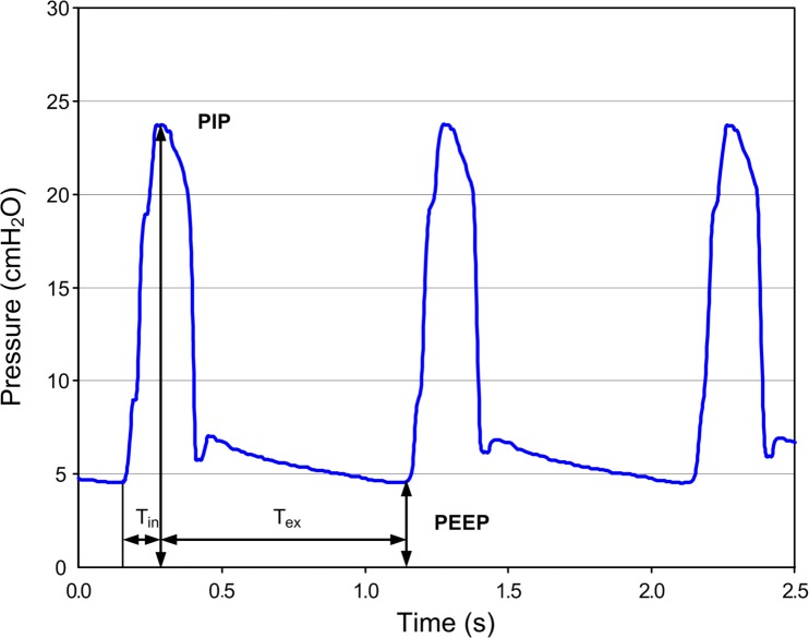
The pressure curves that were recorded for each inflation cycle showed a continuous decrease of the delivered pressure during the expiratory phase of the ventilation cycle until the final PEEP was reached just before the subsequent inflation started. This pressure profile is illustrated in Fig 3.
Fig 3. Pressure profile of a self-inflating bag with 8 l/min flow supply.
In contrast to the serial measurements, the variability of the delivered PEEP between the different parameter settings was significantly higher, as shown in Table 2. Again, valve 3 was excluded from the analysis because no relevant PEEP could be delivered. Only in three of the eight combinations of parameter settings statistically significant differences (p<0.05) in the delivered PEEP could be shown between the manufacturers. However, these differences were weak and near the limit of statistical significance.
Table 2. Delivered PEEP.
| PEEP Setting | PIP Setting | VR Setting | PEEP measured (cmH2O) | p-value | |||
|---|---|---|---|---|---|---|---|
| (cmH2O) | (cmH2O) | (1/min) | Valve 1 | Valve 2 | Valve 4 | Valve 5 | |
| 5 | 20 | 40 | 3.44 (2.81–4.17) | 2.89 (2.15–3.12) | 2.16 (1.85–2.89) | 2.67 (2.34–3.28) | 0.049 |
| 5 | 20 | 60 | 4.19 (3.41–4.80) | 3.30 (3.14–3.73) | 2.84 (2.36–3.66) | 3.04 (2.56–3.50) | 0.021 |
| 5 | 40 | 40 | 1.64 (1.09–1.91) | 1.60 (1.52–1.95) | 1.35 (1.13–1.71) | 1.52 (1.47–1.65) | 0.515 |
| 5 | 40 | 60 | 2.70 (1.19–3.00) | 2.39 (2.30–2.63) | 1.87 (1.62–2.24) | 2.43 (2.30–2.59) | 0.246 |
| 10 | 20 | 40 | 6.44 (4.39–7.91) | 4.53 (3.26–6.11) | 4.06 (3.31–5.21) | 5.42 (3.46–6.33) | 0.118 |
| 10 | 20 | 60 | 7.10 (5.20–8.63) | 6.06 (5.10–6.33) | 5.04 (3.60–5.66) | 6.38 (4.81–7.34) | 0.039 |
| 10 | 40 | 40 | 3.03 (1.88–3.52) | 2.72 (2.40–4.00) | 2.05 (1.38–3.10) | 2.91 (2.55–4.48) | 0.083 |
| 10 | 40 | 60 | 4.57 (2.34–5.29) | 4.35 (4.29–4.63) | 3.07 (2.89–3.28) | 4.65 (4.11–4.90) | 0.055 |
Comparison of the delivered PEEP by the resuscitation bag using 2 PEEP valves each of 5 different manufacturers and two parameter settings each for PEEP, peak inspiratory pressure (PIP) and ventilation rate (VR). Valve 3 did not generate any PEEP and was excluded from the evaluation. (Presented are median and 25th and 75th percentile in brackets. Statistically significant p-values are printed in bold).
The measured PEEP was lower than the preset PEEP for all parameter settings. For standard settings the median (25th and 75th percentile) PEEP was 3.4 (2.7–3.8) cmH2O and 6.1 (4.9 -7.1) cmH2O for a set PEEP of 5 and 10 cmH2O, respectively. Using the pooled data of all measurements the relative deviation from the set PEEP was only slightly higher for 10 cmH2O (-55.6% (-69.9%—-40.8%)) than for 5 cmH2O (-52.7% (-65.8%—-36.6%)). As shown in Table 2, the influence of PIP and VR on the delivered PEEP was distinctly higher than the differences between the manufacturers.
Effect of PIP and VR
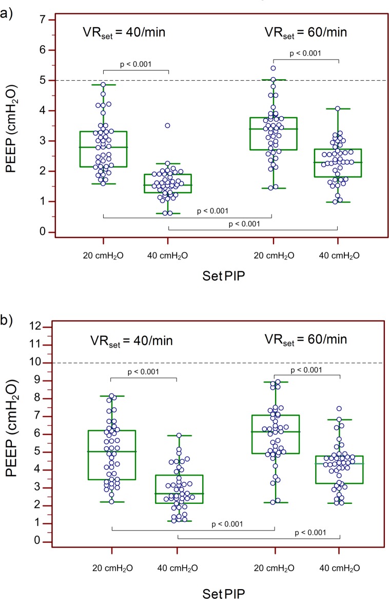
The effect of PIP and VR on the measured PEEP is shown in Fig 4, using pooled data of valves 1, 2, 4 and 5. For both PEEP settings, there was a statistically significant decrease (p<0.001) of the delivered PEEP with increasing PIP. An opposing trend was seen for VR. With increasing VR and a shortening of the expiratory time the delivered PEEP increased significantly (p<0.001). For standard settings an increase of the PIP from 20 to 40 cmH2O lead to a decrease (p<0.001) in PEEP to 2.3 (1.8–2.7) cmH2O and 4.3 (3.2–4.8) cmH2O; a decrease of VR from 60 to 40/min also lead to a PEEP decrease (p<0.05) to 2.8 (2.1–3.3) cmH2O and 5.0 (3.5–6.2) cmH2O for a set PEEP of 5 and 10 cmH2O, respectively.
Fig 4. Effect of PIP and VR.
Effect of PIP and VR on the delivered PEEP for a PEEP setting of a) 5 cmH2O and b) 10 cmH2O.
Delivered tidal volume
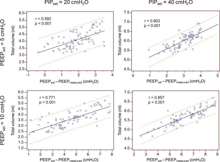
For both PIP and PEEP settings there was a statistically significant positive correlation between the delivered tidal volume (Vt) and the difference between the set and the delivered PEEP (p<0.001; Fig 5). Hence, insufficient PEEP generation led to an increase of the delivered Vt, as shown in Fig 5. The strength of this correlation increased with higher PIP and PEEP settings.
Fig 5. Effect of PEEP on tidal volume.
Increase of the delivered tidal volume(Vt) with increasing difference between the set and delivered PEEP for a PEEP setting of 5 cmH2O (top) and 10 cmH2O (bottom) and PIP of 20 cmH2O (left) and 40 cmH2O (right). Presented is the regression line with 95% prediction interval.
Discussion
This in vitro study shows that the tested single-use PEEP valves do not reliably deliver the set PEEP and some valves did not generate any clinically relevant pressure. In contrast to T-piece resuscitators, the PEEP delivered by SIBs and PEEP valves decreased during the expiratory phase and the measured end-expiratory pressure was always lower than the set PEEP. The detected PEEP increased with increasing VR and decreased with increasing PIP. The impact of VR and PIP on the delivered PEEP was higher than the differences between manufactures.
PEEP provision using a SIB
Several studies have shown that PEEP valves did not reliably deliver the set PEEP.[11–13] We have demonstrated in a previous study that even factory-new multi-use PEEP valves did not generate the PEEP to which they were set and repeated thermo-sterilization led to further functional impairment.[14]
In contrast to PEEP generation using T-piece resuscitators,[12, 15] during our measurements with the SIB and PEEP valve the pressure constantly decreased during the expiratory phase. This difference is due to the T-piece resuscitator being a constant flow device that can generate a continuous preset pressure. The driving flow can even compensate for small air leaks. Furthermore, the flow through the PEEP valve is nearly constant which improves the stability of PEEP generation. As opposed to that, using a SIB the remaining PEEP during expiration is influenced by the highly variable patient flow and even small air leaks (e.g. by the PEEP valve itself) can lead to a pressure decrease as visible in our study. However, T-piece devices cannot be operated without a constant gas flow, so in out-of-hospital settings or situations in which a gas flow is not readily available a SIB can be applied instead.
Effect of PIP and VR on the delivered PEEP
PEEP provision was influenced by the VR, as previously shown by Morley et al.[13] They demonstrated that no sufficient PEEP could be delivered at low VRs, particularly at less than 40 inflations per minute.[13] In our study measurements with VR of 40 and 60 /min were conducted to demonstrate that higher VRs lead to more reliable PEEP provision. As mentioned above, PEEP decreases continuously during expiration with the SIB. Consequently, with lower VRs time between inflations extends and PEEP decreases to lower levels.
Unexpectedly, increasing PIP led to lower PEEP levels. We suppose that this is due to increasing leak flow at higher pressures. As the model lung and intratracheal tube were firmly connected and leak-free, we speculate, that there may have been an air leak in the SIB or PEEP valve. In a previous study we could demonstrate a significant decrease of PEEP in the presence of leak when using a SIB, despite the use of a PEEP valve.[16] Furthermore, the targeted PIP of 20 cmH2O was slightly exceeded as soon as a PEEP valve was attached and 40 cmH2O could not be reached during most of the measurements, as the pressure relief valve opened early and prevented a further rise of the pressure. This may have had an impact on PEEP as well.
Effect of lower PEEP on delivered tidal volume
The delivered Vt during manual ventilation with a SIB depends on the compliance of the lung and the pressure difference between PIP and PEEP. In our experimental setup the compliance of the model lung was constant and PIP could be preset by adjustment of the indentation depth of the compressing device. Consequently, insufficient generation of PEEP increased the pressure difference and resulted in higher delivered Vt. The impact of insufficient PEEP on Vt is illustrated in Fig 5. A similar effect could be observed in a previous study using multi-use PEEP valves, where increasing leak resulted in lower PEEP levels and consequently higher Vt.[16] To which extent the demonstrated effects might be observed in vivo remains unclear, as we were unable to sufficiently simulate the changing respiratory mechanics of a preterm neonate in our laboratory setting.
Clinical implications
The tested SIBs and PEEP valves could not deliver the set PEEP. Newborn infants suffering from respiratory distress may benefit from the provision of an adequate PEEP, as it can help them establish a functional residual capacity [5, 6, 17] and improves oxygenation.[17–19] Insufficient PEEP levels during resuscitation in the delivery room should cautiously be avoided as they unnecessarily impede establishment of a functional residual capacity and can lead to atelectrauma.[20–22] There is even evidence suggesting that insufficient PEEP provision can contribute to a higher incidence of bronchopulmonary dysplasia.[23] Despite the fact that single-use PEEP valves are not subject to damage caused by repeated thermo-sterilization procedures or material fatigue due to long-term use, they do not reliably deliver the set PEEP. Operators should be aware of their unreliable PEEP provision and test the valves before clinical use as described elsewhere.[14] They should also be aware that a lower PEEP will generate a higher VT, which can contribute to potentially dangerous lung overdistension[8, 20, 24] and even brain injury.[25] In order to deliver an adequate PEEP level, operators should bear the effect of different VR and PIP on the delivered PEEP in mind and adjust the PEEP valve as appropriate.
Limitations
The experimental setup allowed for standardized PIP and VR settings during the measurements, so the effect of each particular PIP, VR, PEEP adjustment and PEEP valve could be studied separately. However, our study has several limitations. As the measurements were conducted in the described laboratory setting, several factors that may influence PEEP provision during a real resuscitation scenario could not be taken into consideration. Using an intubated and leak-free manikin, flow leak with an undesirable effect on PIP and PEEP could be prevented. However, the effect of potential flow leak that is common during initial resuscitation applying bag and mask ventilation could not be assessed. Furthermore, we used the same lung model for all the measurements, hence the effect of different respiratory mechanics due to various diseases, gestational ages or compliance changes during the first minutes after birth could not be studied. As only two valves per manufacturer were tested, we cannot make general assumptions regarding the valves’ quality or extrapolate our results to the valves of other manufacturers.
Conclusion
In conclusion, the single-use PEEP valves tested in our study delivered less than the set PEEP with all combinations of set PIP, PEEP and VR. PEEP valves should be tested before clinical use and substituted if necessary. Awareness of the valves’ characteristics and influencing factors is crucial for the provision of adequate PEEP during manual ventilation of the neonate.
Acknowledgments
We thank the manufacturers of the tested PEEP valves and SIB for their kind and unconditional provision of these.
Abbreviations
- CV
coefficient of variation
- PEEP
positive end expiratory pressure
- PIP
positive inflation pressure
- SD
standard deviation
- SIB
self-inflating bag
- VR
ventilation rate
- Vt
tidal volume
Data Availability
All data files are available from the Dryad database (accession number http://dx.doi.org/10.5061/dryad.dt43f).
Funding Statement
The authors have no support or funding to report.
References
- 1.O'Donnell CP, Davis PG, Morley CJ. Positive pressure ventilation at neonatal resuscitation: review of equipment and international survey of practice. Acta Paediatr. 2004;93(5):583–8. Epub 2004/06/04. . [DOI] [PubMed] [Google Scholar]
- 2.Roehr CC, Grobe S, Rudiger M, Hummler H, Nelle M, Proquitte H, et al. Delivery room management of very low birth weight infants in Germany, Austria and Switzerland—a comparison of protocols. Eur J Med Res. 2010;15(11):493–503. Epub 2010/12/17. . [DOI] [PMC free article] [PubMed] [Google Scholar]
- 3.Ersdal HL, Singhal N. Resuscitation in resource-limited settings. Seminars in fetal & neonatal medicine. 2013;18(6):373–8. 10.1016/j.siny.2013.07.001 . [DOI] [PubMed] [Google Scholar]
- 4.Thallinger M, Ersdal HL, Ombay C, Eilevstjonn J, Stordal K. Randomised comparison of two neonatal resuscitation bags in manikin ventilation. Arch Dis Child Fetal Neonatal Ed. 2015. 10.1136/archdischild-2015-308754 . [DOI] [PubMed] [Google Scholar]
- 5.Siew ML, Te Pas AB, Wallace MJ, Kitchen MJ, Lewis RA, Fouras A, et al. Positive end-expiratory pressure enhances development of a functional residual capacity in preterm rabbits ventilated from birth. J Appl Physiol. 2009;106(5):1487–93. Epub 2009/03/28. 91591.2008 [pii] 10.1152/japplphysiol.91591.2008 . [DOI] [PubMed] [Google Scholar]
- 6.te Pas AB, Siew M, Wallace MJ, Kitchen MJ, Fouras A, Lewis RA, et al. Establishing functional residual capacity at birth: the effect of sustained inflation and positive end-expiratory pressure in a preterm rabbit model. Pediatr Res. 2009;65(5):537–41. 10.1203/PDR.0b013e31819da21b . [DOI] [PubMed] [Google Scholar]
- 7.Vento M, Lista G. Managing Preterm Infants in the First Minutes of Life. Paediatric respiratory reviews. 2015;16(3):151–6. 10.1016/j.prrv.2015.02.004 . [DOI] [PubMed] [Google Scholar]
- 8.Wyckoff MH. Initial resuscitation and stabilization of the periviable neonate: the Golden-Hour approach. Seminars in perinatology. 2014;38(1):12–6. 10.1053/j.semperi.2013.07.003 . [DOI] [PubMed] [Google Scholar]
- 9.Perlman JM, Wyllie J, Kattwinkel J, Wyckoff MH, Aziz K, Guinsburg R, et al. Part 7: Neonatal Resuscitation: 2015 International Consensus on Cardiopulmonary Resuscitation and Emergency Cardiovascular Care Science With Treatment Recommendations. Circulation. 2015;132(16 Suppl 1):S204–41. 10.1161/CIR.0000000000000276 . [DOI] [PubMed] [Google Scholar]
- 10.Wyllie J, Bruinenberg J, Roehr CC, Rudiger M, Trevisanuto D, Urlesberger B. European Resuscitation Council Guidelines for Resuscitation 2015: Section 7. Resuscitation and support of transition of babies at birth. Resuscitation. 2015;95:249–63. 10.1016/j.resuscitation.2015.07.029 . [DOI] [PubMed] [Google Scholar]
- 11.Bennett S, Finer NN, Rich W, Vaucher Y. A comparison of three neonatal resuscitation devices. Resuscitation. 2005;67(1):113–8. Epub 2005/08/06. S0300-9572(05)00172-3 [pii] 10.1016/j.resuscitation.2005.02.016 . [DOI] [PubMed] [Google Scholar]
- 12.Kelm M, Proquitte H, Schmalisch G, Roehr CC. Reliability of two common PEEP-generating devices used in neonatal resuscitation. Klin Padiatr. 2009;221(7):415–8. Epub 2009/09/05. 10.1055/s-0029-1233493 . [DOI] [PubMed] [Google Scholar]
- 13.Morley CJ, Dawson JA, Stewart MJ, Hussain F, Davis PG. The effect of a PEEP valve on a Laerdal neonatal self-inflating resuscitation bag. J Paediatr Child Health. 2010;46(1–2):51–6. JPC1617 [pii] 10.1111/j.1440-1754.2009.01617.x . [DOI] [PubMed] [Google Scholar]
- 14.Hartung JC, Schmolzer G, Schmalisch G, Roehr CC. Repeated thermo-sterilisation further affects the reliability of positive end-expiratory pressure valves. J Paediatr Child Health. 2013;49(9):741–5. 10.1111/jpc.12258 . [DOI] [PubMed] [Google Scholar]
- 15.Schmolzer GM, Roehr CC. Use of respiratory function monitors during simulated neonatal resuscitation. Klin Padiatr. 2011;223(5):261–6. 10.1055/s-0031-1275696 . [DOI] [PubMed] [Google Scholar]
- 16.Hartung JC, te Pas AB, Fischer H, Schmalisch G, Roehr CC. Leak during manual neonatal ventilation and its effect on the delivered pressures and volumes: an in vitro study. Neonatology. 2012;102(3):190–5. 10.1159/000339325 . [DOI] [PubMed] [Google Scholar]
- 17.Dinger J, Topfer A, Schaller P, Schwarze R. Effect of positive end expiratory pressure on functional residual capacity and compliance in surfactant-treated preterm infants. J Perinat Med. 2001;29(2):137–43. Epub 2001/05/10. 10.1515/JPM.2001.018 . [DOI] [PubMed] [Google Scholar]
- 18.Probyn ME, Hooper SB, Dargaville PA, McCallion N, Crossley K, Harding R, et al. Positive end expiratory pressure during resuscitation of premature lambs rapidly improves blood gases without adversely affecting arterial pressure. Pediatr Res. 2004;56(2):198–204. Epub 2004/06/08. 10.1203/01.PDR.0000132752.94155 13 01.PDR.0000132752.94155.13 [pii]. . [DOI] [PubMed] [Google Scholar]
- 19.Michna J, Jobe AH, Ikegami M. Positive end-expiratory pressure preserves surfactant function in preterm lambs. Am J Respir Crit Care Med. 1999;160(2):634–9. Epub 1999/08/03. . [DOI] [PubMed] [Google Scholar]
- 20.Schmolzer GM, Te Pas AB, Davis PG, Morley CJ. Reducing lung injury during neonatal resuscitation of preterm infants. J Pediatr. 2008;153(6):741–5. Epub 2008/11/19. S0022-3476(08)00689-6 [pii] 10.1016/j.jpeds.2008.08.016 . [DOI] [PubMed] [Google Scholar]
- 21.Vento M. Improving fetal to neonatal transition of the very preterm infant: novel approaches. Chinese medical journal. 2010;123(20):2924–8. . [PubMed] [Google Scholar]
- 22.Hooper SB, Polglase GR, Roehr CC. Cardiopulmonary changes with aeration of the newborn lung. Paediatric respiratory reviews. 2015;16(3):147–50. 10.1016/j.prrv.2015.03.003 [DOI] [PMC free article] [PubMed] [Google Scholar]
- 23.Szyld E, Aguilar A, Musante GA, Vain N, Prudent L, Fabres J, et al. Comparison of devices for newborn ventilation in the delivery room. J Pediatr. 2014;165(2):234–9 e3. 10.1016/j.jpeds.2014.02.035 . [DOI] [PubMed] [Google Scholar]
- 24.Vento M, Cheung PY, Aguar M. The first golden minutes of the extremely-low-gestational-age neonate: a gentle approach. Neonatology. 2009;95(4):286–98. 10.1159/000178770 . [DOI] [PubMed] [Google Scholar]
- 25.Barton SK, Tolcos M, Miller SL, Roehr CC, Schmolzer GM, Davis PG, et al. Unraveling the Links Between the Initiation of Ventilation and Brain Injury in Preterm Infants. Frontiers in pediatrics. 2015;3:97 10.3389/fped.2015.00097 [DOI] [PMC free article] [PubMed] [Google Scholar]
Associated Data
This section collects any data citations, data availability statements, or supplementary materials included in this article.
Data Availability Statement
All data files are available from the Dryad database (accession number http://dx.doi.org/10.5061/dryad.dt43f).


