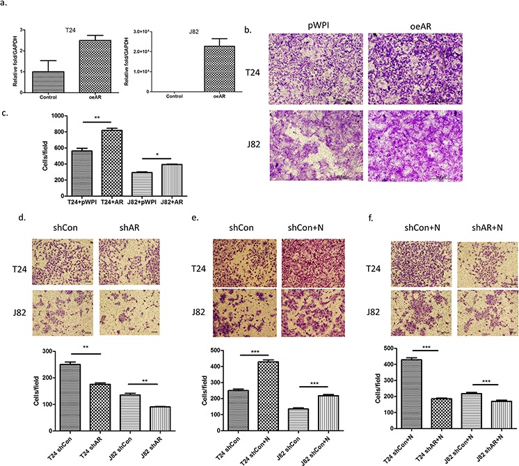
Figure 4. AR is involved in the invasion-promoting effect induced by neutrophils.

a. AR mRNA expression after using lentivirus to overexpress AR (oeAR) in BCa cells. b. Microscopic images of invasion assay of cells in a. (scale bar = 20 μm). c. Quantitation of the result of invasion assay of Figure 5b. d. Microscopic images of BCa invasion assay after knocking down AR (shAR). (scale bar = 20 μm). e. Microscopic images of shControl (shCon) BCa invasion assay after co-culturing with neutrophils. f. Microscopic images of HL-60N cells co-cultured with BCa cells after BCa after knocking down AR (shAR). Quantitation is below images in d–f. **p < 0,01; ***p < 0.001; pWP1 = vector control; N = Neutrophils.
