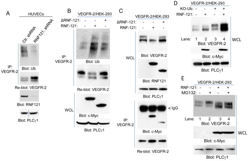
Figure 5. RNF121 ubiquitinates VEGFR-2.
(A) Human primary endothelial cells (HUVECs) were infected with control shRNA or RNF121 shRNA. Cells were lysed and whole cell lysates (WCL) was subjected to immunoprecipitation assay using anti-VEGFR-2 antibody followed by immunoblotting with anti-ubiquitin antibody (Ub). The same membrane was blotted for VEGFR-2. An aliquot of whole cell lysates from the same group were blotted for RNF121 and for loading control protein, PLCγ1. (B) HEK293 cells expressing VEGFR-2 transfected with control vector, myc-RNF121 or RING Finger domain truncated RNF121 (ΔRNF121). After 48 hours, cells were lysed and whole cell lysates were subjected to immunoprecipitation using anti-VEGFR-2 antibody followed by immunoblotting with anti-ubiquitin antibody. Aliquot of whole cell lysates were also blotted for RNF121 and loading control protein, PLCγ1. Also shown is a graph quantification of ubiquitination of VEGFR-2 by RNF121 or ΔRNF121. It is representative of three independent experiments. *p< 0.05. (C) HEK293 cells expressing VEGFR-2 transfected with control vector, Myc-RNF121 or RING domain truncated RNF121 as panel B, and whole cell lysates (WCL) were blotted for VEGFR-2 and RNF121. Whole cell lysates from the same group were subjected to immunoprecipitation using anti-VEGFR-2 antibody followed by immunoblotting with anti-c-Myc antibody for RNF121. All the blots presented here are representative of at least three independent experiments.

