Abstract
Objectives
Enamel fluorosis is a hypomineralization caused by chronic exposure to high levels of fluoride during tooth development. Previous research on the relationship between enamel fluoride content and fluorosis severity has been equivocal. The current study aimed at comparing visually and histologically assessed fluorosis severity with enamel fluoride content.
Methods
Extracted teeth (n=112) were visually examined using the Thylstrup and Fejerskov Index for fluorosis. Eruption status of each tooth was noted. Teeth were cut into 100 μm slices to assess histological changes with polarized light microscopy. Teeth were categorized as sound, mild, moderate, or severe fluorosis, visually and histologically. They were cut into squares (2×2 mm) for the determination of fluoride content (microbiopsy) at depths of 30, 60 and 90 μm from the external surface.
Results
Erupted teeth with severe fluorosis had significantly greater mean fluoride content at 30, 60 and 90 μm than sound teeth. Unerupted teeth with mild, moderate and severe fluorosis had significantly greater mean fluoride content than sound teeth at 30 μm; unerupted teeth with mild and severe fluorosis had significantly greater mean fluoride content than sound teeth at 60 μm, while only unerupted teeth severe fluorosis had significantly greater mean fluoride content than sound teeth at 90 μm.
Conclusions
Both erupted and unerupted severely fluorosed teeth presented higher mean enamel fluoride content than sound teeth.
Clinical Significance
Data on fluoride content in enamel will further our understanding of its biological characteristics which play a role in the management of hard tissue diseases and conditions.
Keywords: Enamel fluorosis, Dental Enamel, Fluoride
Introduction
Enamel fluorosis is a hypomineralization of dental enamel characterized by chronic exposure to high levels of fluoride during tooth development [1, 2]. Fluoride interacts with mineralized tissues and when present in excess disturbs dental enamel development. As severity of fluorosis increases, changes in the porosity of subsurface enamel extend deeper into the tissue resulting in hypomineralized areas covered by a defined zone of highly mineralized tissue that may affect the entire enamel surface [3].
An analysis comparing fluorosis prevalence data in the United States from the 1930s and 1980s indicated an increase in enamel fluorosis in children with a prevalence 27% in children residing in areas with optimal fluoride (0.7 to 1.2 μg/ml) and 15% in children from areas with suboptimal fluoride (< 0.7 μg/ml) in the 1980s versus a prevalence of 12 to 25% for those living in areas with optimal natural fluoride content (ranging from 0.09 to 1.3 μg/ml) and 7% in children in areas with suboptimal fluoride (< 0.7 μg/ml) in the 1930s [4]. Comparison of the data from 1986–1987 and a survey conducted in 1999–2002 identified a 9% increase of fluorosis prevalence in children and adolescents aged 6–19 years (from 22.8% in 1986–1987 to 32% in 1999–2002. Although the increase in prevalence has occurred primarily in very mild and mild forms of enamel fluorosis, between 3 and 4% of children and adolescents had moderate or severe fluorosis in 1999–2002 [5].
Studies into the relationship between enamel fluorosis severity and fluoride content have been equivocal. Some studies have reported a positive correlation between the clinical severity of enamel fluorosis and enamel fluoride content [6–8]. Brudevold et al. [9] reported a positive trend between enamel fluorosis severity and fluoride content within rat incisors. However, each fluorosis category had a large standard deviation and overlapping fluoride content. Therefore, teeth that were categorized visually as sound may have had the same fluoride content as those teeth that presented with severe forms of fluorosis. Furthermore, it has been reported that enamel fluoride content vary depending on the location within the oral cavity with central incisors having significantly lower fluoride [10].
On the other hand, some studies have reported that enamel fluorosis severity is independent of enamel fluoride content. Olsen and Johansen [11] studied human teeth and found that enamel surface appearance was independent of the enamel fluoride content. Furthermore, Vieira et al. [12] reported a correlation between dentin fluoride concentration and the presence of enamel fluorosis in unerupted third molars. With regards to enamel; however, the authors found no such correlation.
Despite conflicting results, no studies have thoroughly examined the correlation between enamel fluorosis severity and enamel fluoride content. Additionally, to the authors’ knowledge, no studies have been reported investigating fluoride content at different enamel depths in relation to fluorosis severity. Previous studies have relied on visual examinations to classify the severity of enamel fluorosis’ clinical signs and none have related the fluoride content of enamel to the histological severity of fluorosed enamel changes. Therefore, the aim of this study was to investigate the relationship between fluoride content in dental enamel at different depths with the presence of enamel fluorosis detected visually and using histological assessments.
Materials and Methods
Extracted teeth with fluorosis, free of caries, including incisors, premolars and molars (total of 120, 44 erupted – 8 incisors, 5 premolars and 31 molars; and 76 unerupted – 11 incisors, 9 premolars and 56 molars), were collected after approval was obtained from the Indiana University Institutional Review Board. Teeth were stored in deionized water saturated with 0.1% thymol. The teeth were collected from dental offices and transported to the study site in the saturated 0.1% thymol solution. Upon receipt, the teeth were sorted, cleaned and the root removed.
The teeth were visually examined using the Thylstrup and Fejerskov Index for fluorosis (TFI) by two trained and calibrated examiners (AESR and EAMM). In those cases where there was disagreement, the examiners discussed their findings and reached an agreement [13]. Using the buccal and occlusal aspects of posterior permanent teeth and labial aspects of anterior permanent teeth, in vitro scores were assigned to each aspect. Fluorosis severity in this study was rated as mild, moderate, or severe. Thirty sound teeth were included as negative controls. Four categories were therefore created: mild, moderate, severe and sound. Thirty teeth from each of the four categories, as determined visually, were analyzed in this study.
After visual scores were assigned, specimens were cut into halves. From one half of each tooth, bucco-lingual tooth sections of 100 ± 20 μm were cut longitudinally in the midline of the fluorosis area using a Series 1000 Deluxe Hard Tissue Microtome (SciFab, Lafayette, CO, USA), and an average of 2 sections per tooth were obtained. Sections were imbibed in water and examined using a polarized light microscope (Orthoplan, Leitz, Wetzlar, Germany). Digital images were taken for qualitative evaluation of the hypomineralized area. A demineralized area in enamel or dentin with positive birefringence was defined as a lesion. Using the results from polarized light microscopy, each tooth was assigned as sound, mildly fluorosed, moderately fluorosed, or severely fluorosed based on histological characteristics described by Fejerskov et al. [14].
The other half of each tooth was mounted onto an acrylic plate (Total Plastics, Indianapolis, IN, USA) using sticky wax (Kerr Corporation, Orange, CA, USA) with an area of flat enamel facing up. Using an Isomet low speed saw (Buehler, Lake Bluff, IL, USA) with a 2 mm spacer between two blades, 2 × 2 mm square pieces were cut from each tooth. Each piece was then waxed with enamel facing down to another acrylic block, and the dentin was sanded away using a RotoPol-31 machine (Struers, Cleveland, OH, USA) with 1200 grit laboratory grade SiC abrasive paper (Struers, Cleveland, OH, USA) until a flat dentin surface remained. Pieces were then glued onto steel drill rods (Grainger, Inc., Indianapolis, IN, USA) measuring ~3.0 mm in diameter using Duro Quick Gel No-Run Super Glue (Loctite, Plainfield, IL, USA) with enamel facing up and parallel to top of drill rod. Clear, fluoride-free fingernail polish (Del Laboratories, Inc., Farmingdale, NY, USA) was applied to the rods to prevent rust, and they were individually stored in vials (7 mL-vials, Fisher Scientific Co., Itasca, IL, USA) with enough water to cover each specimen. Finally, 4.0 × 5.0 mm pieces of 1200 grit laboratory grade fluoride-free SiC abrasive paper were cut and attached to ¾ × 1 × 1 in acrylic blocks (Total Plastics, Indianapolis, IN, USA) with a light application of sticky wax.
Fluoride Microbiopsy
Fluoride microbiopsies were conducted using a modification of the procedure described by Hellwig et al. [15]. Each drill rod with the attached specimen was inserted into the chuck of a microdrill machine (Stellar Systems, Vienna, VA, USA). Acrylic blocks were secured onto the microdrill stage, and the spinning specimen was slowly lowered until initial contact with the abrasive paper was made. A digital micrometer attached to the microdrill was zeroed, and the specimen was lowered in small increments while using the stage controls to navigate over the entire abrasive surface until 30 μm of enamel had been removed. The acrylic block was removed from the stage, and an open vial was placed on top of the abrasive paper. The block was turned upside-down, and the bottom was tapped to remove the lose powder into the vial. Next, the vial was placed under the specimen in the microdrill, and the rod was tapped to remove lose powder attached to the specimen. The abrasive paper with enamel powder was removed from the acrylic block and placed in the vial. Finally, the vial was capped tightly, and the specimen was brushed to prevent carry-over of enamel powder to succeeding samples. This process was repeated twice on each specimen, resulting in samples at depths of 30 μm, 60 μm, and 90 μm for each tooth.
Due to the tooth morphology, the employed abrasion technique did not always collect powder from 100% of the enamel surface within the first 30 μm. Therefore, after collection of the 30 μm sample, a picture of the 2 × 2 mm specimen was taken on a Nikon SMZ1500 Stereomicroscope (Nikon Instruments Inc., Elgin, IL, USA) with attached NI-150 High Intensity Illuminator (Nikon Instruments Inc., Elgin, IL, USA). Using this photograph, a percent volume of abrasion was calculated. This percent volume was used to adjust observed fluoride concentrations to an estimated fluoride concentration value if 100% of the 2 × 2 mm surface was subjected to abrasion within the first 30 μm sample.
Fluoride Content Analysis
Eighty μL of 0.5 M perchloric acid (HClO4 Fisher Scientific Co., Itasca, IL, USA) were added to a vial containing the enamel powder collected though the microbiopsies. Vials were capped tightly, vortexed for approximately 10 s, and centrifuged in a micro 18R microcentrifuge (VWR International, Germany) at 15000 rpm for 5 min to ensure settling of enamel powder and abrasive paper. Vials were then placed in a rack and secured to a CH-4103 rotary shaker (Infors HT, Bottminger, Switzerland). Samples were shaken at approximately 50 rpm for 16–18 h. Analyzer caps (Curtis Matheson Scientific/Fisher Scientific Co., Itasca, IL, USA) were prepared using 20 μl of the contents of each vial, 40 μl of DI water, and 40 μl of Citrate-EDTA buffer (Fisher Scientific Co., Itasca, IL, USA). Fluoride content of each sample was analyzed using a combination fluoride ion-specific electrode (Orion #96-909-00/Fisher Scientific Co., Itasca, IL, USA) and an Orion EA940 pH/ion meter (Fisher Scientific Co., Itasca, IL, USA). Standard fluoride solutions of 0.02; 0.04; 0.1; 0.2; 0.4; 1.0; and 4.0 μg/ml were used to create a standard curve. Fluoride content of samples were calculated in units of μg/cm3.
Statistical Analysis
Results were analyzed via an analysis of variance (ANOVA) model to compare mean fluoride content between visual exams and histology group assignment. Fluoride content was log transformed to satisfy model assumptions, and a Sidak adjustment was used to adjust for multiple comparisons. Furthermore, subgroup analyses based on tooth eruption pattern were conducted using similar statistical methods. At each depth and by tooth eruption pattern, Pearson correlation coefficients (r2) were reported to assess strength of correlations between histology and visual examinations.
Results
Of the original 120 selected teeth, only 112 were analyzed throughout the entire process. The eight rejected teeth were excluded from analysis either due to cracks that caused the tooth to break or due to chipping off of the enamel surface during the cutting process. Of these, one tooth was sound, one had mild fluorosis, two were moderate and four were severe. The loss of these samples resulted in 36 erupted teeth and 76 unerupted teeth. Of the remaining teeth, 29 teeth were from the sound group, 29 teeth were visually assessed as TFI group I (mild fluorosis), 28 teeth in TFI group II (moderate fluorosis), and 26 teeth scored as TFI group III (severe fluorosis). Based on histology, a second set of blinded examiners categorized 24 teeth as sound, 50 were scored as mild, 17 were characterized as moderate, and 12 were scored as severe. Kappa values for inter- and intra-examiner agreement for histological assessments varied from 0.83 to 0.92.
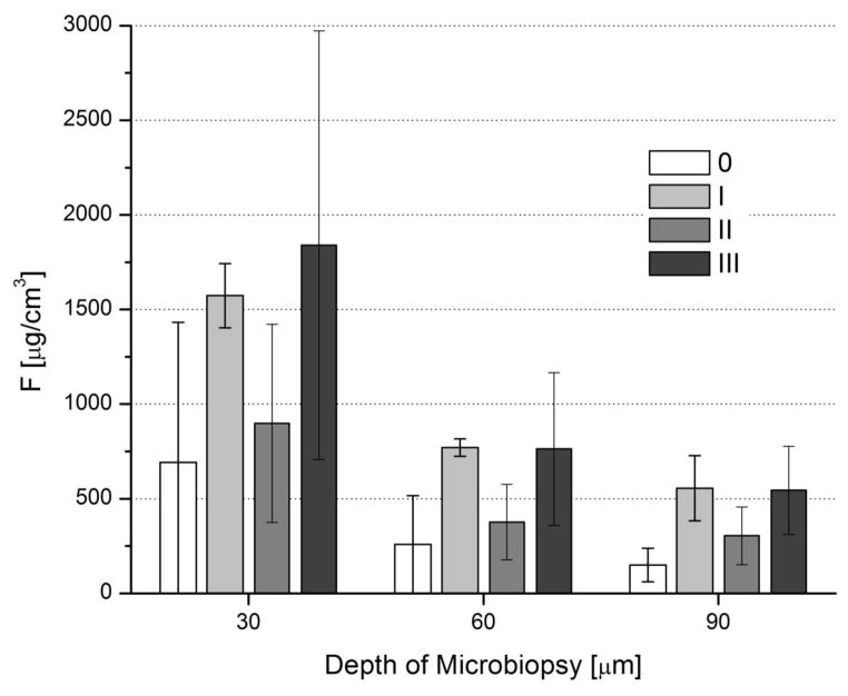
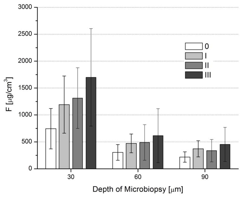
When the fluoride content of erupted teeth was compared to that of unerupted teeth, significant differences were found only for TFI group I, with a larger fluoride content found in erupted teeth at 30 μm (p=0.0006), but a smaller content at 60 and 90 μm (p=0.0006 and 0.0008). The average fluoride content for both erupted (Figure 1) and unerupted (Figure 2) teeth decreased as depth into enamel increased.
Figure 1.
Mean fluoride content with standard deviations as a function of microbiopsy depth for each TFI group based on visual analysis for erupted teeth
Figure 2.
Mean fluoride content with standard deviations as a function of microbiopsy depth for each TFI group based on visual analysis for unerupted teeth
For erupted teeth, TFI group III teeth had significantly higher values than sound teeth at all depths of 30 μm (p=0.009), 60 μm (p=0.0007), and 90 μm (p<0.001). Meanwhile, TFI group II had significantly larger mean fluoride values (p=0.009) than sound teeth at a depth of 90 μm. In addition, TFI group I had significantly larger mean fluoride values than sound teeth on at depths of 60 μm (p=0.009) and 90 μm (p<0.001).
For unerupted teeth, it was observed that TFI group III teeth had significantly larger mean fluoride values than those for sound teeth at 30 μm (p<0.001), 60 μm (p=0.018), and 90 μm (p<0.001). TFI group II teeth had significantly larger values (p=0.005) than sound teeth only at the depth of 30 μm. Finally, TFI group I teeth had larger mean fluoride values when compared to sound teeth at 30 μm (p=0.019) and 90 μm (p=0.022).
When both erupted and unerupted teeth were analyzed together, each fluorosis category had a large standard deviation and overlapping fluoride content (Table 1). It was observed that on average teeth characterized as having some form of fluorosis (TFI groups I–III) had significantly larger mean fluoride content at 30 μm (p=0.0026), 60 μm (p=0.0168), and 90 μm (p=0.0015) than those characterized as sound. Finally, it was observed that teeth from TFI group III had significantly higher values (p=0.0442) than those from TFI group II at a depth of 30 μm.
Table 1.
Summary statistics for fluoride content based on visual grouping
| Depth (μm) | Visual Grouping | N | Summary Statistics (μg/cm3)
|
Max | Overall | p-values
|
vs. II | ||||
|---|---|---|---|---|---|---|---|---|---|---|---|
| Mean | SD | Min | Median | vs. 0 | vs. I | ||||||
| 30 | 0 (Sound) | 29 | 717.8 | 582.8 | 117.4 | 556.1 | 2507.0 | <0.0001 | |||
| I (Mild) | 29 | 1231.2 | 517.8 | 406.7 | 1324.6 | 2226.9 | 0.0001 | ||||
| II (Moderate) | 28 | 1150.4 | 576.4 | 167.5 | 1209.7 | 2446.6 | 0.0026 | 0.9698 | |||
| III (Severe) | 26 | 1736.5 | 949.6 | 646.6 | 1397.7 | 3955.8 | <0.0001 | 0.2603 | 0.0442 | ||
| 60 | 0 (Sound) | 29 | 280.7 | 208.8 | 66.6 | 219.9 | 1003.9 | <0.0001 | |||
| I (Mild) | 29 | 503.0 | 189.1 | 212.4 | 554.4 | 823.5 | 0.0001 | ||||
| II (Moderate) | 28 | 445.1 | 287.3 | 116.3 | 343.5 | 1263.0 | 0.0168 | 0.5919 | |||
| III (Severe) | 26 | 655.0 | 473.7 | 219.3 | 489.0 | 2358.0 | < 0.0001 | 0.9191 | 0.1047 | ||
| 90 | 0 (Sound) | 29 | 182.8 | 97.1 | 49.9 | 147.9 | 426.1 | <0.0001 | |||
| I (Mild) | 29 | 390.9 | 159.7 | 125.5 | 402.7 | 742.8 | <0.0001 | ||||
| II (Moderate) | 28 | 324.4 | 186.1 | 82.6 | 259.0 | 839.0 | 0.0015 | 0.393 | |||
| III (Severe) | 26 | 478.2 | 295.0 | 164.7 | 367.4 | 1262.5 | <0.0001 | 0.9569 | 0.072 | ||
Moderate to weak but significant (all p<0.05) linear associations were found between fluoride content and visual scores. Fluoride content showed a fair amount of overlap between categories, hence no clear linear increase or decrease was observed: for erupted teeth, r2 = 0.42 at 30 μm, r2 = 0.50 at 60 μm, and r2 = 0.62 at 90 μm, and for unerupted teeth, r2 = 0.56 at 30 μm, r2 = 0.46 at 60 μm, and r2 = 0.46 at 90 μm. As depth increased, mean fluoride content in all four groups (TFI I; II; III; sound) decreased.
When the fluoride content of teeth was compared among the different severities of fluorosis assessed histologically, no significant differences were found between erupted and unerupted teeth. Therefore, comparisons were made for all teeth as a single group (Table 2). As observed when the teeth were assessed visually when the depth of sample taken increased, mean fluoride content of each group decreased. Mean fluoride content increased between each severity category at all three depths. In addition, teeth characterized as having fluorosis [TFI group I (mild), TFI group II (moderate), TFI group III (severe)] had significantly larger mean fluoride content than those characterized as sound at 30 μm (p=0.031), 60 μm (p=0.009), and 90 μm (p<0.001). Furthermore, teeth characterized through histology to have severe fluorosis were observed to have significantly higher mean fluoride content than teeth characterized as having mild fluorosis (TFI group II) at depths of 30 μm (p=0.004), 60 μm (p=0.002), and 90 μm (p=0.005). Finally, teeth with severe fluorosis had significantly larger mean fluoride content (p=0.024) than teeth with moderate fluorosis at a depth of 30 μm.
Table 2.
Summary statistics for fluoride content based on histological grouping
| Depth (μm) | Histology | N | Summary Statistics (μg/cm3)
|
Max | Overall | p-values
|
vs. Moderate | ||||
|---|---|---|---|---|---|---|---|---|---|---|---|
| Mean | SD | Min | Median | vs. Sound | vs. Mild | ||||||
| 30 | Sound | 24 | 813.3 | 649.9 | 117.4 | 556.4 | 2507 | <0.0001 | |||
| Mild | 50 | 1146.0 | 507.1 | 406.7 | 1105.9 | 2446.6 | 0.0032 | ||||
| Moderate | 17 | 1215.7 | 481.4 | 167.5 | 1357.2 | 1917.8 | 0.0307 | 0.9999 | |||
| Severe | 12 | 2261.7 | 1117.7 | 1055.6 | 1878.0 | 3955.8 | <0.0001 | 0.0041 | 0.0243 | ||
| 60 | Sound | 24 | 316.2 | 264.8 | 66.6 | 231.2 | 1086.1 | <0.0001 | |||
| Mild | 50 | 437.9 | 210.8 | 143.2 | 419.2 | 1081.1 | 0.0086 | ||||
| Moderate | 17 | 499.1 | 180.9 | 116.3 | 531.6 | 823.5 | 0.0051 | 0.9064 | |||
| Severe | 12 | 907.0 | 586.6 | 390.6 | 830.1 | 2358 | <0.0001 | 0.0018 | 0.0887 | ||
| 90 | Sound | 24 | 207.5 | 156.5 | 49.9 | 155.4 | 834.8 | <0.0001 | |||
| Mild | 50 | 338.6 | 170.8 | 82.6 | 275.5 | 839.0 | 0.0004 | ||||
| Moderate | 17 | 371.6 | 150.8 | 97.6 | 383.2 | 742.8 | 0.0008 | 0.9643 | |||
| Severe | 12 | 603.5 | 303.4 | 220.8 | 608.6 | 1262.5 | <0.0001 | 0.0052 | 0.1235 | ||
The correlation among histological categories and fluoride content by eruption pattern were assessed at all three enamel depths. For erupted teeth, moderate to weak but significant linear associations were found between fluoride content and histologically assessed severity categories (r2 = 0.45 at 30 μm, r2 = 0.54 at 60 μm, and r2 = 0.67 at 90 μm). Similar results were found for unerupted teeth (r2 = 0.54 at 30 μm, r2 = 0.44 at 60 μm, and r2 = 0.40 at 90 μm). No differences were observed between molars, premolars and incisors.
Discussion
The present study has shown that teeth with fluorosis exhibit higher enamel fluoride content than sound teeth at depths up to 90 μm below the anatomical surface and regardless of fluorosis severity. There was a clear trend for teeth with increasing fluorosis severities exhibiting higher fluoride content, and especially for the outermost enamel layer, although not all comparisons were of statistical significance. These findings are in agreement with previous investigations [6,7] and highlight that excess systemic fluoride can be incorporated into the dental hard tissues.
Our results are also similar to those of Vieira et al. [12] in their assessment of 99 third molars for mean fluoride content. They determined that enamel fluoride content were not strongly correlated to increasing fluorosis severity. Furthermore, similar observations were made by Olsen and Johansen [11] in their study of human enamel which showed no relationship between fluoride content and surface appearance.
When analyzing the present data by eruption status, both erupted and unerupted teeth with severe fluorosis presented higher mean fluoride content than sound teeth at all three enamel depths for this current study. No other significant differences between sound and the other fluorosis severity categories were consistent among all enamel depths for the erupted and unerupted subgroups. Also, there were few significant differences between fluorosis categories in the subgroup analyses. However, it was not possible to establish cut-off values that distinguished fluorosis severity categories. These results are in disagreement with those of Richards et al. [7] who found that mean fluoride content increase in unerupted teeth as fluorosis severity increases and were able to statistically differentiate all severity categories. However, in their study, they used the entire TFI, and the authors predicted TFI scores as if the teeth had erupted based on surface appearance and pore volume distribution when viewed by transmitted light. In this current study, the mean fluoride content were determined without predicting TFI scores for unerupted teeth.
Other factors may need to be considered when attempting to correlate fluorosis severity to mean fluoride content. For instance, one study by Everett et al. [16] showed that factors such as genetic history may affect presence of enamel fluorosis in different individuals. In their study, mice were inbred in order to create populations with similar genotypes. They found that the severity of enamel fluorosis ranged widely between different mice populations despite similar conditions such as age, gender, housing, food, and fluoride levels in drinking water. Furthermore, Vieira et al. [12] showed that genetics may also play a role in the severity of fluorosis experienced within a population. Other external factors, such as altitude, have been shown to influence the presence of fluorosis as well [17]. Finally, ten Cate [18] determined that the final fluoride content is dependent somewhat on the amount of time between enamel formation and eruption. Therefore, teeth that take longer to erupt could have larger mean fluoride content. These genetic and environmental differences between the populations in which teeth for different studies have been collected may be contributors to the absence of a correlation between enamel fluoride content and enamel fluorosis severity. However, all the studies discussed in this paper have been conducted in vitro, making it difficult to determine the origin of teeth and how these factors may influence results.
In conclusion, there is a significant difference in mean fluoride content between teeth with severe fluorosis and sound teeth in diagnosed visually or histologically in both unerupted and erupted teeth. However, different severity categories presented overlapping fluoride content.
Acknowledgments
The study was supported by a National Institute of Dental and Craniofacial Research grant (NIDCR – R21DE016034-02). The funders had no role in study design, data collection and analysis, decision to publish, or preparation of the manuscript.
Footnotes
Declaration of Interests
No potential conflicts of interest are reported for any of the authors.
Publisher's Disclaimer: This is a PDF file of an unedited manuscript that has been accepted for publication. As a service to our customers we are providing this early version of the manuscript. The manuscript will undergo copyediting, typesetting, and review of the resulting proof before it is published in its final citable form. Please note that during the production process errors may be discovered which could affect the content, and all legal disclaimers that apply to the journal pertain.
References
- 1.Den Besten PK. Dental fluorosis: its uses as a biomarker. Advances in Dental Research. 1994;8:105–10. doi: 10.1177/08959374940080010201. [DOI] [PubMed] [Google Scholar]
- 2.Vieira APGF, Hancock R, Eggertson H, Everett ET, Grynpas MD. Tooth quality in dental fluorosis: genetic and environmental factors. Calcified Tissue International. 2005;76:17–25. doi: 10.1007/s00223-004-0075-3. [DOI] [PubMed] [Google Scholar]
- 3.Aoba T, Fejerskov O. Dental fluorosis: chemistry and biology. Critical Reviews in Oral Biology and Medicine. 2002;13:155–70. doi: 10.1177/154411130201300206. [DOI] [PubMed] [Google Scholar]
- 4.Beltran-Aguilar ED, Griffin S, Lockwood S. Prevalence and trends in enamel fluorosis in the United States from the 1930s to the 1980s. Journal of the American Dental Association. 2002;133:157–65. doi: 10.14219/jada.archive.2002.0139. [DOI] [PubMed] [Google Scholar]
- 5.Beltran-Aguilar ED, Barker LK, Canto MT, Dye BA, Gooch BF, Griffin SO, et al. Surveillance for dental caries, dental sealants, tooth retention, edentulism, and enamel fluorosis-United States, 1988–1994 and 1999–2002. Morbidity and Mortality Weekly Report Surveillance Summaries. 2005;54:1–43. [PubMed] [Google Scholar]
- 6.Retief DH, Bradley EL, Barbokow FH, Friedman M, Van der Merwe EH, Bischoff JI. Relationships among fluoride concentration in enamel, degree of fluorosis and caries incidence in a community residing in a high fluoride area. Journal of Oral Pathology. 1979;8:224–36. doi: 10.1111/j.1600-0714.1979.tb01889.x. [DOI] [PubMed] [Google Scholar]
- 7.Richards A, Fejerskov O, Baelum V. Enamel fluoride in relation to severity of human dental fluorosis. Advances in Dental Research. 1989;3:147–53. doi: 10.1177/08959374890030021301. [DOI] [PubMed] [Google Scholar]
- 8.Richard A, Likimani S, Baelum V, Fejerskov O. Fluoride concentrations in unerupted fluorotic human enamel. Caries Research. 1992;26:328–32. doi: 10.1159/000261463. [DOI] [PubMed] [Google Scholar]
- 9.Brudevold F, Bakhos Y, Aasenden R. Dental fluorosis as related to the concentration of fluoride in teeth and bone. Journal of the American Dental Association. 1978;96:459–63. doi: 10.14219/jada.archive.1978.0097. [DOI] [PubMed] [Google Scholar]
- 10.Aasenden R, Moreno EC, Brudevold F. Fluoride levels in the surface enamel of different types of human teeth. Archives of Oral Biology. 1973;18:1403–10. doi: 10.1016/0003-9969(73)90114-3. [DOI] [PubMed] [Google Scholar]
- 11.Olsen T, Johansen E. Fluoride content of human fluorosed enamel (abstract) Journal of Dental Research. 1978;57:281. [Google Scholar]
- 12.Vieira APGF, Hancock R, Limeback H, Maia R, Grynpas MD. Is fluoride concentration in dentin and enamel a good indicator of dental fluorosis? Journal of Dental Research. 2004;83:76–80. doi: 10.1177/154405910408300115. [DOI] [PubMed] [Google Scholar]
- 13.Thylstrup A, Fejerskov O. Clinical appearance and surface distribution of dental fluorosis in permanent teeth in relation to histological changes. Community Dentistry and Oral Epidemiology. 1978;6:315–28. doi: 10.1111/j.1600-0528.1978.tb01173.x. [DOI] [PubMed] [Google Scholar]
- 14.Fejerskov O, Johnson NW, Silverstone LM. The ultrastructure of fluorosed human dental enamel. Scandinavian Journal of Dental Research. 1974;82:357–72. doi: 10.1111/j.1600-0722.1974.tb00389.x. [DOI] [PubMed] [Google Scholar]
- 15.Hellwig E, Klimek J, Albert G. In vivo retention of KOH soluble and firmly bound fluoride in demineralized dental enamel. Deutsche zahnarztliche Zeitschrift. 1989;44:173–6. [PubMed] [Google Scholar]
- 16.Everett ET, McHenry MA, Reynolds N, Eggertsson H, Sullivan J, Kantmann C, et al. Dental fluorosis: variability among different inbred mouse strains. Journal of Dental Research. 2002;81:794–8. doi: 10.1177/0810794. [DOI] [PubMed] [Google Scholar]
- 17.Manji F, Baelum V, Fejerskov O. Fluoride, altitude, and dental fluorosis. Caries Research. 1986;20:473–80. doi: 10.1159/000260977. [DOI] [PubMed] [Google Scholar]
- 18.Nanci A. Ten Cate’s Oral histology: development, structure, and function. 7. St. Louis: Mosby Elsevier; 2008. [Google Scholar]


