Abstract
In this paper, we report on our pilot evaluation of a prototype foot/ankle prosthesis. This prototype has been designed and fabricated with the intention of providing decreased ankle joint stiffness during the middle portion of the stance phase of gait, and increased (i.e., more normal) knee range of motion during stance. Our evaluation involved fitting the existing prototype foot/ankle prosthesis, as well as a traditional solid ankle cushioned heel (SACH) foot, to an otherwise healthy volunteer with a below-knee (BK) amputation. We measured this individual’s lower extremity joint kinematics and kinetics during walking using a video motion analysis system and force platform. These measurements permitted direct comparison of prosthetic ankle joint stiffness and involved side knee joint motion, as well as prosthetic ankle joint moment and power.
Index Terms: Prosthetic foot, prototype, stiffness
I. Introduction
Over the last several years the prosthetics industry has developed many new foot/ankle prostheses, several of which have been reviewed or studied by previous investigators [1]–[17]. A majority of these prosthetic components, for which the term “dynamic elastic response” (DER) feet has been introduced, have been designed with the intention of increasing the energy returned during late stance phase of gait. Little apparent attention, however, has been afforded to minimizing loads transmitted from prosthesis to residual limb. While axial loads are largely related to body weight and walking speed, nonaxial loads, particularly those transmitted to anterior and posterior aspects of residual limbs, result primarily from sagittal plane rotational loads (i.e., moments) [18]. The common supposition that DER feet are often contraindicated for geriatric and/or dysvascular amputees [13] is likely related, in part, to their lower tolerances for excessive socket/stump load transmission that may occur with DER feet.
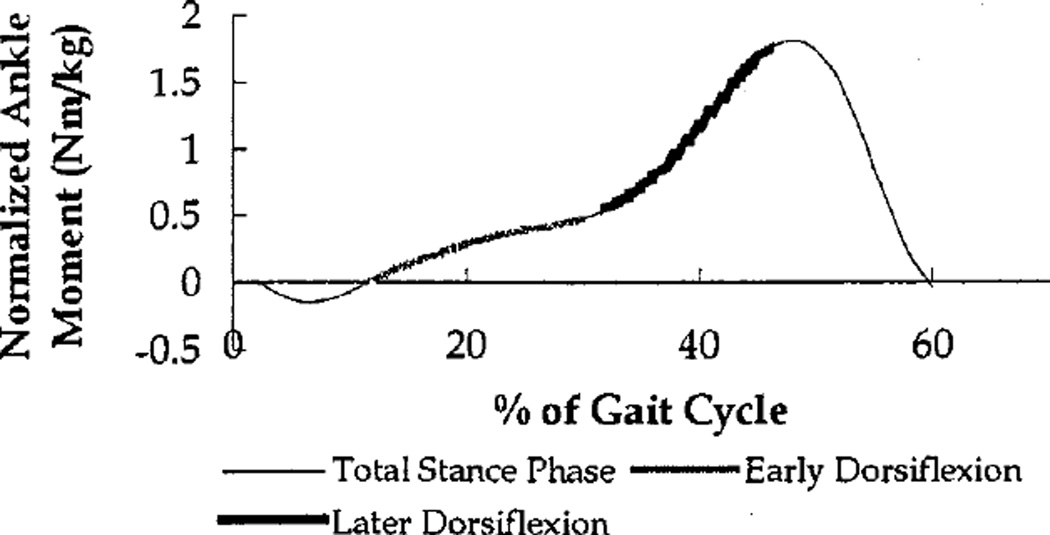
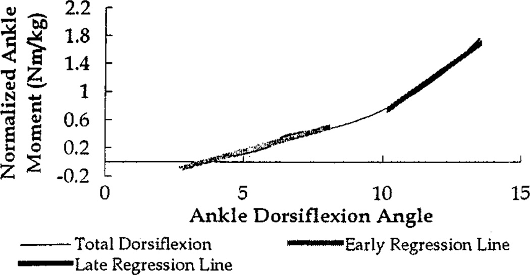
While prosthetic energy return has garnered much attention, the middle and late stance phase relationship between sagittal plane ankle joint moment and ankle joint rotation, is an aspect of foot/ankle prosthetic function that merits greater consideration. Examination of anatomic ankle function is instructive for understanding better the aspect of foot/ankle prosthetic function that is addressed in the present study. Through early dorsiflexion during middle stance, anatomic ankle plantar flexion moment rises more moderately than during later stance dorsiflexion (Fig. 1). The ratio of joint moment increase to ankle angle change is also lower during middle stance than during later stance (Fig. 2), as active plantar flexor opposition to dorsflexion is lower during middle stance, and increases substantially later in stance. This pattern is consistent with the presence of more rapid ankle moment rise during later stance than during middle stance. Thus, effective joint stiffness (defined here as local slope of joint moment versus joint rotation) of the anatomic ankle joint is less during middle stance than during later stance (Fig. 2).
Fig. 1.
Normalized, sagittal plane, ankle moment versus normalized time for a nonamputated individual, during stance phase of walking. Shaded portions of curve represent dorsiflexing portions of stance phase.
Fig. 2.
Normalized, sagittal plane, ankle moment versus sagittal plane, ankle angle for a nonamputated individual, during the dorsiflexing portion of stance phase of walking. Slopes of shaded regression lines represent ankle joint stiffness during early and later dorsiflexing portions of stance phase.
In contrast to anatomic ankles, typical prosthetic foot/ankle components demonstrate more constant effective joint stiffness, during middle and later stance loading [18]. This behavior is related to the lack of varying active plantar flexor activity to distinguish later from middle stance. Prosthetic foot/ankle systems generally contain passive deforming elements that support increasing load as deformation increases. Typical stiffness of these elements often induce plantar flexion moments during middle stance that are comparable to, if not greater than, those for anatomic ankles. Such moments are transmitted through prosthetic sockets to residual limbs, and can contribute to discomfort, pain, and skin breakdown.
The prototype foot/ankle prosthesis that has been evaluated here, is a version of a design concept, dubbed the rolling joint foot/ankle (RJFA) [18]. The RJFA was conceived with a primary intention of providing decreased sagittal plane, ankle joint stiffness during midstance. It was further anticipated that lower prosthetic ankle stiffness would permit greater dorsiflexion during middle stance, which could facilitate greater forward tibial rotation and, thus, affect stance phase knee flexion [19], [20]. Such knee flexion is considered to serve as a shock absorption mechanism. Early literature also suggests that stance knee flexion reduces energy expenditure associated with vertical body displacement [21], although this supposition has been questioned more recently [22], [23]. Expected characteristics of the RJFA could substantially mitigate pain, discomfort and ulceration risk associated with the stump/socket interface during amputee walking.
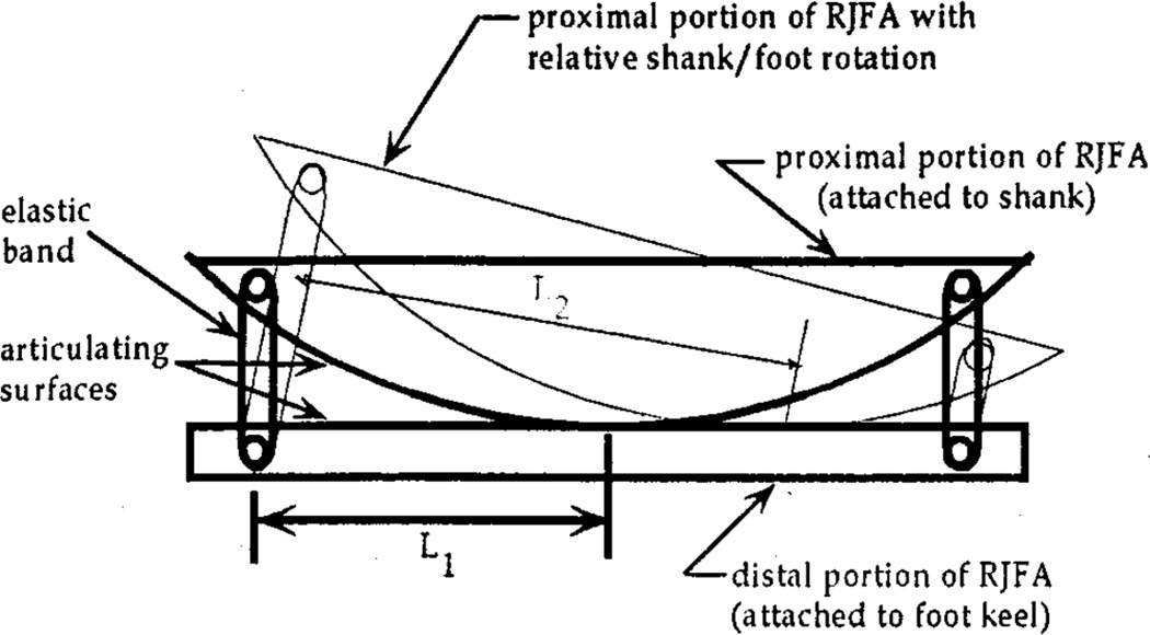
To obtain the desired dynamics, the RJFA prototypes proximal portion was attached rigidly to the shank and was terminated with a curved surface, whose geometry has been previously described [19]. This curved surface articulated with the distal portion’s upper surface, which was rigidly attached to the foot keel and cover (Fig. 3). Elastic bands, at the articulating surfaces’ anterior and posterior ends, connected the proximal and distal portions. The elastic bands functioned to generate moments opposing relative rotation between foot and shank (e.g., posterior bands lengthen during dorsiflexion resulting in posterior band forces, and causing moments acting to oppose dorsiflexion). The bands also maintained contact between shank and foot articulating surfaces when no external loads were applied (e.g., during swing).
Fig. 3.
Schematic diagram of rolling joint foot/ankle design concept (L1 represents the effective moment arm at the neutral position and L2 represents the moment arm following rotation of the rolling joint foot/ankle).
With the articulation described above, the instant center of rotation progresses forward as the shank rotates forward relative to the foot (i.e., as dorsiflexion angle increases) (Fig. 3), and progresses backward as plantar flexion angle increases. Consequently, the moment arms, through which elastic band forces generate moments to oppose rotation, increase as sagittal plane ankle joint angle moves away from neutral (i.e., ankle dorsiflexes or plantar flexes). Thus, ankle joint stiffness (i.e., rate of moment change with respect to angular displacement) is conceptually lower at smaller angular displacements. This design was anticipated to result in lower midstance and overall ankle joint stiffness, with smaller and more gradually increasing ankle moments around midstance and more rapidly increasing moments during later stance.
In the present investigation we evaluated a RJFA prototype in comparison with a traditional SACH foot to determine whether the prototype demonstrated lower dynamic ankle joint stiffness, as its design intended, and to evaluate effects on stance knee flexion. These assessments involved both video-based kinematic recording and ground reaction force measurements during walking for a volunteer below-knee amputee.
II. Methodology
The existing RJFA prototype was fitted to a socket and endoskeletal shank pylon of a below-knee amputee who volunteered to participate in this prototype evaluation. Our subject was an otherwise healthy, male with a left side below-knee amputation that resulted from a motor vehicle accident. The subject’s age, height and mass were 36 years, 1.88 m, and 105 kg, respectively. Prior to data collection the volunteer had been permitted a few limited periods (up to approximately an hour) of wearing the RJFA prototype, but did not leave the development environment with the prototype.
Kinematic and kinetic data were collected, in accord with an IRB approved gait testing protocol, using a six-camera VICON motion analysis system (Oxford, England) and an AMTI (Watertown, MA) force platform. Reflective markers were placed on the subject’s lower extremities using a Helen Hayes [24] type marker set. For each trial the subject walked across the laboratory walkway at a self-selected, comfortable speed. The subject was also instructed to direct his attention straight ahead and not to target the force platform. The volunteer’s starting position was adjusted until he struck the force platform in his natural stride. Trials without clean force plate footfalls were not retained. The VICON Motion Analysis system collected video data at 50 Hz, while the force platform sampled ground reaction data synchronously at 1000 Hz. Trials were collected with the subject wearing, first, the RJFA prototype and, then, a traditional SACH foot, and with the same shoe used with each. Three or more acceptable trials were obtained for each condition.
Raw video data were processed initially with the AMASS software package to reconstruct and identify marker coordinate trajectories, during representative walking trials selected for each foot/ankle component. AMASS output files were further processed using the VCM software package to compute the lower extremity joint motions, moments, and powers. This software package computed three dimensional joint rotations based upon a multilink segment model with imbedded joint coordinate system axes, as well as three dimensional joint moments and powers based upon inverse dynamic techniques. The VCM software package also smoothed the marker coordinate data using techniques that are commonly applied in the motion biomechanics field.
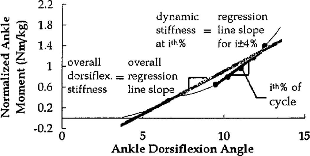
Last, ankle joint moment and motion data from VCM were used to quantify dynamic ankle joint stiffness and overall ankle joint stiffness, during stance phase dorsiflexion. Dynamic ankle joint stiffness was computed, at each 2 percent of walking cycle, as the linear regression slope of joint moment versus joint rotation, across a ±4% window (Fig. 4). Overall ankle joint stiffness was computed as the regression slope of the joint moment versus joint rotation curve, during the entire dorsiflexion portion of stance phase.
Fig. 4.
Graphical representation of overall and dynamic ankle joint stiffness. Stiffness is represented by slopes of the shaded regression lines.
III. Results
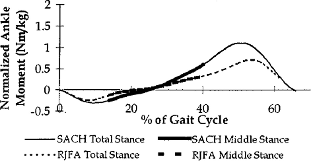
During the middle portion of stance phase, the ankle joint moment pattern of the RJFA prototype had a more gradual rise, with respect to time (expressed as % gait cycle), than did the ankle joint moment pattern of the SACH foot (Fig. 5). The overall maximum ankle joint moment was also lower for the RJFA prototype than for the SACH foot (0.72 N-m/kg for the RJFA versus 1.09 N-m/kg for the SACH foot).
Fig. 5.
Normalized, sagittal plane, ankle moment versus normalized time for the SACH foot (solid line) and RJFA (dashed line), during stance phase of walking.
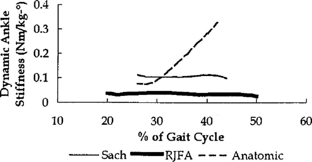
The RJFA demonstrated a dynamic joint stiffness that was generally less than half of the SACH foot dynamic stiffness, throughout middle stance phase (Fig. 6). The overall middle stance stiffness of the RJFA was 0.034 N-m/kg/°, while the overall SACH foot stiffness was 0.107 N-m/kg/°.
Fig. 6.
Dynamic, ankle joint stiffness versus normalized time for the SACH foot (thin line), RJFA (thick line), and anatomic foot (thin dashed line) during dorsiflexing portion of stance phase.
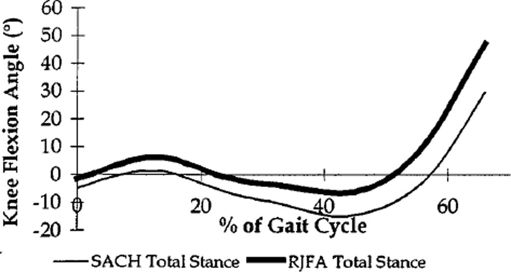
Sagittal knee joint rotations suggest a possible tendency toward increased stance knee flexion and alleviation of later stance hyperextension, associated with prototype use (Fig. 7). Differences between sagittal knee motions with the RJFA and the SACH foot, however, may be within measurement variation, particularly variation that may be due to possible static offset.
Fig. 7.
Sagittal plane, knee flexion angle versus normalized time for the SACH foot (thin line) and RJFA (thick line), during stance phase of walking.
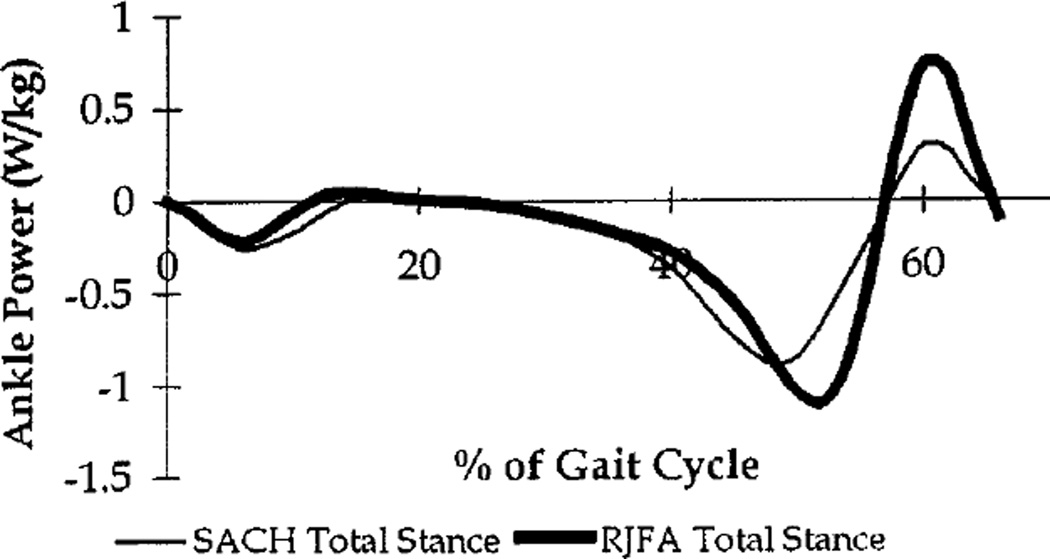
Although not a specific RJFA objective, the prototype also demonstrated greater energy storage (i.e., area between the x-axis and the midstance, negative portion of the ankle power curve) and energy return (i.e., area between the x-axis and the late stance, positive portion of the ankle power curve) than did the SACH foot (Fig. 8). This greater energy return occurred, even though the RJFA displayed lower sagittal plane ankle joint moments.
Fig. 8.
Normalized, sagittal plane, ankle power versus normalized time for the SACH foot (solid line) and RJFA (dashed line), during stance phase of walking.
IV. Discussion
The RJFA prototype’s primary objective, to provide lower dynamic ankle joint stiffness during middle stance phase, differed from the intent of other foot/ankle components, particularly DER feet. Various DER feet have constituted the bulk of recent prosthetic foot/ankle designs. Despite their proliferation, most reports on DER feet have not conclusively established a quantitative relationship, during walking, between increased mechanical energy return and decreased metabolic energy cost [4], [7], [8], [10], [14]. One report involving running has suggested substantial benefits of DER feet with regard to mechanical energy and power, during this activity; however, metabolic energy effects were not reported [25]. Conversely, a treadmill-based study did suggest potentially lower metabolic energy costs with a particular new foot design, but did not assess mechanical energy [13].
While qualitative and anecdotal descriptions of improved walking with DER feet have continued to appear, suggestions that many of these DER feet may not be suitable for geriatric and/or dysvascular amputees are also common [13]. It seems reasonable to speculate that excessive socket to residual limb load transmission may have a substantial role in the disfavor, for DER components, among such individuals. Such reports have contributed to many prosthetists’ recommendations that these feet be prescribed with care and with regard to such issues as tolerance for discomfort and pain, and risk of ulceration. Many clinicians are, subsequently, hesitant to advocate DER feet for geriatric or vascularly impaired amputees. A goal of RJFA prototype development was, thus, to reduce potential pain, discomfort, and ulceration risk by decreasing dynamic ankle joint stiffness during middle stance phase of walking.
Reservations regarding prescription of DER feet are not without a biomechanical basis. Energy available for late stance return from typical prosthetic foot/ankle components is limited to that which is passively stored from the middle stance instant when the ankle passes through its unloaded neutral position until the latter stance instant when elastic elements are maximally deformed. To strive for greater late stance energy return, the designs of DER feet generally involve deforming elements with considerably higher stiffness than those of standard prosthetic feet. Consequently, midstance ankle moments with DER feet can be even greater than those described previously for standard prosthetic feet, and further increase the potential for discomfort, pain, and skin breakdown.
The data obtained in this initial investigation, although preliminary, suggest that the RJFA prototype may meet its objective of reducing ankle joint stiffness during the middle portion of stance phase when the ankle is near its neutral position. Such ankle stiffness reduction could diminish nonaxial loads transmitted to the residual limb [20]. Reduced nonaxial load transmission could in turn diminish pain and discomfort, which have been indicated by amputee surveys to be among the primary subjective concerns of lower extremity amputees [17], [26]. Reduced load transmission could also decrease risk of residual limb breakdown and/or ulceration.
The prototype, however, did not completely realize its intended stiffness pattern in that stiffness did not tend to increase for later stance. While our volunteer did not express any particular difficulties with regard to later stance stiffness, the lack of later stance stiffenning could render the RJFA somewhat unstable for some individuals. Additional prototype development is proceeding to address this issue, as well as others beyond the scope of this report (e.g., adjustability, durability, manufacturability, etc.).
Kinematic data indicate a potential for more normal knee motion range with the prototype foot/ankle prosthesis; however, typical variability of knee angle data, particularly with regard to static offset, render it difficult to express much confidence in such a conclusion at this time. It was further noted, with regard to early stance knee flexion, that the peak of this wave occurs with foot/ankle components generally close to their neutral positions. Consequently, facilitation of greater dorsiflexion via reduced ankle stiffness should, perhaps, not be expected to have substantial influence on peak early stance knee flexion. Ankle stiffness, however, may more directly relate to middle stance hyperextension, since external moments tending to dorsiflex the ankle can result in reaction moments, tending to rotate the shank posteriorly, and lead to hyperextension. With regard to this potential mechanism, the influence of ankle stiffness on knee hyperextension may have more merit.
It was interesting to note that, although increased energy return was not a particular objective of the prototype’s design, the prototype demonstrated greater energy return at late stance than did the SACH foot. While the SACH foot makes no attempt to be a high energy return foot, it can still serve, by virtue of being so commonly prescribed, as useful point of comparison.
The results of this initial investigation raise a number of issues that should be considered in future work. The potential for reduced residual limb load transmission during middle stance should be evaluated with experimental measurements. A number of previous studies have described techniques that could potentially be applied to obtain such measurements [27]–[30]. Assessment of residual limb loading should, likely, focus on the distal anterior and posterior aspects of residual limbs.
The recent suggestion, that reduced prosthetic ankle stiffness could improve early stance stability by facilitating earlier full foot ground contact, indicates the potential for an additional benefit, not specifically considered in the RJFA’s development. Dynamic stability during walking with prostheses can be compromised by such factors as terrain. Consequently, experimental testing involving varying terrain may be useful for evaluating potential stability improvements associated with the RJFA.
As development of the RJFA progresses, continued evaluation should include longer duration subject trials outside of the laboratory environment. Quantitative measurments can provide strong indicators of potential benefits of prosthetic components; however, ultimate success of the RJFA will require acceptance of prosthesis users. Performance of subject trials should specifically include amputee populations, such as elderly and vascularly impaired groups, that have been more prone to residual limb problems.
Acknowledgments
The authors wish to thank N. Denniston, M. Miller, and G. Rutherford for their assistance with this effort.
Contributor Information
P. M. Quesada, Department of Mechanical Engineering, University of Louisville, Louisville, KY 40292 USA.
M. Pitkin, Department of Physical Medicine and Rehabilitation, Tufts University, Boston, MA 02111 USA.
J. Colvin, Ohio Willow Wood Co., Mt. Sterling, OH 43143 USA.
References
- 1.Edelstein JE. Prosthetic feet state of the art. Phys. Ther. 1988;68(12):1874–1881. doi: 10.1093/ptj/68.12.1874. [DOI] [PubMed] [Google Scholar]
- 2.Wing DC, Hittenberger DA. Energy-storing prosthetic feet. Arch. Phys. Med. Rehab. 1989;70(4):330–335. [PubMed] [Google Scholar]
- 3.Torburn L, Perry J, Ayyappa E, Shanfield SL. Below-knee amputee gait with dynamic elastic response prosthetic feet: A pilot study. J Rehab. Res. Dev. 1990;27(4):369–384. doi: 10.1682/jrrd.1990.10.0369. [DOI] [PubMed] [Google Scholar]
- 4.Gitter A, Czerniecki JM, DeGroot DM. Biomechanical analysis of the influence of prosthetic feet on below-knee amputee walking. Amer. J. Phys. Med. Rehab. 1991;70(3):142–148. doi: 10.1097/00002060-199106000-00006. [DOI] [PubMed] [Google Scholar]
- 5.Barr AE, Siegel KL, Danoff JV, McGarvey CL, III, Tomasko A, Sable I, Stanhope SJ. Biomechanical comparison of the energystoring capabilities of SACH and Carbon Copy II prosthetic feet during the stance phase of gait in a person with below-knee amputation. Phys. Ther. 1992;72(5):344–354. doi: 10.1093/ptj/72.5.344. [DOI] [PubMed] [Google Scholar]
- 6.Ehara Y, Beppu M, Nomura S, Kunimi Y, Takahashi S. Energy storing property of so-called energy-storing prosthetic feet. Arch. Phys. Med. Rehab. 1993;74(1):68–72. [PubMed] [Google Scholar]
- 7.Lehmann JF, Price R, Boswell-Bessette S, Dralle A, Questad K. Comprehensive analysis of dynamic elastic response feet: Seattle ankle/lite foot versus SACH foot. Arch. Phys. Med. Rehab. 1993;74(8):853–861. doi: 10.1016/0003-9993(93)90013-z. [DOI] [PubMed] [Google Scholar]
- 8.Perry J, Shanfield S. Efficiency of dynamic elastic response prosthetic feet. J Rehab. Res. Dev. 1993;30(1):137–143. [PubMed] [Google Scholar]
- 9.Alaranta H, Lempinen VM, Haavisto E, Pohjolainen T, Hurri H. Subjective benefits of energy storing prostheses. Prosthet. Orthot. Int. 1994;18(2):92–97. doi: 10.3109/03093649409164390. [DOI] [PubMed] [Google Scholar]
- 10.Lehmann JF, Price R, Boswell-Bessette S, Dralle A, Questad K, deLateur BJ. Comprehensive analysis of energy storing prosthetic feet: Flex foot and Seattle foot versus standard SACH foot. Arch. Phys. Med. Rehab. 1993;74(11):1225–1231. [PubMed] [Google Scholar]
- 11.Torburn L, Schweiger GP, Perry J, Powers CM. Below-knee amputee gait in stair ambulation. A comparison of stride characteristics using five different prosthetic feet. Clin. Orthop. Rel. Res. 1994;303:185–192. [PubMed] [Google Scholar]
- 12.Powers CM, Torburn L, Perry J, Ayyappa E. Influence of prosthetic foot design on sound limb loading in adults with unilateral below-knee amputations. Arch. Phys. Med. Rehab. 1994;75(7):825–829. [PubMed] [Google Scholar]
- 13.Casillas JM, Dulieu V, Cohen M, Marcer I, Didier JP. Bioenergetic comparison of a new energy-storing foot and SACH foot in traumatic below-knee vascular amputations. Arch. Phys. Med. Rehab. 1995;76(1):39–44. doi: 10.1016/s0003-9993(95)80040-9. [DOI] [PubMed] [Google Scholar]
- 14.Torburn L, Powers CM, Guiterrez R, Perry J. Energy expenditure during ambulation in dysvascular and traumatic below-knee amputees: A comparison of five prosthetic feet. J Rehab. Res. Dev. 1995;32(2):111–119. [PubMed] [Google Scholar]
- 15.Perry J, Boyd LA, Rao SS, Mulroy SJ. Prosthetic weight acceptance mechanics in transtibial amputees wearing the single axis, Seattle lite and flex foot. IEEE Trans. Rehab. Eng. 1997 Dec.5:283–289. doi: 10.1109/86.650279. [DOI] [PubMed] [Google Scholar]
- 16.Postema K, Hermens HJ, de Vries J, Koopman HF, Eisma WH. Energy storage and release of prosthetic feet—Part 1: Biomechanical analysis related to user benefits. Prosthet. Orthot. Int. 1997;21(1):17–27. doi: 10.3109/03093649709164526. [DOI] [PubMed] [Google Scholar]
- 17.Postema K, Hermens HJ, de Vries J, Koopman HF, Eisma WH. Energy storage and release of prosthetic feet—Part 2: Subjective ratings of 2 energy storing and 2 conventional feet, user choice of foot and deciding factor. Prosth. & Orthotics Inter. 1997;21(1):28–34. doi: 10.3109/03093649709164527. [DOI] [PubMed] [Google Scholar]
- 18.Pitkin MR. Mechanical outcomes of a rolling joint prosthetic foot and its performance in the dorsiflexion phase of transtibial amputee gait. J Prosthet. Orthot. 1995;7(4):114–123. doi: 10.1097/00008526-199507040-00003. [DOI] [PMC free article] [PubMed] [Google Scholar]
- 19.Pitkin MR. Synthesis of a cycloidal mechanism of the prosthetic ankle. Prosthet. Orthot. Int. 1996;20:159–171. doi: 10.3109/03093649609164438. [DOI] [PMC free article] [PubMed] [Google Scholar]
- 20.Pitkin MR. Effects of design variants in lower limb prostheses on gait synergy. J Prosthet. Orthot. 1997;9(3):113–122. doi: 10.1097/00008526-199700930-00005. [DOI] [PMC free article] [PubMed] [Google Scholar]
- 21.Saunders JB, Inman VT, Eberhart HD. The major determinants in normal and pathological gait. J Bone Joint Surg. 1953;35-A(3):543–558. [PubMed] [Google Scholar]
- 22.Chidress DS, Gard SA. Investigation of vertical motion of the human body during normal walking. Gait Posture. 1997;6(2):161. [Google Scholar]
- 23.Quesada PM, Rash GS. Simulation of walking without stance phase knee flexion. Gait Posture. 1998;7(2):151–152. [Google Scholar]
- 24.Kadaba MP, Ramakrishnan HK, Wootten ME. Measurement of lower extremity kinematics during level walking. J Orthop. Res. 1990;8(3):383–392. doi: 10.1002/jor.1100080310. [DOI] [PubMed] [Google Scholar]
- 25.Czerniecki JM, Gitter A, Munro C. Joint moment and muscle power output characteristics of below knee amputees during running: The influence of energy storing prosthetic feet. J Biomechan. 1991;24(1):63–75. doi: 10.1016/0021-9290(91)90327-j. [DOI] [PubMed] [Google Scholar]
- 26.Hoaglund FT, Jergesen HE, Wilson L, Lamoreux LW, Roberts R. Evaluation of problems and needs of veteran lower-limb amputees in the San Francisco Bay Area during the period 1977–1980. J Rehab. Res. Dev. 1983;20(1):57–71. [PubMed] [Google Scholar]
- 27.Zhang M, Turner-Smith AR, Tanner A, Roberts VC. Clinical investigation of the pressure and shear stress on the trans-tibial stump with a prosthesis. Med. Eng. Phys. 1998;20(3):188–198. doi: 10.1016/s1350-4533(98)00013-7. [DOI] [PubMed] [Google Scholar]
- 28.Sanders JE, Lam D, Dralle AJ, Okumura R. Interface pressures and shear stresses at thirteen socket sites on two persons with transtibial amputation. J Rehab. Res. Dev. 1997;34(1):19–43. [PubMed] [Google Scholar]
- 29.Sanders JE. Interface mechanics in external prosthetics: Review of interface stress measurement techniques. Med. Biol. Eng. Comput. 1995;33(4):509–516. doi: 10.1007/BF02522507. [DOI] [PubMed] [Google Scholar]
- 30.Sanders JE, Daly CH, Burgess EM. Interface shear stresses during ambulation with a below-knee prosthetic limb. J Rehab. Res. Dev. 1992;28(4):1–8. doi: 10.1682/jrrd.1992.10.0001. [DOI] [PMC free article] [PubMed] [Google Scholar]


