A 30-year-old female presented with progressively increasing dysphagia associated with loss of appetite and weight. Upper gastrointestinal endoscopy revealed a polypoidal and ulcerated lesion in the mid esophagus with endoscopic biopsies being inconclusive [Figure 1]. An endoscopic ultrasound (EUS) revealed asymmetrical thickening of the esophageal wall with loss of the wall stratification [Figure 2] as well as loss of fat planes with right pulmonary artery [Figure 3]. No mediastinal lymphadenopathy was noted. EUS guided fine-needle aspiration (FNA) [Figure 4] yielded caseous material and cytology revealed epithelioid cell granuloma with a giant cell and caseation necrosis [Figure 5] with presence of acid-fast bacilli [Figure 6]. The patient was initiated on weight based four drug anti-tubercular therapy (rifampin, isoniazid, pyrazinamide, and ethambutol). At 1 month of follow-up the patient had gained 5 kg of weight with complete resolution of dysphagia.
Figure 1.
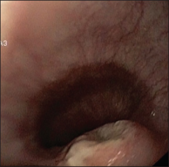
Polypoidal and ulcerated lesion in mid esophagus
Figure 2.
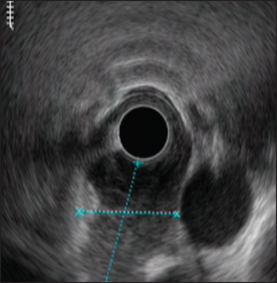
Endoscopic ultrasound: Asymmetrical thickening of the mid esophagus
Figure 3.
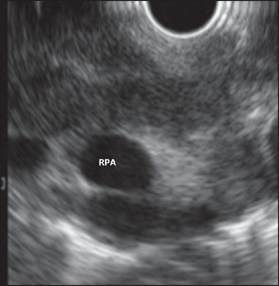
Loss of fate planes with right pulmonary artery
Figure 4.
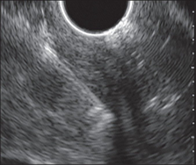
Endoscopic ultrasound fine-needle aspiration of the lesion
Figure 5.
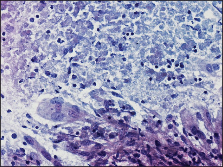
Epithelioid cell granuloma with a giant cell and caseation necrosis (Pap, ×40)
Figure 6.
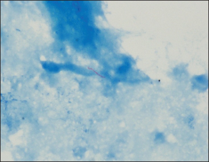
Ziehl–Neelsen stain showing acid-fast bacillus (×100)
Esophageal tuberculosis is usually secondary to mediastinal lymphadenopathy causing extrinsic narrowing or secondarily due to infiltration of the esophageal wall.[1,2] Primary esophageal tuberculosis, as in our case, is uncommon. Except a few, most such reports are from the pre-EUS era where the mediastinal lymphadenopathy may have been missed.[3] It is unusual for esophageal tuberculosis to result in vascular involvement although this has been described in relation to the pancreatic tuberculosis.[4] EUS-FNA has emerged as an important tool to diagnose the esophageal tuberculosis.[1,2]
Footnotes
Source of Support: Nil.
Conflicts of Interest: None declared.
REFERENCES
- 1.Rana SS, Bhasin DK, Rao C, et al. Tuberculosis presenting as dysphagia: Clinical, endoscopic, radiological and endosonographic features. Endosc Ultrasound. 2013;2:92–5. doi: 10.4103/2303-9027.117693. [DOI] [PMC free article] [PubMed] [Google Scholar]
- 2.Rana SS, Bhasin DK, Sharma V, et al. Dysphagia as the first manifestation of tuberculosis. Endoscopy. 2011;43(Suppl 2 UCTN):E300–1. doi: 10.1055/s-0030-1256456. [DOI] [PubMed] [Google Scholar]
- 3.Huang YK, Wu YC, Liu YH, et al. Esophageal tuberculosis mimicking submucosal tumor. Interact Cardiovasc Thorac Surg. 2004;3:274–6. doi: 10.1016/j.icvts.2003.11.016. [DOI] [PubMed] [Google Scholar]
- 4.Rana SS, Sharma V, Sampath S, et al. Vascular invasion does not discriminate between pancreatic tuberculosis and pancreatic malignancy: A case series. Ann Gastroenterol. 2014;27:395–8. [PMC free article] [PubMed] [Google Scholar]


