
Figure 7. The miR-214-mediated EMT process in LAD cells depends on Sufu inhibition.
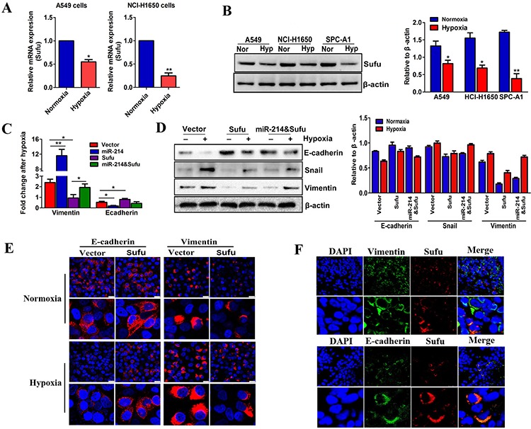
A–B. Real-time PCR (A) and western blot (B) analyses of Sufu expression in three LAD cells under normoxia (21% O2) or hypoxia (0.5% O2) treatment for 24 hours. β-actin was used as an internal loading control. C–D. The fold changes in the EMT markers detected by real-time PCR or western blots in Sufu-over-expressed or Sufu and miR-214-co-overexpressed A549 cells. E. The immunofluorescence detection of EMT markers changed in Sufu-over-expressed cells under normoxia or hypoxia treatment. Scale bar = 25 μm. F. Confocal microcopy of LAD patient tissues co-stained for E-cadherin, or vimentin with Sufu. The nuclei were staining with DAPI. Scale bar = 25 μm. All experiments were performed at least three times, and the data are expressed as means ± SD. The statistical significance of differences was measured by unpaired student's t-test. *P < 0.05, **P < 0.01.