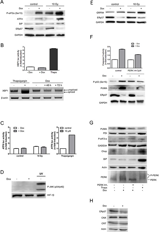
Figure 3. Knockdown of ERp57 in HCT116 cells activates the UPR exclusively via PERK.

A. Inducible HCT116 shERp57 cells were irradiated with 10 Gy 24 h after induction of ERp57 knockdown. 72 h after irradiation the cells were lysed and subjected to Western blotting. P-eIF2α (Ser51), ATF4 and BiP protein levels were monitored as indicators of ER-stress, GAPDH served as a loading control. B. XBP1 splicing as an indicator for IRE1 activation was analysed with a luciferase reporter gene assay (upper panel). Cells were transfected with the pXBP1u-FLuc reporter plasmid 24 h after induction of ERp57 knockdown and harvested 48 h later. Firefly luciferase activity was normalized to renilla luciferase activity. As a positive control for ER stress, cells were treated with 10 μM thapsigargin for 24 h. Lower panel: total RNA was subjected to RT-PCR and analysed for XBP1 splicing. β-actin served as a loading control. C. 24 h after knockdown induction, the cells were transiently transfected with an ATF6-luciferase reporter gene construct. After 48 h lysates were prepared and analysed by luciferase activity detection. D. 96 h after induction of ERp57 knockdown, P-JNK was detected by Western blotting as an indicator of IRE1 activation. Hif-1β was used as a loading control and UV-treated cells as a positive control for JNK activation. A representative Western blot from two independent experiments is shown. E. Cells were treated as in (A) and tested for GRP94 as an indicator of ERAD, GAPDH served as a loading control. F. After ERp57 knockdown induction and treatment with 3 μM PERK inhibitor for 96 h, cell extracts were tested for caspase-3 activity (upper panel). Representative data of two experiments are shown. In parallel, cell lysates were subjected to Western blotting (lower panel). Representative Western blots from two experiments are shown. G. Cells were treated as in (F) Cell lysates were analysed by Western blotting. Phosphorylated PERK is detected as a higher molecular weight smear. Western blots from two independent experiments are shown. H. After ERp57 knockdown induction for 96 h, cell lysates were analysed by Western blotting. A representative Western blot from two independent experiments is shown.
