Abstract
Among the various scientific fields covered in the area of hygiene such as environmental medicine, epidemiology, public health and preventive medicine, we are investigating the immunological effects of fibrous and particulate substances in the environment and work surroundings, such as asbestos fibers and silica particles. In addition to these studies, we have attempted to construct health-promoting living conditions. Thus, in this review we will summarize our investigations regarding the (1) immunological effects of asbestos fibers, (2) immunological effects of silica particles, and (3) construction of a health-promoting living environment. This review article summarizes the 2014 Japanese Society for Hygiene (JSH) Award Lecture of the 85th Annual Meeting of the JSH entitled “Environmental health effects: immunological effects of fibrous and particulate matter and establishment of health-promoting environments” presented by the first author of this manuscript, Prof. Otsuki, Department of Hygiene, Kawasaki Medical School, Kurashiki, Japan, the recipient of the 2014 JSH award. The results of our experiments can be summarized as follows: (1) asbestos fibers reduce anti-tumor immunity, (2) silica particles chronically activate responder and regulatory T cells causing an unbalance of these two populations of T helper cells, which may contribute to the development of autoimmune disorders frequently complicating silicosis, and (3) living conditions to enhance natural killer cell activity were developed, which may promote the prevention of cancers and diminish symptoms of virus infections.
Keywords: Asbestos, Silica, Living environment, NK cell, T cell
Introduction
This review article summarizes the 2014 Japanese Society for Hygiene (JSH) Award Lecture of the 85th Annual Meeting of the JSH entitled “Environmental health effects: immunological effects of fibrous and particulate matter and establishment of health-promoting environments” presented by the first author of this manuscript, Prof. Otsuki, Department of Hygiene, Kawasaki Medical School, Kurashiki, Japan, the recipient of the 2014 JSH award. Since we feel the award was given as a result of the scientific achievements of all our department as well as various contributions to the JSH, the present members of our department are listed as authors. We sincerely appreciate all the members of the JSH as well as the selection commissioners and board members. In addition, all authors thank the former members of our department (listed in the Acknowledgments section of this manuscript).
Among the various scientific fields covered in the area of hygiene such as environmental medicine, epidemiology, public health and preventive medicine, we are investigating the immunological effects of fibrous and particulate substances in the environment and work surroundings, such as asbestos fibers and silica particles. In addition to these studies, we have attempted to construct health-promoting living conditions. Thus, in this review we summarize our investigations regarding the (1) immunological effects of asbestos fibers, (2) immunological effects of silica particles, and (3) construction of a health-promoting living environment.
All research using biological samples from patients such as pleural plaque (PP), malignant mesothelioma (MM), silicosis (SIL), and healthy volunteers (HV) was performed in accordance with the principles of the 1983 Declaration of Helsinki and was approved by the ethical committee of the Kawasaki Medical School and other organizations involved in the collection of patient samples.
The immunological effects of asbestos fibers
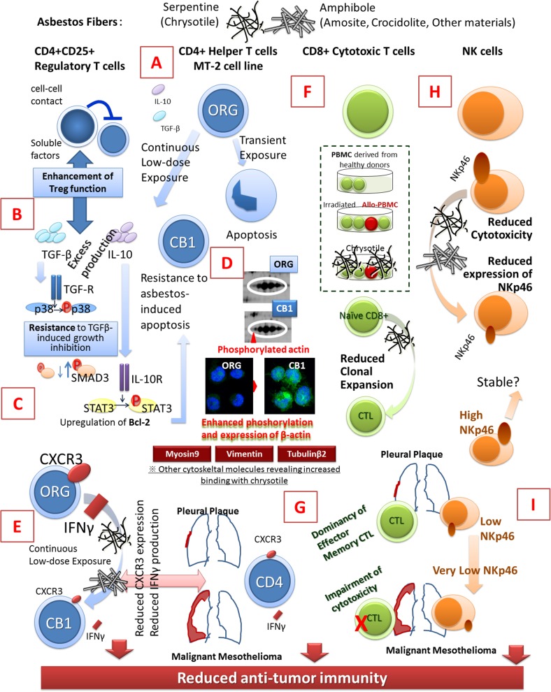
Figure 1 shows a schematic summary of the immunological effects of asbestos fibers and details the effects on CD4+ helper T cells, CD8+ cytotoxic lymphocytes (CTL) and natural killer (NK) cells. Since similar summaries were reported previously [1–9], it would be better to refer to these previous reports in addition to this review. Basically, we hypothesized that asbestos may reduce tumor immunity by affecting immune cells due to malignant complications occurring in asbestos-exposed patients such as mesothelioma and lung cancer [10–13].
Fig. 1.
Schematic presentation showing the immunological effects of asbestos fibers on regulatory T cells, CD4+ helper T cells, CD8+ cytotoxic T and natural killer cells. ORG MT-2 original cell line, CB1 one of the sublines of the MT-2 cell line continuously exposed to chrysotile B asbestos, IL interleukin, TGF transforming growth factor, SMAD signal transducing adaptor proteins, STAT3 signal transducer and activator of transcription 3, R receptor, CXCR3 chemokine (C-X-C motif) receptor 3, IFN interferon, CTL cytotoxic T lymphocyte, PBMC peripheral blood mononuclear cells, NK natural killer. a Excess production of IL-10 and TGF-β in MT-2 subline (CB1) continuously exposed to asbestos, b resistance to TGF-β-induced growth inhibition in MT-2 subline (CB1), c acquisition of asbestos-induced apoptosis in MT-2 subline (CB1) via excess production of IL-10, phosphorylation of STAT3 subsequent overexpression of Bcl-2, d enhanced β-actin phosphorylation in MT-2 subline (CB1), e reduction of CXCR3 and IFN-γ-producibility inMT-2 subline (CB1) meaning reduction of anti-tumor immunity, f reduction of CTL clonal expansion in co-culture with asbestos, g aberrant CTL population and function in patients with pleural plaque (PP) or malignant mesothelioma (MM), h decrease of NKp46 activating receptor by asbestos exposure in NK cells and i progressive reduction of NKp46 in patients with PP–MM
Effects on helper T cells
To investigate the effects of asbestos fibers on human T cells, a human T cell leukemia virus type-1 (HTLV-1) immortalized polyclonal T cell line, MT-2, was utilized [14, 15], since this cell line was the most sensitive to transient exposure to chrysotile asbestos proceeding to apoptosis among various human lymphoid cell lines derived from T cell acute leukemia, T and B cell lymphomas, myeloma, as well as Epstein–Barr virus immortalized lymphoblastoid cell lines. Apoptosis occurred in MT-2 cells when chrysotile was exposed transiently and with a relatively high dose and led to the production of reactive oxygen species (ROS), activation–phosphorylation of pro-apoptotic signaling molecules such as p38 and JNK, activation of the mitochondrial apoptotic pathway with release of cytochrome c from mitochondria to the cytoplasm, increase of the Bax and Bax/Bcl-2 ratio, and cleavage of caspase 9 and 3 [14].
However, when MT-2 (ORG: original cell line which was never exposed to asbestos) cells were continuously exposed to chrysotile asbestos with concentrations that resulted in less than half of the cells being killed following transient exposure for more than 8 months, the MT-2 cells acquired resistance to asbestos-induced apoptosis [15]. This subline was designated as CB1 (exposure to chrysotile B, subline #1). Thereafter, we established the sublines CB1–3, CA (exposure to chrysotile A) 1–3, and CR (crocidolite exposure) [16]. The CB1 subline showed overproduction of interleukin (IL)-10 and transforming growth factor (TGF)-β (Fig. 1a). The excess TGF-β caused resistance to TGF-β-induced growth inhibition found in MT-2 ORG with phosphorylation of p38 and signal transducing adaptor proteins (SMAD)-3 (Fig. 1b) [16]. In addition, excess IL-10 was utilized by an autocrine process and signal transducer and activator of transcription 3 (STAT3) located downstream of the IL-10 receptor (R) and was activated/phosphorylated. Thereafter, the Bcl-2 receiving signal from STAT3 was overexpressed in the CB-1 subline, causing resistance to asbestos-induced apoptosis (Fig. 1c) [15].
Both cytokines IL-10 and TGF-β are typical soluble factors that function in CD4+CD25+ forkhead box P3 (FoxP3)+ regulatory T cells (Treg). In addition, the MT-2 cell line was reported to possess a cellular function similar to Treg by inhibiting the proliferation of responder T cells (Tresp) [17, 18]. If the function and volume of Treg are decreased, excess and prolonged Tresp proliferative reactions against intrinsic and external antigens occur, and allergy and autoimmune diseases may be induced by these conditions. On the other hand, if Treg function and volume are increased, there may be inhibition of the tumor-targeting immune reaction and tumors may result or undergo accelerated progression [19–21]. Thus, the inhibitory function against Treg was compared between MT-2 ORG and the CB1 subline. As expected, MT-2 CB1 showed enhanced inhibitory function compared with that of MT-2 ORG, as well as the overproduction of IL-10 and TGF-β, which were also assumed as part of the inhibitory function [15, 16, 22]. These results supported our hypothesis that exposure of immune cells to asbestos causes reduction of anti-tumor immunity [1–9].
In addition, cellular alterations found in MT-2 CB1 were examined using various proteomics assays [23], and results revealed the excess phosphorylation of β-actin. Furthermore, other cytoskeletal molecules such as myosin 9, vimentin and tubulin β2 showed increased binding capacity to chrysotile fibers (Fig. 1d) [23]. These findings indicated that the cellular and molecular changes caused by continuous exposure of T cells to asbestos may influence cell surface molecules because asbestos fibers do not enter cellular internal spaces [24]. Continuous exposure and recurrent contact occur on the cell surface. The further investigation of these molecules may clarify the interaction between cells and fibers, and the cellular changes caused by these fibers.
Regarding various molecules affecting anti-tumor immunity, we have been focusing on chemokine (C-X-C motif) receptor 3 (CXCR3) and interferon (IFN)-γ (Fig. 1e) [25, 26]. These two molecules were extracted by pathway and network analyses, and IFN-γ signaling canonical pathway analysis employing cDNA microarray examination using MT-2 ORG and six independently established continuously exposed sublines (CB1–3 and CA1–3). These assays revealed that many molecules in the IFN-γ signaling pathway exhibited reduced expressions, and that CXCR3 was closely related to these molecules. Re-examination of CXCR3 expression in the MT-2 ORG line and six continuously exposed sublines by RT-PCR, western blotting, flow cytometrical assay and immunohistochemical analysis revealed that asbestos exposure caused reduced expression of CXCR3 and decreased secretory potential of IFN-γ [25, 26]. Subsequent experiments involving in vitro continuous exposure to chrysotile of freshly isolated CD4+ T helper cells activated via the T cell receptor by anti-CD3 and CD-28 antibodies and IL-2 showed similar reductions. Investigation of CD4+ T helper cells derived from asbestos-exposed patients with PP and MM revealed reduction of CXCR3 and decreased capacity for expression of IFN-γ [25, 26]. Since CXCR3-expressing T cells attract IFN-γ-producing T cells to attack tumor cells [27, 28], these effects of asbestos fibers are sufficient to reduce anti-tumor immunity in asbestos-exposed patients [1–9].
Effects on CTL
To examine the effects of asbestos fibers on CTL, the mixed lymphocyte reaction (MLR) assay was utilized to assess clonal expansion (differentiation and proliferation of CD8+ peripheral blood T cells into CTL possessing cell killing activity) of CD8+ T cells when chrysotile fibers were added in this assay system [29]. Results showed that the activity of specific lysis, granzyme B and IFN-γ expressions, as well as differentiation to CD45RO+ effector-memory cells, was reduced during the asbestos-added MLR assays. Moreover, the production of IL-10, IFN-γ and TNF-α, but not IL-2, decreased in the presence of chrysotile asbestos. These results suggest that exposure to asbestos potentially suppresses differentiation of cytotoxic T lymphocytes, accompanied by decreases in IFN-γ and TNF-α (Fig. 1f) [29].
We then verified the status of CD8+ CTL in peripheral blood of PP and MM patients with control cells from HV [30]. The percentage of CD3+CD8+ cells in PBMCs did not differ among the three groups, although the total number of PBMCs from the PP and MM groups was lower than that of HV. The percentage of IFN-γ+ and CD107a+ cells in phorbol myristate acetate (PMA)/ionomycin-stimulated CD8+ lymphocytes did not differ among the three groups. CD107a was used as the marker for degranulation of T cells upon antigen stimulation. The percentage of perforin+ and CD45RA− cells in fresh CD8+ lymphocytes of the PP and MM groups was higher than that of HV. The percentage of granzyme B+ and perforin+ cells in PMA/ionomycin-stimulated CD8+ lymphocytes was higher in the PP group compared with HV [30]. The MM group showed a decrease of perforin level in CD8+ lymphocytes after stimulation compared to patients with PP [30]. These results indicate that MM patients have characteristics of impairment regarding stimulation-induced cytotoxicity of peripheral blood CD8+ lymphocytes, and that PP and MM patients have a common character of functional alteration in those lymphocytes, namely, an increase in memory cells that is possibly related to asbestos exposure (Fig. 1g) [30].
We have recently been investigating the roles of various cytokines in the reduction of CTL cell killing caused by asbestos exposure, and these findings indicate that asbestos-exposed patients exhibit reduced anti-tumor immunity from the viewpoint of CTL (Fig. 1).
Effects on NK cells
The effects of asbestos exposure on the NK cells and its function have been reported and summarized previously [3, 6, 31, 32]. As shown in Fig. 1h, analyses using the NK cell line exposed continuously to the asbestos fibers, freshly isolated NK cells derived from HV exposed continuously during in vitro activation, and peripheral blood NK cells derived from PP and MM patients showed reduced NK cell killing activity induced by asbestos exposure with downregulation of various NK cell activation receptors. Among these receptors, NKG2D and 2B4 were reduced in cell lines exposed continuously to asbestos, and the NKp46 receptor showed decreased expression in freshly isolated NK cells exposed to asbestos and in peripheral NK cells derived from PP and MM patients. In particular, the expression level of NKp46 on the cell surface of NK cells from patients (PP and MM) and HV was positively related to the cell killing activity of the peripheral blood NK cells. These reductions of NK cell activating receptors were accompanied by reduction of phosphorylation of extracellular signal-regulated kinase (ERK) [31, 32].
Interestingly, the expression level of NKp46 in peripheral NK cells derived from PP patients was divided into two groups, namely, similar to HV as NKp46High and the low expression group as NKp46Low. Although the close follow-up of PP cases will be required, it may be assumed that many PP patients in the NKp46Low group will develop MM or other asbestos-induced cancers, whereas those in the NKp46High group will not exhibit such developments (Fig. 1i) [3, 6, 31, 32].
The overall findings show that asbestos caused reduction of NK activity as part of immunological effects of asbestos leading to the development of reduced anti-tumor immunity [1–9].
The immunological effects of silica particles
Figure 2 summarizes the immunological effects of silica particles, particularly on peripheral blood T cells, and from the viewpoint of dysregulation of autoimmunity. SIL patients exposed continuously to silica particles in the work environment suffer from lung fibrosis and autoimmune disorders such as rheumatoid arthritis (known as Caplan’s syndrome), systemic sclerosis (SSc), and antineutrophil cytoplasmic antibody (ANCA)-related vasculitis as frequent complications [33–35].
Fig. 2.
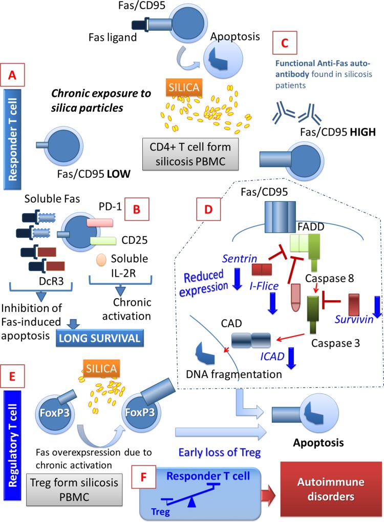
Schematic presentation of the immunological effects of silica particles. Silica particles induced longer survival and resistance to Fas-mediated apoptosis in responder T cells. Silica also induces accelerated Fas-mediated apoptosis in another fraction of peripheral T cells. In addition, chronically activated regulatory T cells induced accelerated apoptosis. Overall, an unbalance of responder T cells and regulatory T cells was produced. This may be the cause for the occurrence of autoimmune diseases as complications of silicosis. PD-1 programed death 1, DcR3 decoy receptor 3, I-Flice inhibitor of FADD-like interleukin-1-beta-converting enzyme, ICAD inhibitor of caspase-activated DNase, FoxP3 forkhead box P3, Treg regulatory T cell, PBMC peripheral blood mononuclear cells. a Reduced Fas-mediated apoptosis in responder T cells from silicosis, b various activation markers in responder T cells from silicosis, c detection of functional anti-Fas autoantibody in serum from silicosis, d reduction of genes which physiologically inhibit Fas-mediated apoptosis found in PBMC from silicosis, e overexpression of Fas in Treg from silicosis as the activation of Treg, and f unbalance between responder T cell and Treg in silicosis
As mentioned above, the function and/or volume of Treg is reduced following asbestos exposure, and the reaction of Tresp against foreign and self-antigens may be prolonged and induced the onset of certain allergic diseases and autoimmune disorders [19–21]. We estimated the Treg function of a peripheral CD4+25+ T helper fraction derived from SIL and compared it with that from HV. Although the strict Treg was defined by FoxP3 expression in the nucleus, the functional assay could not be performed after checking nuclear expression at the timing of flow cytometrical sorting, and the CD4+25+ fraction was collected [19–21]. Results showed that the Treg inhibitory function against the proliferation of auto-CD4+25− T helper cells stimulated by allo-antigen (using allo-PBMC) was reduced in the CD4+25+ fraction derived from SIL [36]. These findings indicated that Treg in SIL may have reduced numbers or function, probably due to the chronic exposure to silica particles.
On the other hand, we reported that peripheral T helper cells in SIL are assumed to have two fractions [1, 2, 37, 38]. One is the fraction in which T cells possess resistance to CD95/Fas-mediated apoptosis by over-production of extracellular inhibitors such as the soluble form of Fas molecules (one of the typical alternatively spliced variants of Fas molecule and known to be elevated in the serum of various autoimmune diseases [39, 40]), and overexpression of decoy receptor 3 (DcR3) [41] and other variant messages of Fas molecules [42]. All these events are assumed to inhibit Fas-mediated apoptosis by the early binding with the Fas-ligand at extracellular sites (Fig. 2a). In addition, soluble IL-2 receptor (sIL-2R) was elevated in SIL serum when compared with that in HV [43]. Experimentally, silica particles activated freshly isolated peripheral T cells derived from HV monitored by the expression of CD69 on the surface [44]. Together with excess expression of PD-1 in T helper cells from SIL (Fig. 2b) [45], a certain fraction of helper T cells in SIL is thought to survive longer with prolonged activation [45]. On the other hand, among the various auto-antibodies, we found in SIL serum [46–50], we detected a functional anti-Fas autoantibody (Fig. 2c) [50]. Thus, some Fas-expressing T cells may proceed to apoptosis when this auto-antibody was formed in SIL. This supposition is supported by the finding that mRNA expression in PBMCs from SIL showed decreased expression of physiological inhibitors for Fas-mediated apoptosis such as sentrin, inhibitor of FADD-like IL-1-beta-converting enzyme (I-flice), survivin, and inhibitor of caspase-activated DNase (ICAD) [51]. In addition, peripheral FoxP3+ Treg from SIL showed higher CD95/Fas expression compared with that from HV as a result of chronic activation [45]. These findings indicated that other parts of peripheral T helper cells in SIL are sensitive to Fas-mediated apoptosis and may include Treg [45]. This fraction may be easily eliminated by Fas-mediated apoptosis and repeatedly recruited from bone marrow (Fig. 2d) [1, 2, 37, 38, 45].
Our in vitro experiments supported these findings. When freshly isolated PBMCs from HV were cultured in vitro with silica particles, the percentage of the CD4+25+ fraction gradually increased during the culture period, although CD4+FoxP3+ cells were reduced [45]. These findings indicated that silica can activate Tresp expressing CD25, although silica also activated Treg-inducing Fas expression and subsequent apoptosis as a result of the activation (Fig. 2e). These results may suggest that the CD4+25+ fraction in SIL showed reduced Treg function, since there might be activated and CD25 expressing Tresp and reduced Treg (Fig. 2f) [45].
As shown in Fig. 2f, the overall findings indicate that an unbalance (decrease of Treg and increase of Tresp) may be present in the peripheral blood of SIL, and this unbalance may be the cause of subsequent autoimmune diseases in SIL [37, 38, 52–56]. In the future, the effects of silica particles on Th17 cells, which are considered to play an important role in the development of autoimmune disease [57–59], should be analyzed to better understand the comprehensive effects of silica particles on immune dysregulation, particularly impairment of autoimmunity.
Construction of health-promoting living conditions
Because of possibilities that various chemicals and other substances in living circumstances cause the health impairment separately or multiply, similar to the immunological effects induced by silica particles or asbestos fibers as mentioned above, it is necessary to construct the health-promoting living environment to adequately activate the human immune system. For this standpoint, we had been investigated the establishment and analyses of health-promoting indoor air condition.
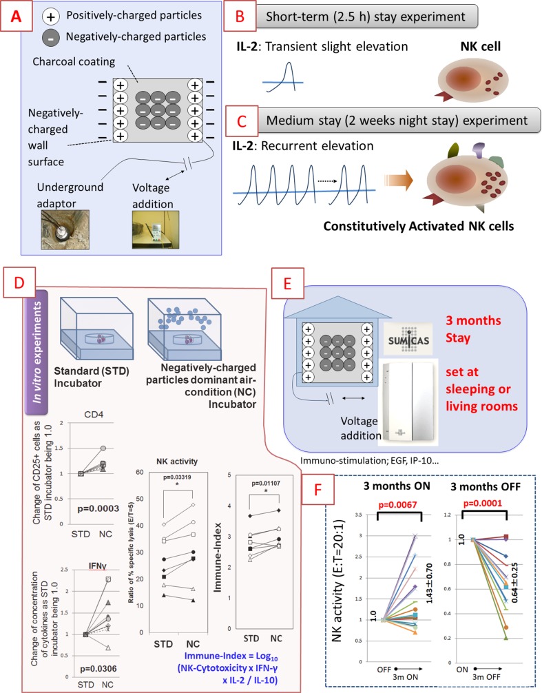
Figure 3 shows one of the approaches that can be used to construct health-promoting living conditions. Our approach succeeded in developing living conditions that enhanced NK cell activity [60]. These series of experiments were performed in collaboration with the research of Sekisui-House Co. Ltd., Yamada SXL Home Co. Ltd. and Artech Kohboh Co. Ltd. The conflict of interest (COI) declaration is shown at the end of this manuscript.
Fig. 3.
Schematic summary of the construction of health-promoting living conditions using negatively charged particle-dominant indoor air conditions. IL interleukin, NK: natural killer. a Mechanisms yielding negatively charged particles dominant indoor air condition (NCPDIAC), b slight but significant elevation of IL-2 by short term (2.5 h stay in CPDIAC), c activation of NK cell killing activity by medium stay (2-week night stay in NCPDIAC), d slight but significant activation of T cell and NK cells examined in vitro experiments resembling NCPDIAC situation, e setting of Sumicas® to produce NCPDIAC at living or sleeping rooms in actual resident homes and experimental trials of 3 months ON or OFF, and f elevation of NK activity in 3 months ON trials and reduction of it in 3 months OFF trials
The basic concept of this approach is to develop negatively charged particle-dominant indoor air conditions (NCPDIAC) [60, 61]. The particles are approximately 20 nm in diameter. NCPDIAC was created by painting a charcoal coating made using fine charcoal powder onto the wall and ceiling of a room. The charcoal coating designated as Health Coat® was produced by Artech Kohboh Co., Ltd. In addition, a forced negatively charged particle-dominant indoor air condition was created by applying an electric voltage (72 V) between the backside of the walls of a room and the ground as shown in Fig. 3a. These mixed devices were designated SUMICAS® and provided by Yamada SXL Home Co. Ltd. and Artech Kohbou Co. Ltd. It is known that a high negatively charged particle-dominant air condition is present in a forest, particularly near a waterfall.
Initial experiments involved the short-term (2.5 h) stay under these air conditions [61]. In the wide underground laboratory of the Comprehensive Housing R&D Institute of Sekisui House Co. Ltd. (Kidugawa, Kyodo, Japan), model rooms were constructed with an NCPDIAC device. The area and volume of the laboratory were approximately 539 and 1,564 m3, respectively, and those of experimental rooms were 9.1 and 22.8 m3, respectively. The negatively charged particles were continuously predominant in experimental rooms at a level of 800 particles/mm3 [61].
After building the experimental (with NCPDIAC) and control (no NCPDIAC) rooms with the same appearance, 60 HV for each condition were recruited and stayed in the rooms for 2.5 h. The various biological parameters listed below were then monitored. (1) As indicators of general conditions, blood chemistry including liver and kidney functions, blood sugar and lactic acid, and peripheral blood count were measured using peripheral blood. Blood pressure and pulse rate were also measured pre- and post-admission. (2) As stress markers, blood cortisol and salivary cortisol, chromogranin A, amylase, and secretary immunoglobulin (Ig) A were measured. (3) The autonomic nervous system was examined using the Flicker test, a stabilometer and heart rate monitoring for 3 min. RR intervals were estimated and the SD was considered an index of the fluctuation of heart rate. (4) As immunological parameters, serum levels of Ig E and Ig A, cytokines related to Th1/Th2 balance, and IFN-γ, TNF-α, IL-2, -4, -6 and -10 were evaluated. (5) Blood viscosity was measured by the Micro-Channel Flow Analyzer MC-FAN (MC Laboratory Inc., Tokyo, Japan).
As shown in Fig. 3b, the most significant and important finding of these experiments was the slight but significant increase of IL-2 after a 2.5-h stay in experimental NCPDIAC rooms compared to control rooms. Additionally, the changes in IL4 (increase), RR interval of heart rate monitor (decrease), and blood viscosity (decrease) was significant when values for NCPDIAC rooms were compared with those for control rooms [61].
Although there were no differences regarding stress markers and emotional mood analysis using the Profile of Mood States (POMS) questionnaire, it was clear that certain effects were induced by NCPDIAC. As a consequence, experiments involving a 2-week night stay were performed as the next step [60, 62]. Fifteen HV were recruited and received 3 months of on-the-job training at the Comprehensive Housing R&D Institute of Sekisui House Co. Ltd. Since HV needed to be familiar with the dormitory and training, and due to the limited number of subjects, they entered dormitory rooms for 4 weeks and then moved into control rooms (without NCPDIAC) without being notified whether or not they had entered rooms with or without the NCPDIAC device. After spending 2 weeks in control rooms, they again moved to rooms with the NCPDIAC without notification. Biological monitoring was then performed before and after entering both control and experimental rooms [60, 62]. The parameters monitored were similar to those used in the initial short-term experiments in addition to other parameters such as NK cell activity and urine 8-hydroxydeoxyguanosine (8-OHdG) corrected by urine creatinine concentration because these may be altered after a 2-week stay, and not a 2.5-h stay. NCPDIAC was utilized in experiments and differences between negatively and positively charged particles were approximately 500 particles/m3 [60, 62]. Although changes of parameters in individual subjects varied, results revealed that the only parameter that showed a significant change was NK cell activity. NCPDIAC induced an increase of NK cell activity (Fig. 3c). This suggested that the recurrent slight elevation of IL-2 as found in short-stay experiments during the 2-week night stay induced activation of NK cells [60, 62].
An in vitro assay was then performed. Since it was very difficult to establish a cell culture incubator with NCPDIAC, negatively charged particles were continuously belched into and sucked out from the culture incubator, and various immunological reactions using lymphocytes derived from HV were investigated (Fig. 3d) [63]. The level of negatively charged particles was approximately 3,000 particles/m3 higher in the experimental incubator compared with the standard control incubator. The immunobiological investigations in the experimental incubator showed upregulation of the CD25 surface marker as the early activation parameter in CD4+ cells, an increase of IFN-γ production in CD4+ cells when stimulated via the T cell receptor and elevation of NK cell activity. In addition to these findings, the Immune-Index as calculated by Log10 {[(NK cytotoxicity) × (concentration of IFN-γ) × (concentration of IL-2)] divided by [concentration of IL-10]} also increased in the experimental incubator when compared with that in the standard control incubator [63]. These findings indicated that NCPDIAC may positively stimulate the human immune system, particularly activation of NK cells, whereas there were no definite adverse effects from immune stimulation because of the slight activation of T cells.
Thus, practically and experimentally, the NCPDIAC device enhanced NK activity with slight immune stimulation. Since there was no critical adverse effect due to NCPDIAC, a final experiment was performed that involved a long-term stay for 3 months [64]. Actually, HV were recruited for this experiment who received information concerning this device, gave their written informed consent, and agreed to add this device to their residential home or condominium in sleeping or living rooms (Fig. 3e). The charcoal coating designated as Health Coat® was set by Artech Kohboh Co. Ltd. In addition, a forced negatively charged particle-dominant indoor air condition was created by applying an electric voltage (72 V) between the backside of the walls of a room and the ground. These mixed devices were designated SUMICAS® and provided by Yamada SXL Home Co. Ltd. The differences between negatively and positively charged particles were approximately 500–800 particles/m3, even in the actual home or condominium setting. Every 3 months HV then switched the NCPDIAC device ON or OFF by themselves, and blood and urine (for 8-OHdG) sampling was performed. In addition to the recording of general conditions, NK cell activity and specific Ig E for various environmental antigens and 29 kinds of cytokines were measured by a Luminex® bead-based multiplex assay. Results showed that the NCPDIAC device enhanced NK activity (Fig. 3f). Specifically, there was a significant increase of NK cell activity after living in NCPDIAC for 3 months. Furthermore, a significant decrease of NK cell activity was detected after living for 3 months in the OFF period for the NCPDIAC device. Moreover, there were slight increases of cytokines such as epidermal growth factor (EGF) and IFN-inducible protein 10 (IP-10) during ON periods. A comparison of cytokine status between ON and OFF periods showed that basic immune status was stimulated slightly during ON periods under NCPDIAC [64].
The overall findings shown in Fig. 3 [60–64] indicate that the NCPDIAC device caused activation of NK activity and stimulated immune status, and, therefore, could be set in the home or office buildings to potentially improve the health of occupants.
Conclusion
Among the various scientific fields covered in the hygiene area such as environmental medicine, epidemiology, public health and preventive medicine, we are investigating the immunological effects of fibrous and particulate substances in the environment and work surroundings such as asbestos fibers and silica particles. In addition to these studies, we have attempted to construct health-promoting living conditions. Thus, in this review we summarize our investigations regarding the (1) immunological effects of asbestos fibers, (2) immunological effects of silica particles, and (3) construction of a health-promoting living environment.
Asbestos fibers cause reduction of anti-tumor immunity in asbestos-exposed patients. To utilize our experimental findings in the clinical fields, we are attempting to establish early diagnostic parameters using various alterations in human immune cells and cytokines, since these cells were easily drawn from peripheral blood and without the need for any irradiation, which is a concern in recent screening radiological examinations for asbestos exposure and individuals with MM.
Silica particles cause dysregulation of autoimmunity and induce various autoimmune diseases in SIL patients as complications. Advances in scientific findings regarding silica-induced autoimmune diseases may support these patients suffering from autoimmune disease and lung fibrosis, particularly in the area of worker compensation. Additionally, there may be certain discoveries that will help us to recognize and understand the pathophysiological mechanisms of etiology-unknown autoimmune disorders.
Finally, the construction of health-promoting living circumstances is important to maintain healthy conditions in all people, particularly the aged members of society. Our device is one of these constructions and may support the prevention of cancers and diminish symptoms from viral infections.
Scientific approaches to examine the impact of environmental factors on human health are necessary to improve the quality of life of all people, and we should continuously make tremendous efforts to establish these health-promoting environments.
Acknowledgments
All authors thank former members of the Department of Hygiene, Kawasaki Medical School, Kurashiki, Japan, including Profs. Yoshio Mochiduki and Ayako Ueki, Drs. Fuminori Hyodoh, Takata-Tomokuni Akiko, Yasuhiko Kawakami, Takaaki Aikoh, Takakazu Matsuki, Yoshie Miura, Shuko Murakami, Ping Wu, Ying Chen, Hiroaki Hayashi and Megumi Maeda. We also thank Ms. Minako Katoh, Naomi Miyahara, Keiko Kimura, Misao Kuroki, Tomoko Sueishi, Yoshiko Yamashita, Satomi Hatada and Haruko Sakaguchi for their technical support. Foundations supporting all the findings described in this review are shown in the articles reported previously and individually. We, therefore, ask that you refer to these publications rather than being shown here a list of the foundations.
Compliance with ethical standards
Conflict of interest
Recently, the first author received a research foundation from Sumitomo Riko Co. Ltd. in 2014. However, this foundation is not related to any of the experiments shown in this manuscript. During experiments involving biological assays of negatively charged particles, Sekisui House Co. Ltd., Yamada SXL Co. Ltd. and Artech Kohbou Co. Ltd. provided the construction fees for experimental rooms, SUMICAS® devices and the experimental incubator as collaboration partners for all experiments. In addition, the first author received a research foundation including purchasing fees for experimental consumable supplies and traffic fees from Sekisui House Co. Ltd. in 2009.
References
- 1.Otsuki T, Maeda M, Murakami S, Hayashi H, Miura Y, Kusaka M, et al. Immunological effects of silica and asbestos. Cell Mol Immunol. 2007;4:261–268. [PubMed] [Google Scholar]
- 2.Maeda M, Nishimura Y, Kumagai N, Hayashi H, Hatayama T, Katoh M, et al. Dysregulation of the immune system caused by silica and asbestos. J Immunotoxicol. 2010;7:268–278. doi: 10.3109/1547691X.2010.512579. [DOI] [PubMed] [Google Scholar]
- 3.Nishimura Y, Kumagai N, Maeda M, Hayashi H, Fukuoka K, Nakano T, et al. Suppressive effect of asbestos on cytotoxicity of human NK cells. Int J Immunopathol Pharmacol. 2011;24(1S):5S–10S. [PubMed] [Google Scholar]
- 4.Kumagai-Takei N, Maeda M, Chen Y, Matsuzaki H, Lee S, Nishimura Y, et al. Asbestos induces reduction of tumor immunity. Clin Dev Immunol. 2011;2011:481439. doi: 10.1155/2011/481439. [DOI] [PMC free article] [PubMed] [Google Scholar]
- 5.Matsuzaki H, Maeda M, Lee S, Nishimura Y, Kumagai-Takei N, Hayashi H, et al. Asbestos-induced cellular and molecular alteration of immunocompetent cells and their relationship with chronic inflammation and carcinogenesis. J Biomed Biotechnol. 2012;2012:492608. doi: 10.1155/2012/492608. [DOI] [PMC free article] [PubMed] [Google Scholar]
- 6.Nishimura Y, Maeda M, Kumagai-Takei N, Lee S, Matsuzaki H, Wada Y, et al. Altered functions of alveolar macrophages and NK cells involved in asbestos-related diseases. Environ Health Prev Med. 2013;18(3):198–204. doi: 10.1007/s12199-013-0333-y. [DOI] [PMC free article] [PubMed] [Google Scholar]
- 7.Matsuzaki H, Nishimura Y, Lee S, Maeda M, Kumagai-Takei N, Hayashi H, et al. Asbestos-induced mesothelioma: Tumor escape and alteration of immune surveillance. In: Pandalai SG, et al., editors. Recent research developments in immunology. Kerala: Research Signpost Publisher; 2012. pp. 13–31. [Google Scholar]
- 8.Nishimura Y, Maeda M, Kumagai-Takei N, Matsuzaki H, Lee S, Fukuoka K, et al. Effect of asbestos on anti-tumor immunity and immunological alteration in patients with mesothelioma. In: Belli C, Anand S, editors. Malignant mesothelioma. Rijeka: InTech Open Access Publisher; 2012. doi:10.5772/33138.
- 9.Otsuki T, Maeda M, Miura Y, Hayashi H, Murakami S, Kumagai N, et al. Immunological effects of asbestos. In: Soto A, Salazar G, et al., editors. Asbestos: risks, environment and impact. New York: Nova Science Publishers, Inc.; 2009. pp. 185–193. [Google Scholar]
- 10.Kamp DW. Asbestos-induced lung diseases: an update. Transl Res. 2009;153:143–152. doi: 10.1016/j.trsl.2009.01.004. [DOI] [PMC free article] [PubMed] [Google Scholar]
- 11.Moolgavkar SH, Anderson EL, Chang ET, Lau EC, Turnham P, Hoel DG. A review and critique of U.S. EPA’s risk assessments for asbestos. Crit Rev Toxicol. 2014;44:499–522. doi: 10.3109/10408444.2014.902423. [DOI] [PubMed] [Google Scholar]
- 12.Heintz NH, Janssen-Heininger YM, Mossman BT. Asbestos, lung cancers, and mesotheliomas: from molecular approaches to targeting tumor survival pathways. Am J Respir Cell Mol Biol. 2010;42:133–139. doi: 10.1165/rcmb.2009-0206TR. [DOI] [PMC free article] [PubMed] [Google Scholar]
- 13.Case BW, Abraham JL, Meeker G, Pooley FD, Pinkerton KE. Applying definitions of “asbestos” to environmental and “low-dose” exposure levels and health effects, particularly malignant mesothelioma. J Toxicol Environ Health B Crit Rev. 2011;14:3–39. doi: 10.1080/10937404.2011.556045. [DOI] [PMC free article] [PubMed] [Google Scholar]
- 14.Hyodoh F, Takata-Tomokuni A, Miura Y, Sakaguchi H, Hatayama T, Hatada S, et al. Inhibitory effects of anti-oxidants on apoptosis of a human polyclonal T-cell line, MT-2, induced by an asbestos, chrysotile-A. Scand J Immunol. 2005;61:442–448. doi: 10.1111/j.1365-3083.2005.01592.x. [DOI] [PubMed] [Google Scholar]
- 15.Miura Y, Nishimura Y, Katsuyama H, Maeda M, Hayashi H, Dong M, et al. Involvement of IL-10 and Bcl-2 in resistance against an asbestos-induced apoptosis of T cells. Apoptosis. 2006;11:1825–1835. doi: 10.1007/s10495-006-9235-4. [DOI] [PubMed] [Google Scholar]
- 16.Maeda M, Chen Y, Hayashi H, Kumagai-Takei N, Matsuzaki H, Lee S, et al. Chronic exposure to asbestos enhances TGF-β1 production in the human adult T cell leukemia virus-immortalized T cell line MT-2. Int J Oncol. 2014;45:2522–2532. doi: 10.3892/ijo.2014.2682. [DOI] [PubMed] [Google Scholar]
- 17.Hamano R, Wu X, Wang Y, Oppenheim JJ, Chen X. Characterization of MT-2 cells as a human regulatory T cell-like cell line. Cell Mol Immunol. 2014 doi: 10.1038/cmi.2014.123. [DOI] [PMC free article] [PubMed] [Google Scholar]
- 18.Chen S, Ishii N, Ine S, Ikeda S, Fujimura T, Ndhlovu LC, et al. Regulatory T cell-like activity of Foxp3+ adult T cell leukemia cells. Int Immunol. 2006;18:269–277. doi: 10.1093/intimm/dxh366. [DOI] [PubMed] [Google Scholar]
- 19.Sakaguchi S. Naturally arising Foxp3-expressing CD25+CD4+ regulatory T cells in immunological tolerance to self and non-self. Nat Immunol. 2005;6:345–352. doi: 10.1038/ni1178. [DOI] [PubMed] [Google Scholar]
- 20.Linehan DC, Goedegebuure PS. CD25+CD4+ regulatory T-cells in cancer. Immunol Res. 2005;32:155–168. doi: 10.1385/IR:32:1-3:155. [DOI] [PubMed] [Google Scholar]
- 21.Yamaguchi T, Sakaguchi S. Regulatory T cells in immune surveillance and treatment of cancer. Semin Cancer Biol. 2006;16:115–123. doi: 10.1016/j.semcancer.2005.11.005. [DOI] [PubMed] [Google Scholar]
- 22.Ying C, Maeda M, Nishimura Y, Kumagai-Takei N, Hayashi H, Matsuzaki H, et al. Enhancement of regulatory T cell-like suppressive function in MT-2 by long-term and low-dose exposure to asbestos. Toxicology. 2015;338:86–94. doi: 10.1016/j.tox.2015.10.005. [DOI] [PubMed] [Google Scholar]
- 23.Maeda M, Chen Y, Kumagai-Takei N, Hayashi H, Matsuzaki H, Lee S, et al. Alteration of cytoskeletal molecules in a human T cell line caused by continuous exposure to chrysotile asbestos. Immunobiology. 2013;218:1184–1191. doi: 10.1016/j.imbio.2013.04.007. [DOI] [PubMed] [Google Scholar]
- 24.Nagai H, Toyokuni S. Differences and similarities between carbon nanotubes and asbestos fibers during mesothelial carcinogenesis: shedding light on fiber entry mechanism. Cancer Sci. 2012;103:1378–1390. doi: 10.1111/j.1349-7006.2012.02326.x. [DOI] [PMC free article] [PubMed] [Google Scholar]
- 25.Maeda M, Nishimura Y, Hayashi H, Kumagai N, Chen Y, Murakami S, et al. Reduction of CXC chemokine receptor 3 in an in vitro model of continuous exposure to asbestos in a human T-cell line, MT-2. Am J Respir Cell Mol Biol. 2011;45:470–479. doi: 10.1165/rcmb.2010-0213OC. [DOI] [PubMed] [Google Scholar]
- 26.Maeda M, Nishimura Y, Hayashi H, Kumagai N, Chen Y, Murakami S, et al. Decreased CXCR3 expression in CD4+ T cells exposed to asbestos or derived from asbestos-exposed patients. Am J Respir Cell Mol Biol. 2011;45:795–803. doi: 10.1165/rcmb.2010-0435OC. [DOI] [PubMed] [Google Scholar]
- 27.Hildebrandt GC, Corrion LA, Olkiewicz KM, Lu B, Lowler K, Duffner UA, et al. Blockade of CXCR3 receptor:ligand interactions reduces leukocyte recruitment to the lung and the severity of experimental idiopathic pneumonia syndrome. J Immunol. 2004;173:2050–2059. doi: 10.4049/jimmunol.173.3.2050. [DOI] [PubMed] [Google Scholar]
- 28.Strieter RM, Burdick MD, Mestas J, Gomperts B, Keane MP, Belperio JA. Cancer CXC chemokine networks and tumour angiogenesis. Eur J Cancer. 2006;42:768–778. doi: 10.1016/j.ejca.2006.01.006. [DOI] [PubMed] [Google Scholar]
- 29.Kumagai-Takei N, Nishimura Y, Maeda M, Hayashi H, Matsuzaki H, Lee S, et al. Effect of asbestos exposure on differentiation of cytotoxic T lymphocytes in mixed lymphocyte reaction of human peripheral blood mononuclear cells. Am J Respir Cell Mol Biol. 2013;49:28–36. doi: 10.1165/rcmb.2012-0134OC. [DOI] [PubMed] [Google Scholar]
- 30.Kumagai-Takei N, Nishimura Y, Maeda M, Hayashi H, Matsuzaki H, Lee S, et al. Functional properties of CD8(+) lymphocytes in patients with pleural plaque and malignant mesothelioma. J Immunol Res. 2014;2014:670140. doi: 10.1155/2014/670140. [DOI] [PMC free article] [PubMed] [Google Scholar]
- 31.Nishimura Y, Miura Y, Maeda M, Kumagai N, Murakami S, Hayashi H, et al. Impairment in cytotoxicity and expression of NK cell-activating receptors on human NK cells following exposure to asbestos fibers. Int J Immunopathol Pharmacol. 2009;22:579–590. doi: 10.1177/039463200902200304. [DOI] [PubMed] [Google Scholar]
- 32.Nishimura Y, Maeda M, Kumagai N, Hayashi H, Miura Y, Otsuki T. Decrease in phosphorylation of ERK following decreased expression of NK cell-activating receptors in human NK cell line exposed to asbestos. Int J Immunopathol Pharmacol. 2009;22:879–888. doi: 10.1177/039463200902200403. [DOI] [PubMed] [Google Scholar]
- 33.Iannello S, Camuto M, Cantarella S, Cavaleri A, Ferriero P, Leanza A, et al. Rheumatoid syndrome associated with lung interstitial disorder in a dental technician exposed to ceramic silica dust. A case report and critical literature review. Clin Rheumatol. 2002;21:76–81. doi: 10.1007/s100670200019. [DOI] [PubMed] [Google Scholar]
- 34.Parks CG, Conrad K, Cooper GS. Occupational exposure to crystalline silica and autoimmune disease. Environ Health Perspect. 1999;107(S5):793–802. doi: 10.1289/ehp.99107s5793. [DOI] [PMC free article] [PubMed] [Google Scholar]
- 35.Mayes MD. Epidemiologic studies of environmental agents and systemic autoimmune diseases. Environ Health Perspect. 1999;107(S5):743–748. doi: 10.1289/ehp.99107s5743. [DOI] [PMC free article] [PubMed] [Google Scholar]
- 36.Wu P, Miura Y, Hyodoh F, Nishimura Y, Hatayama T, Hatada S, et al. Reduced function of CD4+25+ regulatory T cell fraction in silicosis patients. Int J Immunopathol Pharmacol. 2006;19:357–368. doi: 10.1177/039463200601900212. [DOI] [PubMed] [Google Scholar]
- 37.Otsuki T, Miura Y, Nishimura Y, Hyodoh F, Takata A, Kusaka M, et al. Alterations of Fas and Fas-related molecules in patients with silicosis. Exp Biol Med (Maywood) 2006;231:522–533. doi: 10.1177/153537020623100506. [DOI] [PubMed] [Google Scholar]
- 38.Lee S, Hayashi H, Maeda M, Chen Y, Matsuzaki H, Takei-Kumagai N, et al. Environmental factors producing autoimmune dysregulation—chronic activation of T cells caused by silica exposure. Immunobiology. 2012;217:743–748. doi: 10.1016/j.imbio.2011.12.009. [DOI] [PubMed] [Google Scholar]
- 39.Tomokuni A, Aikoh T, Matsuki T, Isozaki Y, Otsuki T, Kita S, et al. Elevated soluble Fas/APO-1 (CD95) levels in silicosis patients without clinical symptoms of autoimmune diseases or malignant tumours. Clin Exp Immunol. 1997;110:303–309. doi: 10.1111/j.1365-2249.1997.tb08332.x. [DOI] [PMC free article] [PubMed] [Google Scholar]
- 40.Otsuki T, Sakaguchi H, Tomokuni A, Aikoh T, Matsuki T, Kawakami Y, et al. Soluble Fas mRNA is dominantly expressed in cases with silicosis. Immunology. 1998;94:258–262. doi: 10.1046/j.1365-2567.1998.00509.x. [DOI] [PMC free article] [PubMed] [Google Scholar]
- 41.Otsuki T, Tomokuni A, Sakaguchi H, Aikoh T, Matsuki T, Isozaki Y, et al. Over-expression of the decoy receptor 3 (DcR3) gene in peripheral blood mononuclear cells (PBMC) derived from silicosis patients. Clin Exp Immunol. 2000;119:323–327. doi: 10.1046/j.1365-2249.2000.01132.x. [DOI] [PMC free article] [PubMed] [Google Scholar]
- 42.Otsuki T, Sakaguchi H, Tomokuni A, Aikoh T, Matsuki T, Isozaki Y, et al. Detection of alternatively spliced variant messages of Fas gene and mutational screening of Fas and Fas ligand coding regions in peripheral blood mononuclear cells derived from silicosis patients. Immunol Lett. 2000;72:137–143. doi: 10.1016/S0165-2478(00)00177-2. [DOI] [PubMed] [Google Scholar]
- 43.Hayashi H, Maeda M, Murakami S, Kumagai N, Chen Y, Hatayama T, et al. Soluble interleukin-2 receptor as an indicator of immunological disturbance found in silicosis patients. Int J Immunopathol Pharmacol. 2009;22:53–62. doi: 10.1177/039463200902200107. [DOI] [PubMed] [Google Scholar]
- 44.Wu P, Hyodoh F, Hatayama T, Sakaguchi H, Hatada S, Miura Y, et al. Induction of CD69 antigen expression in peripheral blood mononuclear cells on exposure to silica, but not by asbestos/chrysotile-A. Immunol Lett. 2005;98:145–152. doi: 10.1016/j.imlet.2004.11.005. [DOI] [PubMed] [Google Scholar]
- 45.Hayashi H, Miura Y, Maeda M, Murakami S, Kumagai N, Nishimura Y, et al. Reductive alteration of the regulatory function of the CD4(+)CD25(+) T cell fraction in silicosis patients. Int J Immunopathol Pharmacol. 2010;23:1099–1109. doi: 10.1177/039463201002300414. [DOI] [PubMed] [Google Scholar]
- 46.Ueki A, Isozaki Y, Kusaka M. Anti-caspase-8 autoantibody response in silicosis patients is associated with HLA-DRB1, DQB1 and DPB1 alleles. J Occup Health. 2005;47:61–67. doi: 10.1539/joh.47.61. [DOI] [PubMed] [Google Scholar]
- 47.Ueki A, Isozaki Y, Tomokuni A, Hatayama T, Ueki H, Kusaka M, et al. Intramolecular epitope spreading among anti-caspase-8 autoantibodies in patients with silicosis, systemic sclerosis and systemic lupus erythematosus, as well as in healthy individuals. Clin Exp Immunol. 2002;129:556–561. doi: 10.1046/j.1365-2249.2002.01939.x. [DOI] [PMC free article] [PubMed] [Google Scholar]
- 48.Tomokuni A, Otsuki T, Sakaguchi H, Isozaki Y, Hyodoh F, Kusaka M, et al. Detection of anti-topoisomerase I autoantibody in patients with silicosis. Environ Health Prev Med. 2002;7:7–10. doi: 10.1007/BF02898059. [DOI] [PMC free article] [PubMed] [Google Scholar]
- 49.Ueki H, Kohda M, Nobutoh T, Yamaguchi M, Omori K, Miyashita Y, et al. Antidesmoglein autoantibodies in silicosis patients with no bullous diseases. Dermatology. 2001;202:16–21. doi: 10.1159/000051578. [DOI] [PubMed] [Google Scholar]
- 50.Takata-Tomokuni A, Ueki A, Shiwa M, Isozaki Y, Hatayama T, Katsuyama H, et al. Detection, epitope-mapping and function of anti-Fas autoantibody in patients with silicosis. Immunology. 2005;116:21–29. doi: 10.1111/j.1365-2567.2005.02192.x. [DOI] [PMC free article] [PubMed] [Google Scholar]
- 51.Otsuki T, Tomokuni A, Sakaguchi H, Hyodoh F, Kusaka M, Ueki A. Reduced expression of the inhibitory genes for Fas-mediated apoptosis in silicosis patients. J Occup Health. 2000;42:163–168. doi: 10.1539/joh.42.163. [DOI] [Google Scholar]
- 52.Lee S, Matsuzaki H, Kumagai-Takei N, Yoshitome K, Maeda M, Chen Y, et al. Silica exposure and altered regulation of autoimmunity. Environ Health Prev Med. 2014;19:322–329. doi: 10.1007/s12199-014-0403-9. [DOI] [PMC free article] [PubMed] [Google Scholar]
- 53.Lee S, Maeda M, Hayashi H, Matsuzaki H, Kumagai-Takei N, Nishimura Y, et al. Immunostimulation by silica particles and the development of autoimmune dysregulation. In: Duc GHT, editor. Immunostimulation. Rijeka: InTech Open Access Publisher; 2014. doi:10.5772/57544.
- 54.Takei-Kumagai N, Lee S, Matsuzaki H, Hayashi H, Maeda M, Nishimura Y, Otsuki T. Immunological effects of silica. In: Uversky VN, Kretsinger RH, Permyakov EA, editors. Encyclopedia of metalloproteins. New York: Springer Science + Business Media; 2013. pp. 1965–1971. [Google Scholar]
- 55.Kumagai N, Hayashi H, Maeda M, Miura Y, Nishimura Y, Matsuzaki H, et al. Immunological effects of silica and related dysregulation of autoimmunity. In: Mavragani CP, editor. Autoimmune disorders—pathogenetic aspects. Rijeka: InTech Open Access Publisher; 2011. p. 157–74. doi:10.5772/19218.
- 56.Hayashi H, Nishimura Y, Hyodo F, Maeda M, Kumagai N, Miura Y, et al. Dysregulation of autoimmunity caused by silica exposure: Fas-mediated apoptosis in T lymphocytes derived from silicosis patients. In: Petro ME, et al., editors. Autoimmune disorders: symptoms, diagnosis and treatment. New York: Nova Science Publishers, Inc.; 2011. pp. 293–301. [Google Scholar]
- 57.Maddur MS, Miossec P, Kaveri SV, Bayry J. Th17 cells: biology, pathogenesis of autoimmune and inflammatory diseases, and therapeutic strategies. Am J Pathol. 2012;181:8–18. doi: 10.1016/j.ajpath.2012.03.044. [DOI] [PubMed] [Google Scholar]
- 58.Bolon B. Cellular and molecular mechanisms of autoimmune disease. Toxicol Pathol. 2012;40:216–229. doi: 10.1177/0192623311428481. [DOI] [PubMed] [Google Scholar]
- 59.Ghoreschi K, Laurence A, Yang XP, Hirahara K, O’Shea JJ. T helper 17 cell heterogeneity and pathogenicity in autoimmune disease. Trends Immunol. 2011;32:395–401. doi: 10.1016/j.it.2011.06.007. [DOI] [PMC free article] [PubMed] [Google Scholar]
- 60.Otsuki T, Takahashi K, Mase A, Kawado T, Kotani M, Nishimura Y, et al. Establishment of negatively-charged indoor air conditions and their biological effects. In: Nemecek J, Schulz O, et al., editors. Buildings and the environment. New York: Nova Science Publishers, Inc.; 2009. pp. 201–214. [Google Scholar]
- 61.Takahashi K, Otsuki T, Mase A, Kawado T, Kotani M, Ami K, et al. Negatively-charged air conditions and responses of the human psycho-neuro-endocrino-immune network. Environ Int. 2008;34:765–772. doi: 10.1016/j.envint.2008.01.003. [DOI] [PubMed] [Google Scholar]
- 62.Takahashi K, Otsuki T, Mase A, Kawado T, Kotani M, Nishimura Y, et al. Two weeks of permanence in negatively-charged air conditions causes alteration of natural killer cell function. Int J Immunopathol Pharmacol. 2009;22:333–342. doi: 10.1177/039463200902200210. [DOI] [PubMed] [Google Scholar]
- 63.Nishimura Y, Takahashi K, Mase A, Kotani M, Ami K, Maeda M, et al. Exposure to negatively charged-particle dominant air-conditions on human lymphocytes in vitro activates immunological responses. 2015. doi:10.1016/j.imbio.2015.07.006. [DOI] [PubMed]
- 64.Nishimura Y, Takahashi K, Mase A, Kotani M, Ami K, Maeda M, et al. Enhancement of NK cell cytotoxicity induced by long-term living in negatively charged-particle dominant indoor air-conditions. PLoS One. 2015. doi:10.1371/journal.pone.0132373. [DOI] [PMC free article] [PubMed]


