

Abstract
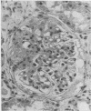
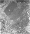
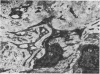
Renal biopsy material from seven cases of the nephrotic syndrome due to focal glomerular sclerosis has been studied by light, electron, and immunofluorescent microscopy. The nature of glomerular basement membrane changes and the scar tissue was also studied. It was found that the glomerular basement membrane and mesangial matrix formed the major components of scar tissue. On the basis of a short history in some of our cases, a poor response to steroid therapy in the early stages, and the distinct morphological changes, it is suggested that focal glomerular sclerosis has an independent origin and is not a stage of minimal change lesion.
Full text
PDF


Images in this article


Selected References
These references are in PubMed. This may not be the complete list of references from this article.
- Churg J., Habib R., White R. H. Pathology of the nephrotic syndrome in children: a report for the International Study of Kidney Disease in Children. Lancet. 1970 Jun 20;760(1):1299–1302. doi: 10.1016/s0140-6736(70)91905-7. [DOI] [PubMed] [Google Scholar]
- JONES D. B. The nature of scar tissue in glomerulonephritis. Am J Pathol. 1963 Feb;42:185–199. [PMC free article] [PubMed] [Google Scholar]
- RICH A. R. A hitherto undescribed vulnerability of the juxtamedullary glomeruli in lipoid nephrosis. Bull Johns Hopkins Hosp. 1957 Apr;100(4):173–186. [PubMed] [Google Scholar]
- White R. H., Glasgow E. F., Mills R. J. Clinicopathological study of nephrotic syndrome in childhood. Lancet. 1970 Jun 27;1(7661):1353–1359. doi: 10.1016/s0140-6736(70)91268-7. [DOI] [PubMed] [Google Scholar]