Abstract
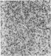
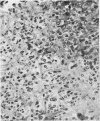
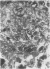
The case of a 42-year-old woman with virilization, hirsutes, and a high level of circulating testosterone is described. A hilar cell tumour of the ovary was found, the histological features of which were typical, with the presence of crystalloids of Reinke, hyaline bodies, lipid, and lipofuscin. Some areas showed appearances similar to those seen in `adrenocortical cell' tumours. In common with other lipid cell tumours of the ovary, hilar cell tumours most probably arise from mesenchymal elements and the group should, therefore, be considered as a subdivision of the gonadal stromal (sex cord-mesenchymal) tumours.
Full text
PDF
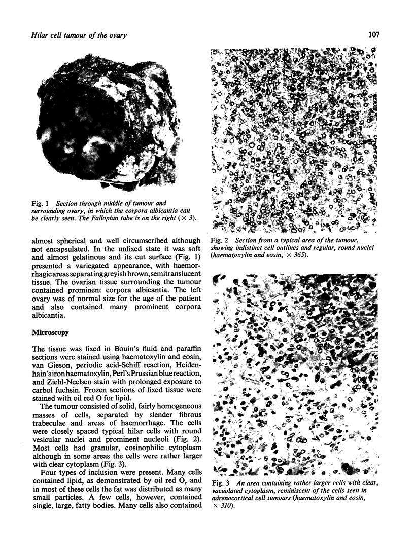
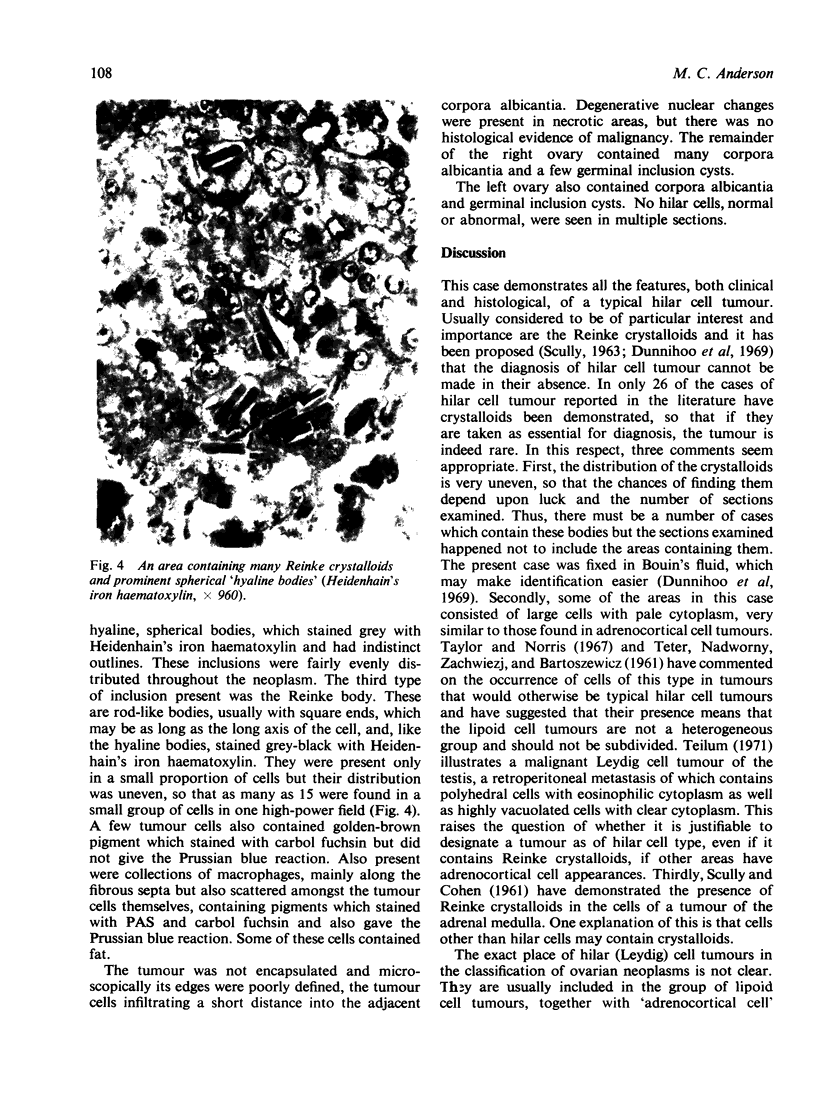
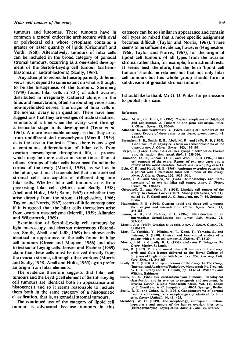

Images in this article
Selected References
These references are in PubMed. This may not be the complete list of references from this article.
- Abell M. R., Holtz F. Ovarian neoplasms in childhood and adolescence. II. Tumors of non-germ cell origin. Am J Obstet Gynecol. 1965 Nov 15;93(6):850–866. doi: 10.1016/0002-9378(65)90085-2. [DOI] [PubMed] [Google Scholar]
- Berendsen P. B., Smith E. B., Abell M. R., Jaffe R. B. Fine structure of Leydig cells from an arrhenoblastoma of the ovary. Am J Obstet Gynecol. 1969 Jan 15;103(2):192–199. doi: 10.1016/s0002-9378(16)34388-5. [DOI] [PubMed] [Google Scholar]
- Dunnihoo D. R., Grieme D. L., Woolf R. B. Hilar-cell tumors of the ovary. Report of 2 new cases and a review of the world literature. Obstet Gynecol. 1966 May;27(5):703–713. [PubMed] [Google Scholar]
- Echt C. R., Hadd H. E. Androgen excretion patterns in a patient with a metastatic hilus cell tumor of the ovary. Am J Obstet Gynecol. 1968 Apr 15;100(8):1055–1061. doi: 10.1016/s0002-9378(15)33403-7. [DOI] [PubMed] [Google Scholar]
- Green J. A., Maqueo M. Histopathology and ultrastructure of an ovarian hilar cell tumor. Am J Obstet Gynecol. 1966 Oct 15;96(4):478–485. doi: 10.1016/s0002-9378(16)34685-3. [DOI] [PubMed] [Google Scholar]
- Hughesdon P. E. Ovarian lipoid and theca cell tumors; their origins and interrelations. Obstet Gynecol Surv. 1966 Apr;21(2):245–288. [PubMed] [Google Scholar]
- Jenson A. B., Fechner R. E. Ultrastructure of an intermediate Sertoli-Leydig cell tumor. A histogenetic misnomer. Lab Invest. 1969 Dec;21(6):527–535. [PubMed] [Google Scholar]
- SCULLY R. E., COHEN R. B. Ganglioneuroma of adrenal medulla containing cells morphologically identical to hilus cells (extraparenchymal Leydig cells). Cancer. 1961 Mar-Apr;14:421–425. doi: 10.1002/1097-0142(196103/04)14:2<421::aid-cncr2820140222>3.0.co;2-z. [DOI] [PubMed] [Google Scholar]
- STERNBERG W. H. The morphology, androgenic function, hyperplasia, and tumors of the human ovarian hilus cells. Am J Pathol. 1949 May;25(3):493–521. [PMC free article] [PubMed] [Google Scholar]
- Salm R. Pure and mixed hilus-cell tumours of ovary. Arris and Gale Lecture delivered at the Royal College of Surgeons of England on 14th November 1966. Ann R Coll Surg Engl. 1967 Oct;41(4):344–363. [PMC free article] [PubMed] [Google Scholar]
- Taylor H. B., Norris H. J. Lipid cell tumors of the ovary. Cancer. 1967 Nov;20(11):1953–1962. doi: 10.1002/1097-0142(196711)20:11<1953::aid-cncr2820201123>3.0.co;2-2. [DOI] [PubMed] [Google Scholar]
- Yoshida Y., Mori T., Nishimura T., Kono T., Yamada S., Tatsumi S. Clinical and biochemical studies of a patient with a hilus cell tumour. J Endocrinol. 1970 May;47(1):13–20. doi: 10.1677/joe.0.0470013. [DOI] [PubMed] [Google Scholar]


