

Abstract
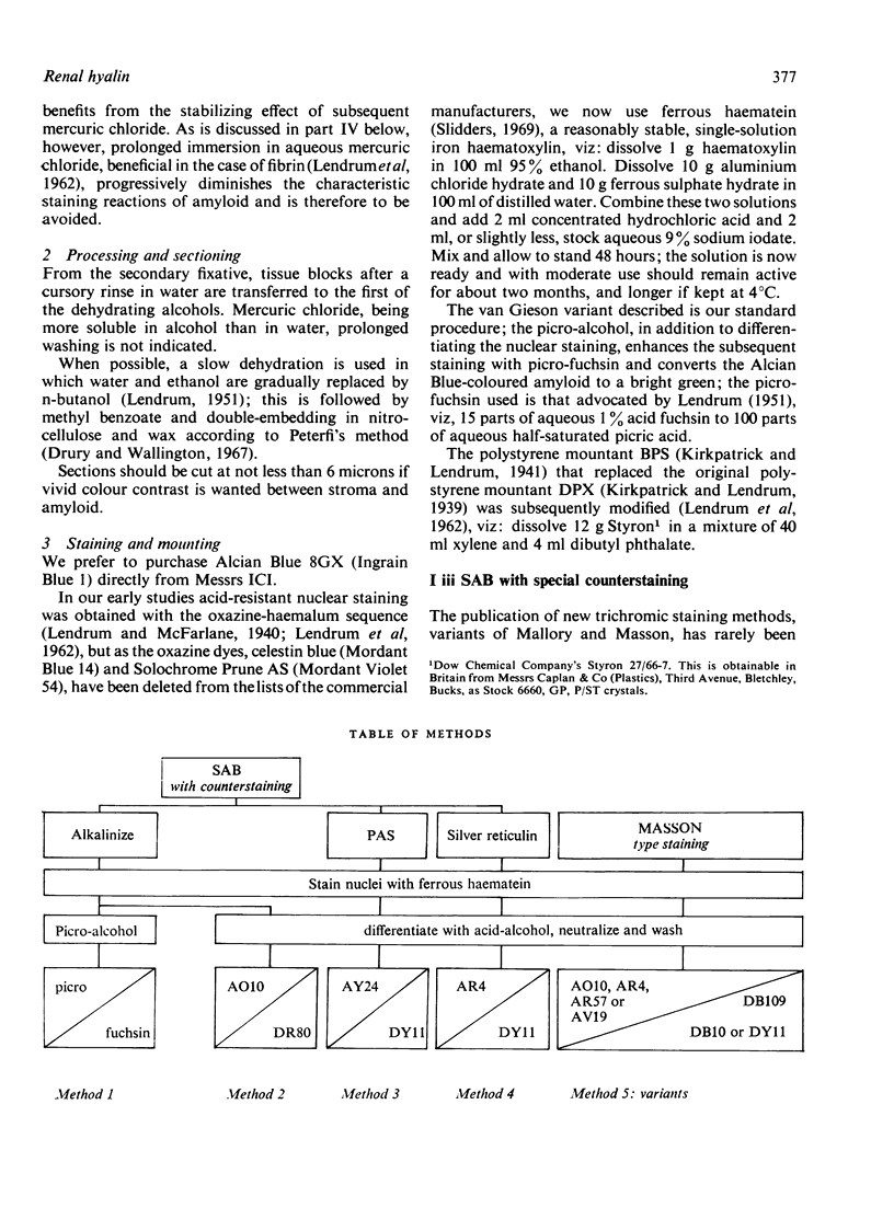
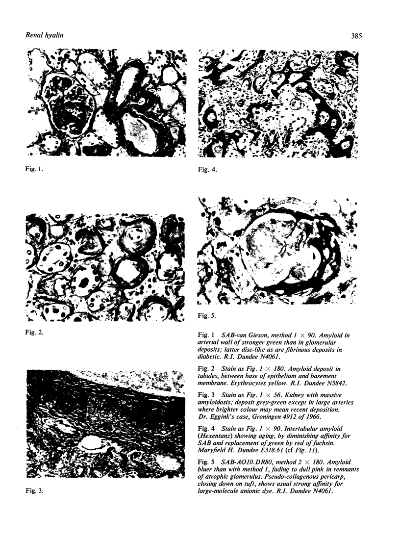
This describes the sodium sulphate-Alcian Blue (SAB) method for staining amyloid in paraffin sections. Its value lies in the possibility of subsequent counterstaining and thus of revealing the structural relationships of amyloid.
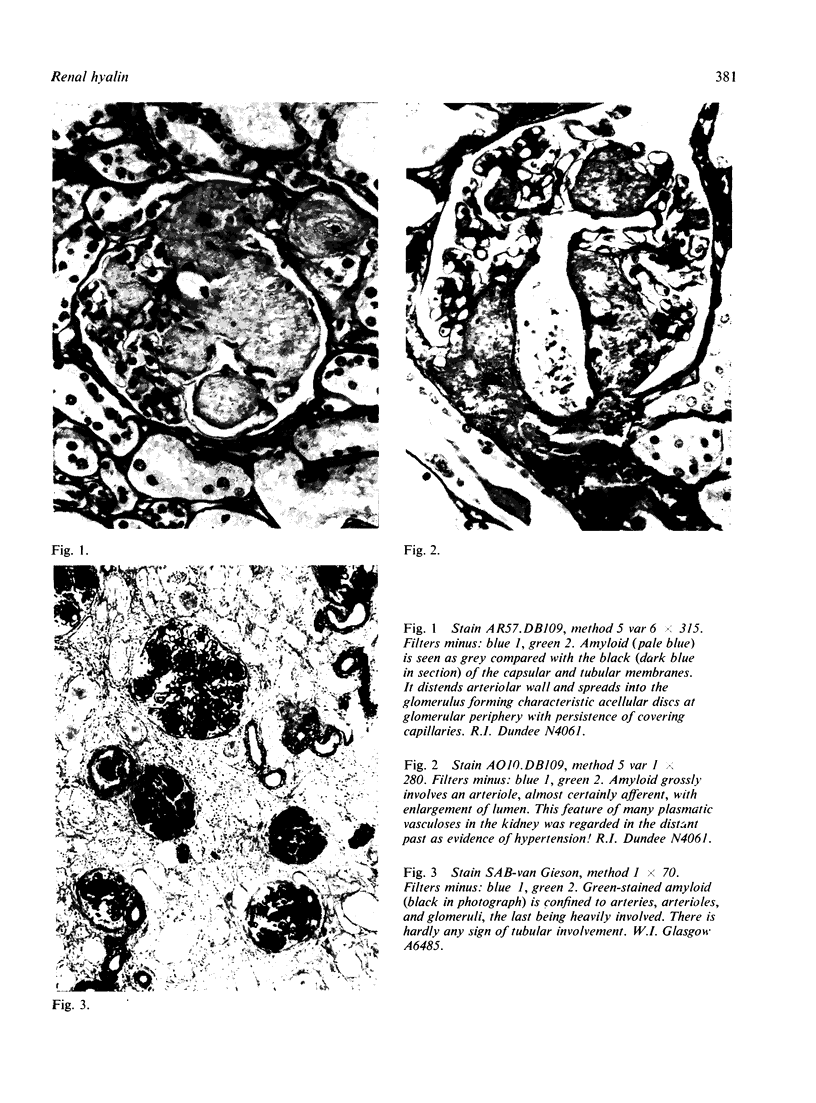
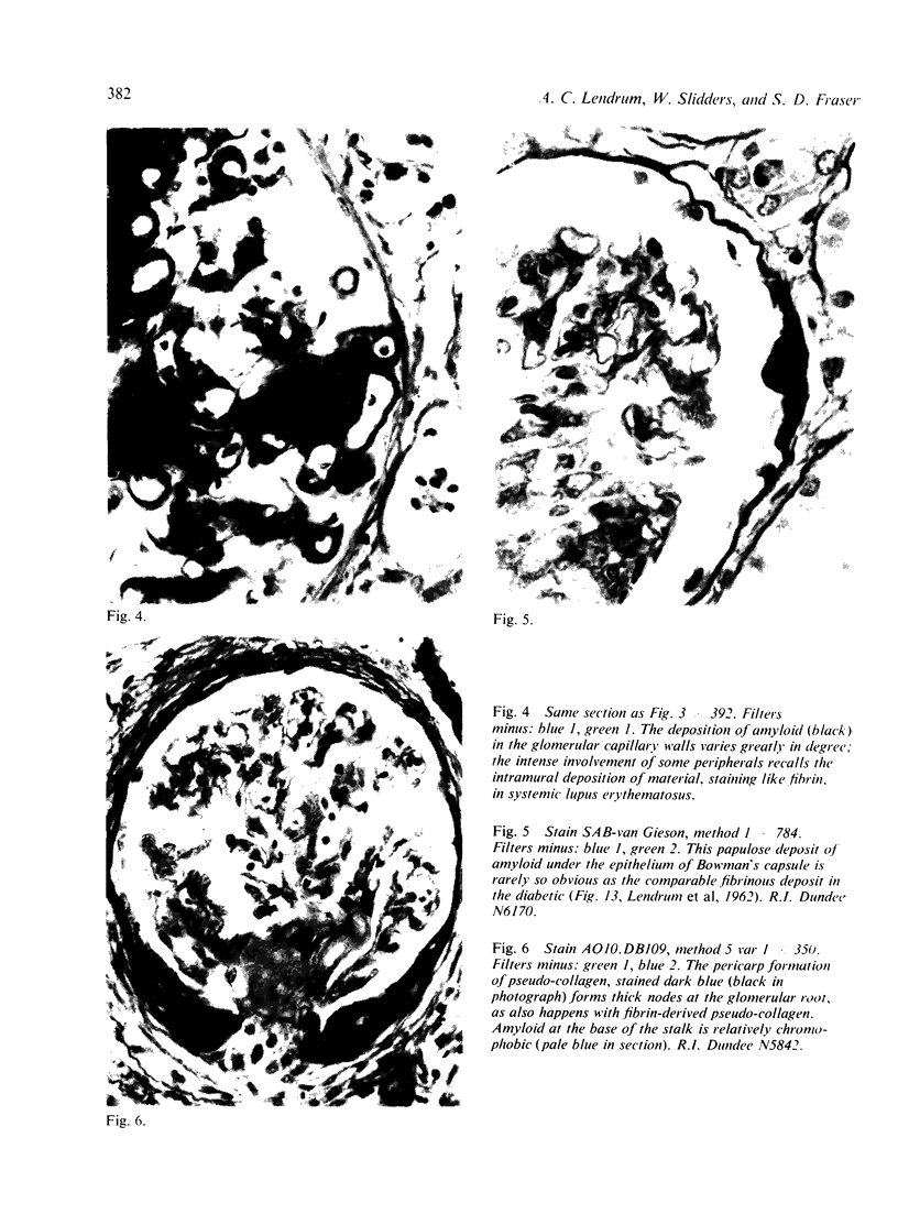
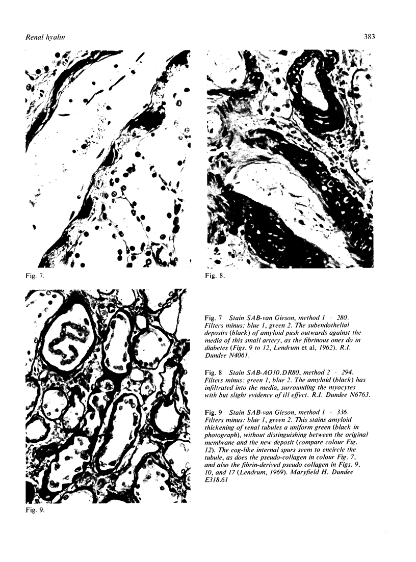
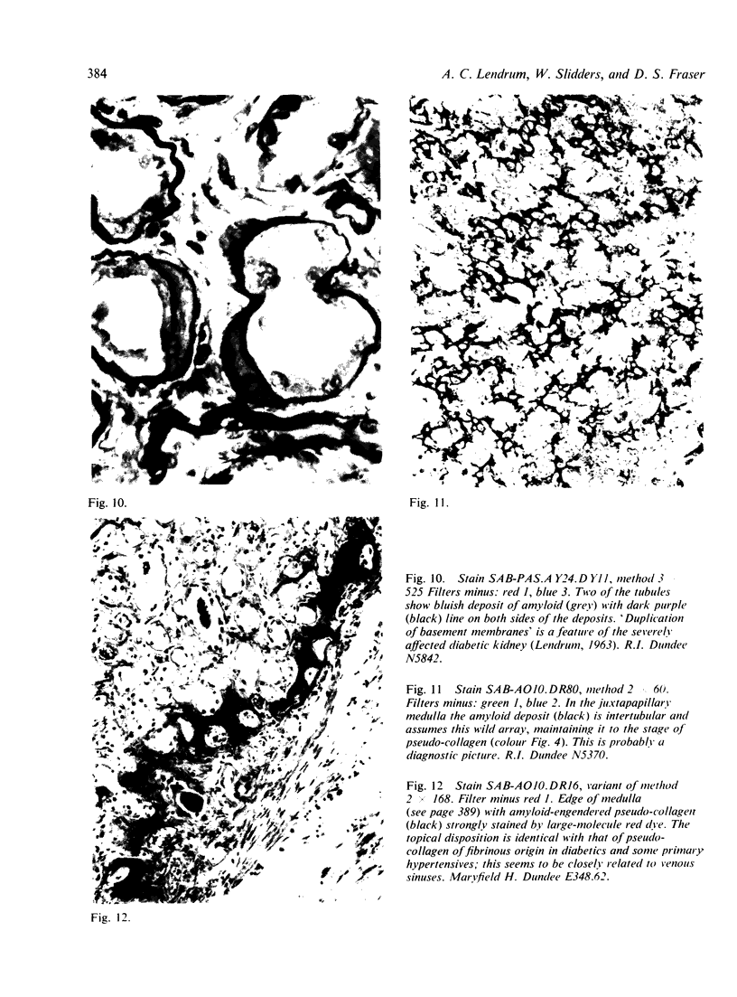
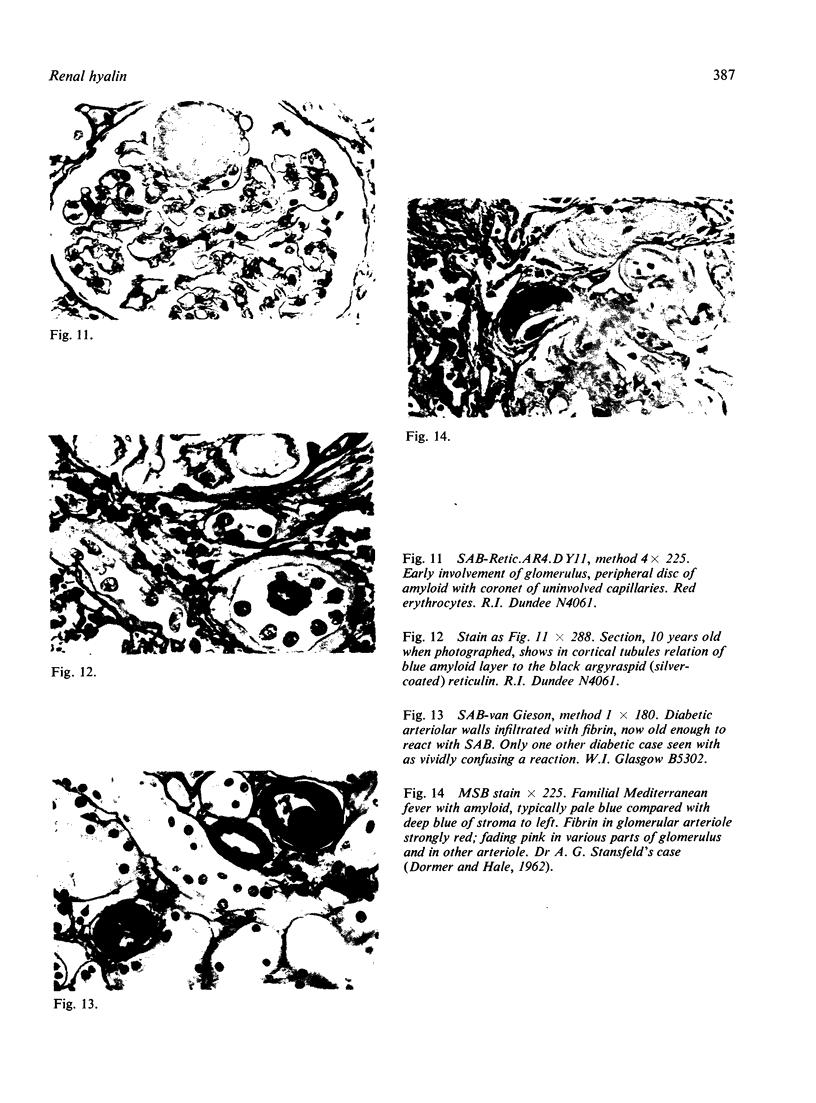
In the kidney the topical disposition of amyloid closely resembles the disposition of fibrin in the kidney of diabetics; this suggests that upset in vascular permeability plays a part in determining the site of the amyloid deposits. Furthermore, an aging process in amyloid can now be envisaged resembling the aging of extraluminal fibrin. Both materials proceed to a hyalin material that, staining like collagen, merits the name pseudo-collagen. This term we apply to a hyalin, staining like collagen, for which, we can postulate a specific precursor.
Full text
PDF

Images in this article
Selected References
These references are in PubMed. This may not be the complete list of references from this article.
- BROWN KELLY H. D., CRAIK J. E. Laryngeal nodes and the so-called amyloid tumour of the cords. J Laryngol Otol. 1952 Aug;66(8):339–358. doi: 10.1017/s0022215100047794. [DOI] [PubMed] [Google Scholar]
- CAESAR R. [Electron microscope studies on kidney amyloidosis in the golden hamster]. Frankf Z Pathol. 1963;72:506–516. [PubMed] [Google Scholar]
- CARONE F. A., EPSTEIN F. H. Nephrogenic diabetes insipidus caused by amyloid disease. Evidence in man of the role of the collecting ducts in concentrating urine. Am J Med. 1960 Sep;29:539–544. doi: 10.1016/0002-9343(60)90050-4. [DOI] [PubMed] [Google Scholar]
- Cohen A. S. The constitution and genesis of amyloid. Int Rev Exp Pathol. 1965;4:159–243. [PubMed] [Google Scholar]
- Cooper J. H. A n evaluation of current methods for the diagnostic histochemistry of amyloid. J Clin Pathol. 1969 Jul;22(4):410–413. doi: 10.1136/jcp.22.4.410. [DOI] [PMC free article] [PubMed] [Google Scholar]
- DAHLIN D. C. Primary amyloidosis, with report of six cases. Am J Pathol. 1949 Jan;25(1):105–123. [PMC free article] [PubMed] [Google Scholar]
- DORMER A. E., HALE J. F. Familial Mediterranean fever: a cause of periodic fever. Br Med J. 1962 Jan 13;1(5271):87–89. doi: 10.1136/bmj.1.5271.87. [DOI] [PMC free article] [PubMed] [Google Scholar]
- Dick G. F., Leiter L. Some factors in the development, localization and reabsorption of experimental amyloidosis in the rabbit. Am J Pathol. 1941 Sep;17(5):741–754.1. [PMC free article] [PubMed] [Google Scholar]
- Druet R. L., Janigan D. T. Experimental amyloidosis. Rates of induction, lymphocyte depletion and thymic atrophy. Am J Pathol. 1966 Nov;49(5):911–929. [PMC free article] [PubMed] [Google Scholar]
- EHRLICH J. C., RATNER I. M. Amyloidosis of the islets of Langerhans. A restudy of islet hyalin in diabetic and non-diabetic individuals. Am J Pathol. 1961 Jan;38:49–59. [PMC free article] [PubMed] [Google Scholar]
- FRENSDORFF A., SOHAR E., HELLER H. Plasma fibrinogen in familial Mediterranean fever. Ann Intern Med. 1961 Sep;55:448–455. doi: 10.7326/0003-4819-55-3-448. [DOI] [PubMed] [Google Scholar]
- Glenner G. G., Terry W., Harada M., Isersky C., Page D. Amyloid fibril proteins: proof of homology with immunoglobulin light chains by sequence analyses. Science. 1971 Jun 11;172(3988):1150–1151. doi: 10.1126/science.172.3988.1150. [DOI] [PubMed] [Google Scholar]
- HOROWITZ R. E., STUYVESANT V. W., WIGMORE W., TATTER D. FIBRINOGEN AS A COMPONENT OF AMYLOID. Arch Pathol. 1965 Mar;79:238–244. [PubMed] [Google Scholar]
- Hüttner I., Jellinek H., Kerényi T. Fibrin formations in vascular fibrinoid change in experimental hypertension: an electron microscopic study. Exp Mol Pathol. 1968 Dec;9(3):309–321. doi: 10.1016/0014-4800(68)90022-1. [DOI] [PubMed] [Google Scholar]
- ITIKAWA O., OGURA Y. Simplified manufacture and histochemical use of the Schiff reagent. Stain Technol. 1954 Jan;29(1):9–11. doi: 10.3109/10520295409115428. [DOI] [PubMed] [Google Scholar]
- JOHANSSON G. A., PFEIFFER H. H. On the amyloid-Congo red complex in histological sections and the genesis of the amyloid substance. Acta Anat (Basel) 1954;20(3):285–290. doi: 10.1159/000140904. [DOI] [PubMed] [Google Scholar]
- KENNEDY J. S. Sulphur-35 in experimental amyloidosis. J Pathol Bacteriol. 1962 Jan;83:165–181. doi: 10.1002/path.1700830120. [DOI] [PubMed] [Google Scholar]
- KING L. S. Atypical amyloid disease, with observations on a new silver stain for amyloid. Am J Pathol. 1948 Sep;24(5):1095–1115. [PMC free article] [PubMed] [Google Scholar]
- Kimmelstiel P., Wilson C. Intercapillary Lesions in the Glomeruli of the Kidney. Am J Pathol. 1936 Jan;12(1):83–98.7. [PMC free article] [PubMed] [Google Scholar]
- LENDRUM A. C., FRASER D. S., SLIDDERS W. FURTHER OBSERVATIONS ON THE AGE CHANGES IN EXTRAVASCULAR FIBRIN. Ned Tijdschr Geneeskd. 1964 Dec 5;108:2373–2373. [PubMed] [Google Scholar]
- LENDRUM A. C., FRASER D. S., SLIDDERS W., HENDERSON R. Studies on the character and staining of fibrin. J Clin Pathol. 1962 Sep;15:401–413. doi: 10.1136/jcp.15.5.401. [DOI] [PMC free article] [PubMed] [Google Scholar]
- LENDRUM A. C. Further observations on fibrinous vasculosis. Ned Tijdschr Geneeskd. 1961 Jul 8;105:1359–1360. [PubMed] [Google Scholar]
- LILLIE R. D. The allochrome procedure; a differential method segregating the connective tissues collagen, reticulum and basement membranes into two groups. Am J Clin Pathol. 1951 May;21(5):484–488. [PubMed] [Google Scholar]
- Mowry R. W., Scott J. E. Observations on the basophilia of amyloids. Histochemie. 1967;10(1):8–32. doi: 10.1007/BF00304372. [DOI] [PubMed] [Google Scholar]
- SCOTT J. E. Aliphatic ammonium salts in the assay of acidic polysaccharides from tissues. Methods Biochem Anal. 1960;8:145–197. doi: 10.1002/9780470110249.ch4. [DOI] [PubMed] [Google Scholar]
- SLIDDERS W., FRASER D. S., LENDRUM A. C. Silver impregnation of reticulin. J Pathol Bacteriol. 1958 Apr;75(2):478–481. doi: 10.1002/path.1700750233. [DOI] [PubMed] [Google Scholar]
- SLIDDERS W. The OFG and BrAB-OFG methods for staining the adenohypophysis. J Pathol Bacteriol. 1961 Oct;82:532–534. doi: 10.1002/path.1700820233. [DOI] [PubMed] [Google Scholar]
- SMITH J. F., BOLTON J. R., TURNBULL A. L. The renal complications of diabetes mellitus. J Pathol Bacteriol. 1955 Oct;70(2):475–493. doi: 10.1002/path.1700700226. [DOI] [PubMed] [Google Scholar]
- Scott J. E., Dorling J., Stockwell R. A. Reversal of protein blocking of basophilia in salt solutions: implications in the localization of polyanions using alcian blue. J Histochem Cytochem. 1968 May;16(5):383–386. doi: 10.1177/16.5.383. [DOI] [PubMed] [Google Scholar]
- Scott J. E., Quintarelli G., Dellovo M. C. The chemical and histochemical properties of Alcian Blue. I. The mechanism of Alcian Blue staining. Histochemie. 1964 Jul 17;4(2):73–85. doi: 10.1007/BF00306149. [DOI] [PubMed] [Google Scholar]
- Shirahama T., Cohen A. S. Ultrastructural studies on renal peritubular amyloid experimentally induced in guinea pigs. I. General aspects. Lab Invest. 1968 Jul;19(1):122–131. [PubMed] [Google Scholar]
- Slidders W. A stable iron-haematoxylin solution for staining the chromatin of cell nuclei. J Microsc. 1969;90(1):61–65. doi: 10.1111/j.1365-2818.1969.tb00694.x. [DOI] [PubMed] [Google Scholar]
- Still W. J., Dennison S. M. The pathogenesis of the glomerular changes in steroid-induced hypertension in the rat. Lab Invest. 1969 Mar;20(3):249–260. [PubMed] [Google Scholar]
- VASSAR P. S., CULLING C. F. Fluorescent stains, with special reference to amyloid and connective tissues. Arch Pathol. 1959 Nov;68:487–498. [PubMed] [Google Scholar]
- Wolman M. Amyloid, its nature and molecular structure. Comparison of a new toluidine blue polarized light method with traditional procedures. Lab Invest. 1971 Aug;25(2):104–110. [PubMed] [Google Scholar]