To the Editor,
Immunoglobulin G4 (IgG4) related thyroiditis is a newly defined disease characterized by extensive infiltration of lymphocytes and IgG4-positive plasmocytes, in combination with diffuse thyroid fibrosis, with or without elevated IgG4 serum levels [1]. Although subacute thyroiditis is the most common cause of thyroid pain, IgG4-related thyroiditis is regarded as a distinct diagnosis, with patients often experiencing severe discomfort. Despite differences in the pathology of the disease, it is often hard to differentiate IgG4-related thyroiditis from other thyroid diseases before thyroidectomy. IgG4 immunostaining is currently regarded as the gold standard for diagnosis of IgG4-related thyroiditis, particularly as the clinical diagnostic criteria of IgG4-related thyroiditis have yet to be established [1]. Pain is not essential for the diagnosis of IgG4-related thyroiditis, although in our case described here, it was a significant concern and the primary factor underlying this study.
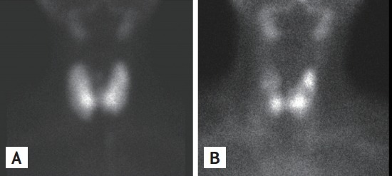
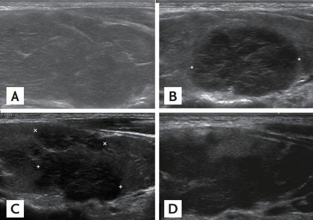
A 35-year-old woman was referred to our hospital from a local clinic due to thyroid dysfunction in October 2011. Her initial thyroid function test in August 2011 showed triiodothyronine (T3) 1.81 ng/mL, free thyroxine (FT4) 2.14 ng/dL, thyroid stimulating hormone (TSH) < 0.01 mIU/mL, anti-thyroid peroxidase antibody (anti-TPO) 694.42 IU/mL (normal range, 0 to 34), and anti-thyroglobulin antibody (anti-Tg) > 2,000 IU/mL (normal range, 0 to 115). The patient was treated with 150 mg/day propylthiouracil (PTU) for 1 month. At the time of follow-up in September 2011, the patient’s T3 and FT4 levels had fallen to 0.29 ng/mL and 0.43 ng/dL, respectively, and TSH was > 150 μIU/mL. PTU therapy was then discontinued for 1 month, after which the patient complained of dizziness, constipation, edema, fatigue, and pantalgia. Upon admission to our hospital, she presented with soft and diffuse goiter conditions with minimal tenderness. Her laboratory results were as follows: T3 0.72 nmol/L, FT4 0.36 ng/dL, TSH > 100 μIU/mL, anti-TPO 258.3 IU/mL, anti-Tg > 4,000 IU/mL, and thyrotropin binding inhibitory immunoglobulin 1.25 IU/L (normal range, 0 to 1.75). Thyroid scans showed diffuse goiter conditions with increased radionuclide uptake (Fig. 1A). Thyroid ultrasounds showed mild goiter conditions with slightly coarse echogenicity, lacking a definite nodule (Fig. 2A). The patient’s thyroid function eventually normalized following treatment with levothyroxine, which relieved her of the symptoms of hypothyroidism; however, significant thyroid pain had returned by March 2012.
Figure 1.

Thyroid scan. (A) Thyroid scan showing diffuse goiter with increased radionuclide uptake in October 2011. (B) Thyroid scan showing multiple hot uptakes with intervening low uptakes in January 2013.
Figure 2.

Ultrasound image of the right thyroid lobe. (A) Thyroid ultrasonography showed only diffuse coarse echogenicity in October 2011. (B) A low-echoic area was observed in March 2012. (C) The low-echoic area encroached in June 2012. (D) The low-echoic area finally expanded to encompass nearly the entire lobe in January 2013.
Upon reexamination, the right lobe of her thyroid was nodular, hard, and tender. Thyroid function was normal, and her white blood cell count and erythrocyte sedimentation rate were 10,100/mm3 and 18 mm/hr, respectively. Thyroid ultrasound showed hypoechoic and hypovascular area in the right lobe (Fig. 2B). Analgesics and 20 mg/day prednisolone were prescribed. After 3 days of steroid treatment, her thyroid nodule started to decrease in size and soften, and pain was alleviated. After 2 weeks of steroid treatment, steroid use was stopped through rapid tapering, but analgesics were maintained. Within 2 weeks of steroid discontinuation, the thyroid gland had again enlarged and become tender, which necessitated continuous administration of 7.5 to 20 mg prednisone for pain relief. In June 2012, FT4 and TSH levels were 1.93 ng/mL and 0.118 μIU/mL, respectively, and levothyroxine was stopped. A follow-up thyroid ultrasound showed progressive and geographic enlargement of the hypoechoic area in both lobes (Fig. 2C). Thyroid fine-needle aspiration cytology revealed follicular cell clusters with oncocytic changes in a lymphoplasma cell background. Her thyroid function test was consistently normal. In January 2013, a follow-up thyroid scan showed multiple hot uptakes with intervening low uptakes (Fig. 1B). A subsequent follow-up ultrasound revealed that the hypoechoic area occupied nearly the entire thyroid (Fig. 2D).
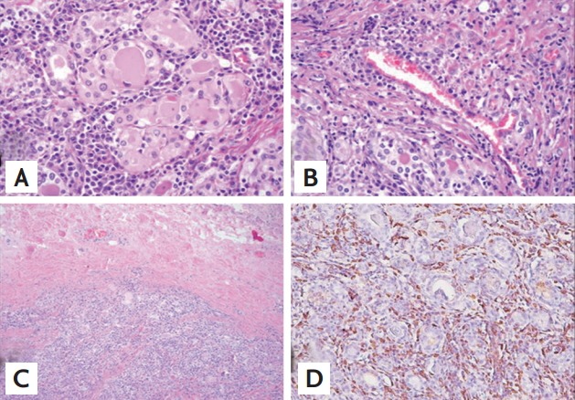
In February 2013, she underwent total thyroidectomy because of steroid tapering failure and an appearance resembling iatrogenic Cushing syndrome features. The tissue pathology revealed severe inflammation and fibrosis. Although the inflammatory infiltrates were predominantly plasmocytes (Fig. 3A), numerous phlebitis foci were also observed (Fig. 3B). The thyroid capsule was thickened with extracapsular fibrosis (Fig. 3C), and IgG4 immunohistochemistry revealed that the infiltrative plasmocytes were mainly IgG4-positive cells (Fig. 3D). In May 2013, she discontinued prednisolone through tapering, and her thyroid function was normal upon treatment with 150 μg/day levothyroxine.
Figure 3.

Pathological findings of the resected thyroid gland. (A) High-power view showing massive lymphocyte and plasmocyte infiltration and fibrosis among destructive follicles, including oncocytic follicular cells (H&E, ×400). (B) Phlebitis is observed (H&E, ×400). (C) Low-power view showing thickening of the thyroid capsule with extracapsular fibrosis (H&E, ×100). (D) Immunohistochemical stain of immunoglobulin G4 (IgG4; 1:2,000 dilution, Abcam, UK) reveals multiple IgG4-positive plasma cells (×200)
IgG4-related thyroiditis is clinically different from non-IgG4-related thyroiditis, characterized by earlier onset, lower female to male ratio, more rapid and aggressive disease course, and higher levels of circulating thyroid autoantibodies compared with non-IgG4-related thyroiditis [1]. Other potential diagnostic factors include anti-Tg and anti-TPO, which are produced by thyroid-infiltrating lymphocytes and plasmocytes [2]. Anti-Tg may be important for differentiating IgG4-related thyroiditis from subacute thyroiditis, which is the major cause of thyroid pain. Anti-TPO is detected in 95% of Hashimoto thyroiditis (HT) cases [2] and consists primarily of IgG1 [2], a subclass known to play an important role in inflammation and a powerful mediator of antibody-dependent cell mediated cytotoxicity [2]. In contrast, the IgG4 subclass seen has a much lower affinity for target antigens and is therefore considered a marker of chronic antigen exposure [3]. Repeated antigenic exposure might induce production of anti-TPO IgG4 antibodies [3]. Proper differentiation between IgG subclasses is particularly important in thyroid diseases. Although serum levels of IgG and IgG4 were not significantly different between IgG4-related thyroiditis and non-IgG4-related thyroiditis, the ratio of IgG4/IgG may help in their differentiation [2]. Unfortunately, such a measurement could not be made in this case, as the patient was diagnosed after thyroidectomy.
IgG4-related systemic diseases appear to respond well to glucocorticoid therapy. In this case, following steroid treatment not only her painful thyroid nodule was rapidly regressed but also her thyroid function was recovered. Therefore, a precise and early diagnosis of IgG4-related thyroiditis is important for adequate follow-up and treatment.
Cytological analysis revealed lymphocyte and plasmocyte infiltrations with Hürthle cell changes, in clear contrast to painful HT, which is traditionally characterized by reactive lymphoid cell infiltrations, with occasional Hürthle cell changes and a few multinucleate histiocytes [4,5]. In subacute thyroiditis, numerous multinucleated giant cells are usually seen, whereas Hürthle cell changes and plasmocytes are usually not.
Despite the incidence and clinical significance of IgG4-related thyroiditis, the clinical diagnostic criteria for this disease have yet to be established. IgG4 immunohistochemistry provides a definitive diagnosis in patients with suspected IgG4-related thyroiditis [2], characterized by a pronounced infiltration of lymphocytes and IgG4-producing plasmocytes (> 20 cells per high powered field), extensive fibrosis, and obliterative phlebitis. Additionally, the thyroid parenchyma of our case revealed islands of micro-follicles that had abundant pale-pink colored cytoplasm and mild nuclear atypia. This phenotype is similar to that of Riedel’s thyroiditis, which exhibits obliterative phlebitis, a mixed infiltrate of lymphocytes, eosinophils, and IgA-positive plasmocytes, and global or partial fibrosis extending beyond the thyroid capsule and into surrounding tissues [4].
In light of this and other cases, painful IgG4-related thyroiditis should be a consideration in the differential diagnosis of painful thyroid disorders and conditions. Measurement of serum IgG4 and IgG levels may help in the diagnosis of IgG4-related thyroiditis before a decision is made to undergo surgical treatment, such as thyroidectomy, which may be necessary for the resolution of symptoms and the establishment of a diagnosis in IgG4-related thyroiditis that does not respond to steroid treatment. The case described here represents the first report in Korea of IgG4-related thyroiditis in a patient suffering from recurrent thyroid pain. The identification of painful IgG4-related thyroiditis offers new insights into not only patient treatment, but also development of new diagnostic and therapeutic approaches for this rapidly progressing destructive subtype of thyroiditis.
Footnotes
No potential conflict of interest relevant to this article was reported.
REFERENCES
- 1.Li Y, Nishihara E, Hirokawa M, Taniguchi E, Miyauchi A, Kakudo K. Distinct clinical, serological, and sonographic characteristics of hashimoto’s thyroiditis based with and without IgG4-positive plasma cells. J Clin Endocrinol Metab. 2010;95:1309–1317. doi: 10.1210/jc.2009-1794. [DOI] [PubMed] [Google Scholar]
- 2.Zhang J, Zhao L, Gao Y, et al. A classification of Hashimoto’s thyroiditis based on immunohistochemistry for IgG4 and IgG. Thyroid. 2014;24:364–370. doi: 10.1089/thy.2013.0211. [DOI] [PubMed] [Google Scholar]
- 3.Xie LD, Gao Y, Li MR, Lu GZ, Guo XH. Distribution of immunoglobulin G subclasses of anti-thyroid peroxidase antibody in sera from patients with Hashimoto’s thyroiditis with different thyroid functional status. Clin Exp Immunol. 2008;154:172–176. doi: 10.1111/j.1365-2249.2008.03756.x. [DOI] [PMC free article] [PubMed] [Google Scholar]
- 4.Papi G, LiVolsi VA. Current concepts on Riedel thyroiditis. Am J Clin Pathol. 2004;121 Suppl:S50–S63. doi: 10.1309/NUU88VAFR9YEHKNA. [DOI] [PubMed] [Google Scholar]
- 5.Kim HK, Shin HJ, Kang HC. A case of painful Hashimoto’s thyroiditis successfully treated with total thyroidectomy. J Korean Endocr Soc. 2008;23:438–443. [Google Scholar]


