Abstract
The radical S-adenosyl-l-methionine (SAM) superfamily is a large and growing group of enzymes that carry out complex radical-mediated transformations. A one-electron reduction of SAM via the +1 state of the cubane [4Fe-4S] cluster generates a 5′-deoxyadenosyl radical, which initiates turnover. The [4Fe-4S] cluster must be reduced from its resting +2 to the catalytically active +1 oxidation state by an electron. In practice, dithionite or the Escherichia coli flavodoxin (EcFldA)/ferredoxin (flavodoxin):NADP+ oxidoreductase (Fpr)/NADPH system is used. Herein, we present a systematic investigation of the reductive activation of the radical SAM enzyme CDG synthase (BsQueE) from Bacillus subtilis comparing biological and chemical reductants. These data show that either of the flavodoxin homologs encoded by the B. subtilis genome, BsYkuN or BsYkuP, as well as a series of small molecule redox mediators, support BsQueE activity. With dithionite as a reductant, activity of BsQueE is ~75-fold greater in the presence of BsYkuN and BsYkuP compared to dithionite alone. By contrast, EcFldA supports turnover to ~10-fold greater levels than dithionite alone under the same conditions. Comparing the ratio of the rate of turnover to the apparent binding constant for the flavodoxin homologs reveals 10- and 240-fold preference for BsYkuN over BsYkuP and EcFldA respectively. The differential activation of the enzyme cannot be explained by the abortive cleavage of SAM. We conclude from these observations that the differential activation of BsQueE by Fld homologs may reside in the details of the interaction between the flavodoxin and the radical SAM enzyme.
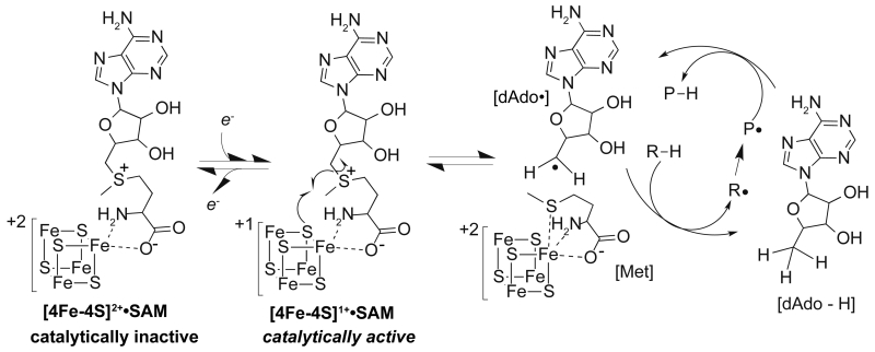
The radical S-adenosyl-l-methionine (SAM) superfamily is a rapidly expanding group of enzymes that catalyze a myriad of biological transformations in primary and secondary metabolism, including cofactor biosynthesis/assembly and RNA modifications. Despite the rich functional diversity, the majority of the members of the radical SAM superfamily engage the three cysteine thiolates in the conserved CX3CX2C motif to coordinate three irons of a catalytically essential [4Fe-4S] cluster.1 The fourth iron is coordinated by the α-carboxylate and α-amino moieties of the SAM. In nearly all radical SAM enzymes described to date, the +1 state of the [4Fe-4S] cluster supplies one electron to SAM generating a 5′-deoxyadenosyl radical (dAdo•) and l-methionine.2,3 (Scheme 1).
Scheme 1.
Reductive cleavage of SAM.
Reduction of the [4Fe-4S] cluster to the +1 oxidation state from the resting +2 form is common to all members of the radical SAM superfamily. This one electron reduction requirement was first reported for pyruvate formate-lyase activating enzyme and lysine 2,3-aminomutase nearly 30 years prior to the unification of the radical SAM superfamily.4,5 Although the biological reductant has never been directly identified, Knappe and co-workers determined that pyruvate formate-lyase activating enzyme isolated from Escherichia coli was reduced by E. coli flavodoxin (EcFldA), suggesting a role for this ubiquitous protein in the reductive activation process.4,6 In addition, EcFldA reductively activates E. coli anaerobic ribonucleotide reductase and biotin synthase by supplying reducing equivalents from NADPH via ferredoxin (flavodoxin):NADP+ reductase (Fpr).7-10 Subsequent studies on additional radical SAM enzymes from E. coli with EcFldA/Fpr and NADPH showed this to be a general feature of these proteins.11,12 Interestingly, Brazeau et al. identified that EcFldA and Fpr were capable of reductively activating lysine 2,3-aminomutase from Porphyromonas gingivalis, demonstrating that the EcFldA and Fpr proteins could reductively activate a radical SAM enzyme from a different species.13 Though EcFldA/Fpr reductively activate radical SAM enzymes from diverse organisms, there has yet to be a systematic study on reductive activation. The closest systematic investigation comparing dithionite and EcFldA/Fpr was reported in two studies by Booker and coworkers on the anaerobic sulfatase maturating enzymes, AtsB and anSMEcpe. The Vmax/[ET] for each enzyme was 10- to 100-fold greater when dithionite was used as the reductant compared to the E. coli biological system.14,15 This observation underscores the need to more closely examine interactions between radical SAM enzymes and in vivo reducing systems.
7-Carboxy-7-deazaguanine (CDG) synthase (QueE) is a member of the radical SAM superfamily that catalyzes the radical-mediated ring contraction of 6-carboxy-5,6,7,8-tetrahydropterin (CPH4) to form CDG.16 Recent investigations on QueE homologs from Burkholderia multivorans and Bacillus subtilis have yielded rich structural and functional insights into the mechanism of the ring contraction.17,18 Additionally, the fact that both dithionite and the E. coli biological reducing system can supply reducing equivalents to B. subtilis QueE (BsQueE) in vitro provides an ideal baseline to systematically investigate reductive activation.17,18 In this paper we compare the activity of BsQueE in the presence of various chemical and biological reductants, highlighting the importance of the reducing system in studying radical SAM enzymes.
EXPERIMENTAL PROCEDURES
Cloning, expression, and protein purification
His6-tagged BsQueE and the His6-tagged E. coli flavodoxin reductase (Fpr) used in this investigation were cloned, expressed, and purified as previously described.16,17 The genes ykuN and ykuP, which encode the two flavodoxin homologs from B. subtilis subsp. subtilis str. 168, were amplified from genomic DNA via PCR using primers (Supplemental Table 1) that incorporated NdeI and XhoI for ykuN or NheI and XhoI for ykuP at the 5′- and 3′-ends of each gene, respectively. The fldA gene was amplified from E. coli W3110 genomic DNA via PCR using primers (Supplemental Table 1) that incorporated NdeI and EcoRI restriction sites at the 5′- and 3′-ends of the gene, respectively. The purified PCR products were A-tailed and ligated into pGEM Easy-T vector (Promega) for blue-white screening and sequencing. The corresponding inserts were digested from the pGEM Easy-T vector with NdeI/XhoI (ykuN) or NdeI/EcoRI (fldA) and ligated into pET29a digested in with the same enzymes. The ykuP gene was digested from the pGEM vector with NheI/XhoI and ligated into similarly digested pET21b (Novagen). Sequencing confirmed the identity of each construct prior to overexpression.
BL21(DE3) cells (Novagen) containing the ykuN-pET29a or fldA-pET29a constructs were grown in LB supplemented with 34 μg/mL kanamycin at 37 °C. HMS-174 (DE3) cells containing the ykuP-pET21b construct were grown in LB supplemented with 100 μg/mL ampicillin at 37 °C. Protein expression was induced with the addition of IPTG to a final concentration of 0.1 mM at OD600~0.5. The media was also supplemented with 0.1 mM riboflavin (final concentration) at the time of induction. Cells were harvested after ~18 h, flash frozen in liquid nitrogen, and stored at −80 °C until purification.
All chromatography was carried out at 4 °C. Frozen cells (1 g) were suspended in ~3 mL lysis buffer containing 50 mM Tris•HCl (pH 7.2), 10 mM DTT, and 1 mM EDTA. PMSF was added to a final concentration of 1 mM. Cells were lysed using via a Branson digital sonifier operated at 50% amplitude. The lysate was centrifuged at 18,000 ×g for 35 min and the supernatant was loaded onto a DEAE-sepharose column (2.6 cm × 12.5 cm, GE Lifesciences) equilibrated in lysis buffer. The column was washed with lysis buffer and protein was eluted using a linear gradient of 0 – 1 M KCl in the same buffer. Fractions containing flavodoxin were identified based on SDS-PAGE analysis, pooled, and dialyzed overnight against buffer containing 50 mM HEPES•NaOH (pH 7.4), 0.1 M KCl and 10 mM DTT. At this stage, EcFldA was >95 % pure and so it was concentrated and aliquots were flash frozen in liquid nitrogen.
BsYkuN and BsYkuP were further purified using hydrophobic interaction chromatography as follows. Fractions containing BsYkuN were pooled and prepared for hydrophobic interaction chromatography by adding solid ammonium sulfate to a final concentration of 1.5 M over 1 h. The solution was loaded onto a butyl sepharose column (2.6 cm × 12.5 cm, GE Lifesciences) equilibrated in 50 mM Tris•HCl (pH 7.2), 2 mM DTT, 1 mM EDTA, and 1.5 M ammonium sulfate. The column was washed with loading buffer and the protein was eluted with a linear gradient of 1.5 – 0 M ammonium sulfate. Fractions containing BsYkuN were pooled and dialyzed overnight at 4 °C against 20 mM HEPES•NaOH (pH 7.5) containing 100 mM KCl, 2 mM DTT. BsYkuP was purified essentially as described above except the ammonium sulfate concentration was 2 M, the column was equilibrated in buffer containing 2 M ammonium sulfate and eluted with a linear gradient from 2 to 0 M.
BsYkuN and BsYkuP were concentrated to a minimal volume and 1 mL fractions were injected onto a HiPrep 16/60 Sephacryl S200 high-resolution size exclusion column (GE Lifesciences) for further purification. The column was equilibrated with dialysis buffer and the flavodoxins were eluted isocractically running at 0.5 mL/min. Fractions containing the proteins were pooled based on color and SDS-PAGE. Each flavodoxin was concentrated to a minimal volume and flash frozen in liquid nitrogen.
EcFldA, BsYkuN, and BsYkuP were quantified based on A467nm and using the extinction coefficient of ε467=8250 M−1cm−1 (EcFldA), ε467=10700 M−1cm−1(BsYkuN), and ε467=9930 M−1cm−1 (BsYkuP).19 The extinction coefficients of BsYkuN and BsYkuP were experimentally determined in triplicate as follows. BsYkuN and BsYkuP were diluted separately into 0.1 M potassium phosphate (pH 7.3) to four different concentrations such that the measured absorbance of each dilution was between 0.1 and 0.8 at 467 nm. The UV-visible spectrum of each sample was measured on an Agilent 8453 diode array spectrophotometer. Next, aliquots (135 μL) of each dilution were treated with 15 μL of 30% (w/v) TCA to acidify the solution and boiled for 10 min to denature the flavodoxin and release the FMN cofactor. The boiled samples were centrifuged at 17,000 ×g for 10 minutes to remove the precipitated protein. The absorbance of the supernatant was measured at 445 nm to determine the amount of free FMN in solutions. The concentration of free FMN was calculated using the ε445nm = 12500 M−1cm−1 factoring in the dilution due to the addition of TCA. The [FMN] measured after the addition of TCA and boiling is equivalent to the [BsYkuN] or [BsYkuP] with bound FMN. The absorbance measured at 467 nm from BsYkuN or BsYkuP in 0.1 M potassium phosphate (pH 7.3) was plotted versus the calculated [BsYkuN] or [BsYkuP] to calculate the molar extinction coefficient of the flavodoxins. FMN from Sigma was diluted in 0.1 M potassium phosphate (pH 7.3) and acid treated and boiled in the same manner to ensure that the extinction coefficient of FMN at 445 nm does not change during the acid treatment and boiling.
Synthesis of substrates
SAM and CPH4 were enzymatically synthesized as described previously.17
Time course using dithionite as reductant
All kinetic experiments described below monitor the turnover of BsQueE by measuring the rate of CDG formation in triplicate. The reactions were setup under anaerobic conditions (95% N2, 5% H2 atmosphere in a Coy anaerobic chamber) and contained 50 mM PIPES•NaOH (pH 7.4), 10 mM DTT, 2 mM MgSO4, 2 mM SAM, 2 mM CPH4, 2 μM BsQueE and 10 mM dithionite. All components except CPH4 were mixed and incubated at room temperature for 10 min. Turnover was initiated upon addition of CPH4. Aliquots (45 μL) were withdrawn at various times and quenched by mixing with 4.5 μL of 30% (w/v) TCA.
Aliquots of the reaction were analyzed by HPLC as follows. A sample from each reaction mixture was injected onto an Eclipse XDB-C18 reverse phase column (Agilent) and eluted with a linear gradient from 0-10% acetonitrile with 0.1% (v/v) TFA. The peak corresponding to CDG was integrated and quantified by comparison with peaks obtained at known concentrations of a CDG standard.
Time course with biological reducing system
The reactions with the biological reducing system were carried out as described above for dithionite with minor modifications as follows. Reactions contained 50 mM PIPES•NaOH (pH 7.4), 10 mM DTT, 2 mM MgSO4, 2 mM SAM, 2 mM CPH4, 1 μM BsQueE, 2 mM NADPH, and either 2 μM Fpr/5 μM BsYkuN, 5 μM Fpr/20 μM BsYkuP, or 5 μM Fpr/20 μM EcFldA. All components except CPH4 were mixed and incubated at room temperature for 10 min. The reaction was initiated upon addition of CPH4. Aliquots (60 μL) were withdrawn at various times and quenched by mixing with 6 μL of 30% (w/v) TCA. CDG was quantified as described above. Negative controls for each flavodoxin were run in the same manner except either NADPH or Fpr was omitted from each reaction.
Initial rate of formation of CDG as a function of [Fld] was fit to Eqn. 1. The Vmax (kCDG) and KFld values from several datasets were averaged to obtain the reported standard deviations.
| (Eqn. 1) |
Flavodoxin dependence of BsQueE activity with Fpr/NADPH as reductant
Reactions contained 50 mM PIPES•NaOH (pH 7.4), 10 mM DTT, 2 mM MgSO4, 2 mM SAM, 2 mM CPH4, 1 μM BsQueE and 2 mM NADPH. The concentrations of Fpr (1 μM or 5 μM for BsYkuN and 5 μM or 20 μM for BsYkuP and EcFldA) and each flavodoxin (0.39 μM, 0.77 μM, 3.9 μM, 7.7 μM, or 23.1 μM for BsYkuN, 0.42 μM, 0.83 μM, 4.2 μM, 8.3 μM, or 24.9 μM for BsYkuP, and 1 μM, 5 μM, 10 μM, 20 μM, and 40 μM for EcFldA) were varied. All components except CPH4 were mixed and incubated at room temperature for 10 min. The reaction was initiated upon addition of CPH4. Each reaction was set up to a final volume of 60 μL and quenched 1 min after addition of CPH4 by mixing with 6 μL of 30% (w/v) TCA. An aliquot of each reaction mixture was analyzed by HPLC for CDG as described above.
Flavodoxin dependence of BsQueE activity with dithionite as reductant
The reactions with flavodoxin using dithionite as reductant were carried out as described above with minor modifications as follows. Reactions mixtures contained 50 mM PIPES•NaOH (pH 7.4), 10 mM DTT, 2 mM MgSO4, 2 mM SAM, 2 mM CPH4, 1-2 μM BsQueE, 2 mM NADPH, 10 mM dithionite, and various concentrations (0 – 30 μM) of flavodoxin. All components except CPH4 were mixed and incubated at room temperature for 10 min. The reaction was initiated upon addition of CPH4. Each reaction was set up to a final volume of 60 μL and quenched 1 min after addition of CPH4 by mixing with 6 μL of 30% (w/v) TCA. CDG was quantified as described above.
Methyl viologen dependence of BsQueE activity with dithionite as reductant
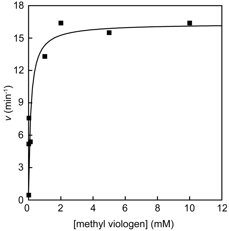
To examine if methyl viologen could replace flavodoxin as a mediator in the reaction BsQueE activity was assayed as follows. Reactions contained 50 mM PIPES•NaOH (pH 7.4), 10 mM DTT, 2 mM MgSO4, 2 mM SAM, 2 mM CPH4, 1-2 μM BsQueE, 10 mM dithionite, and 0 – 10 mM methyl viologen. All components except CPH4 were mixed and incubated at room temperature for 10 min. The reaction was initiated upon addition of CPH4. Each reaction was set up to a final volume of 60 μL and quenched by mixing with 6 μL of 30% (w/v) TCA. CDG was quantified as described above. The initial rate of CDG formation as a function of [methyl viologen] was fit to Eqn. 2 to obtain the Vmax (kCDG) value from triplicate data sets, which was averaged to obtain the reported standard deviation.
| (Eqn. 2) |
Redox mediator dependence of 5′-deoxyadenosine and CDG formation
Reactions contained 50 mM PIPES•NaOH (pH 7.4), 10 mM DTT, 2 mM MgSO4, 2 mM SAM, 2 mM CPH4, 2 μM BsQueE, 10 mM dithionite, and either no mediator, 2 mM methyl viologen (Em=−0.446 V)20, 2 mM benzyl viologen (Em=−0.359 V)20, 1 mM neutral red (Em=−0.325 V)21, or 1 mM lissamine blue BF (Em=−0.276 V, pH 7.5).22 All components except CPH4 were mixed and incubated at room temperature for 10 min. The reaction was initiated by addition of CPH4. Aliquots (65 μL) from the 420 μL total reaction mixture were withdrawn at 0.25, 0.5, 0.75, 1, 1.5 and 2 min and quenched by mixing with 6.5 μL of 30% (w/v) TCA. 5′-Deoxyadenosine formed was quantified with the same HPLC separation method described above for CDG.
RESULTS
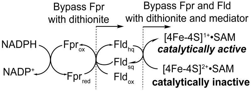
All members of the radical SAM superfamily require the one electron reduction of the [4Fe-4S]2+ cluster to the +1 oxidation state. This can be achieved by using a chemical reductant, such as dithionite, or a biological reducing system where the reducing equivalents are derived from NADPH and transferred to the cluster via flavodoxin reductase and flavodoxin (Scheme 2). Previous investigations on BsQueE showed that reducing equivalents could be derived from either dithionite or NADPH via the E. coli Fpr/FldA reducing system, with the biological system acting as the better reductant.17 While the E. coli Fpr/FldA/NADPH system is capable of reductively activating BsQueE, not all members of the radical SAM superfamily can be activated by this system. The fact that BsQueE is reductively activated by both dithionite and the E. coli biological reducing system affords an opportunity to further investigate the reductive activation step of a radical SAM enzyme.
Scheme 2.
Electron flow for [4Fe-4S] cluster reduction.a
a The scheme shows the flow of electrons and does not imply absolute stoichiometry.
Activation of CDG synthase: biological vs chemical reduction
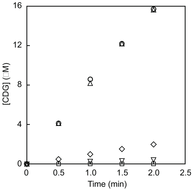
While dithionite and the E. coli FldA/Fpr/NADPH system activated BsQueE, the in vivo reductant for this enzyme has yet to be determined. The B. subtilis genome contains two orfs, ykuN and ykuP, that encode homologs of the EcFldA. We hypothesized that either one or both of these enzymes may support reductive activation of BsQueE, where the reducing equivalents would be derived from NADPH via Fpr.23 To test this hypothesis the genes ykuN and ykuP were cloned, the proteins encoded by each gene were heterologously expressed in E. coli, and BsYkuN and BsYkuP were purified to homogeneity. Initial experiments tested multiple conditions for reductive activation: dithionite alone, NADPH/Fpr/EcFldA, NADPH/Fpr/BsYkuN, and NADPH/Fpr/BsYkuP (Fig. 1); in these experiments, the E. coli Fpr was used to couple NADPH oxidation to cluster reduction.24 The results show that although BsQueE is active with dithionite (0.2 min−1), 75-fold higher rates are observed in assays containing the B. subtilis Fld homologs. Moreover, the BsFld homologs support turnover to 10-fold greater than EcFldA. Control experiments where either NADPH or Fpr was omitted exhibited no detectable activation of BsQueE. Collectively, these results support the notion depicted in Scheme 2 that flavodoxin mediates electron transfer to BsQueE and highlight a differential capacity for Fld homologs to support QueE activity.
Figure 1.
Time course of BsQueE reaction under various reducing conditions. Activity was monitored by measuring CDG formation. BsQueE activity was measured in the presence 10 mM dithionite (upside-down triangle) or 2 μM Fpr/2 mM NADPH with 5 μM BsYkuN (circle), 5 μM Fpr/2 mM NADPH with 20 μM BsYkuP (triangle), or 5 μM Fpr/2 mM NADPH with 20 μM EcFldA (diamond). Assays where Fpr or NADPH (square) were omitted served as negative controls.
Investigation of the flavodoxin-dependent activation of BsQueE
The simplest interpretation of the differential activation of BsQueE by E. coli and B. subtilis Fld homologs shown in Fig. 1 is that the BsFld homologs are optimized for activation of the cognate radical SAM enzyme. However, there are two factors, the Fld•BsQueE protein-protein interaction and the reduction potentials of the FMN cofactor of Fld and the [4Fe-4S]2+ cluster of BsQueE, which can contribute to the flavodoxin-dependent reductive activation. There is only limited data in the literature on the binding of flavodoxin to a protein. In methionine synthase, Matthews and coworkers demonstrated by NMR and cross-linking that flavodoxin interacts with the activation module of the protein.25,26
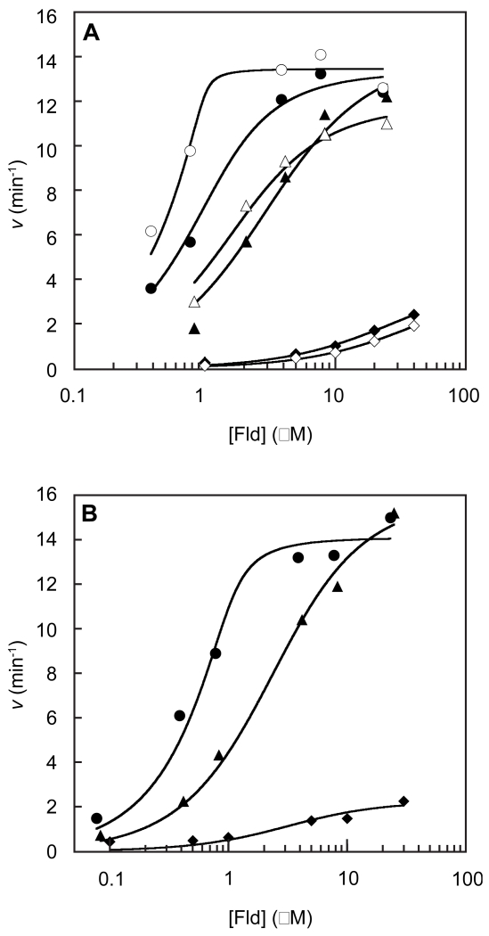
To probe the selectivity of Fld homologs in supporting turnover by BsQueE we examined the concentration-dependence of activation at various Fld/Fpr levels. We reasoned that at low concentrations of Fld the Fld•BsQueE interaction will dominate reductive activation, while at saturating concentrations of Fld the reduction potentials will be limiting. For simplicity, the concentration of Fpr was kept constant at two concentrations (4-5 fold apart), that were both saturating, to isolate the Fld-dependence of BsQueE activity. Very similar saturation profiles were obtained for each Fld homolog at both Fpr concentrations (Fig. 2A). The [Fld] dependence exhibits saturation kinetics and was analyzed accordingly. The fits to the experimental data are summarized in Table 1. Both of the B. subtilis Fld homologs are more effective in supporting turnover by BsQueE than the EcFldA. Moreover, BsYkuN exhibits a KFld for binding to BsQueE that is substantially lower than for BsYkuP and EcFldA. The data with BsYkuN are not fit as well as those with EcFldA and BsYkuP because the lowest concentration of BsYkuN employed (~0.4 μM) was clearly above the half maximal saturation. Nevertheless, we would conservatively place an upper limit of KBsYkuN at <0.4 μM. The ratio of kCDG/KFld obtained by fitting the data for each Fld homolog provides a measure of efficiency of Fld for activating BsQueE (Table 1). The data show that BsYkuN is the most efficient activator of BsQueE by at least 10-fold relative to BsYkuP and greater than 200-fold relative to EcFldA, perhaps indicating the absence of optimized interaction surfaces between the E. coli flavodoxin and BsQueE.
Figure 2.
BsQueE activity is enhanced by increasing the flavodoxin homologs [EcFldA(diamonds)], [BsYkuN (circles)], or [BsYkuP (triangles)] concentration when the electrons are supplied by either NADPH via Fpr (A) or dithionite (B). To confirm Fld reduction by NADPH/Fpr was saturating, the rate of CDG formation dependent on [Fld] was measured at 1 μM (closed) and 5 μM (open) Fpr for BsYkuN and 5 μM (closed) and 20 μM (open) for BsYkuP and EcFldA (A). Depicted are representative data sets for each experiment. The data were fit using Eqn 1 and the Vmax (kCDG) ant KFld are reported in Table 1.
Table 1.
BsQueE kinetic parameters determined using various reduction conditions.
| EcFldA | BsYkuN | BsYkuP | |||||||
|---|---|---|---|---|---|---|---|---|---|
|
|
|||||||||
| Dithionite | 5 μM Fpr | 20 μM Fpr | Dithionite | 1 μM Fpr | 5 μM Fpr | Dithionite | 5 μM Fpr | 20 μM Fpr | |
| KFld (μM) | 3.9 ± 1.4 | 29.0 ± 3.0 | 42.0 ± 9.0 | 0.13 ± 0.05a | 0.3 ± 0.1a | 0.09 ± 0.03a | 2.0 ± 0.2 | 2.1 ± 0.2 | 1.1 ± 0.1 |
| kCDG (min−1) | 1.8 ± 0.5 | 4.2 ± 0.7 | 4.9 ± 0.8 | 14.6 ± 1.0 | 12.4 ± 1.1 | 12.4 ± 1.1 | 16.5 ± 0.7 | 10.7 ± 3.4 | 9.7 ± 2.7 |
| kdAdo (min−1) | 0.28 | n.d.c | n.d.c | 0.26 | n.d.c | n.d.c | 0.26 | n.d.c | n.d.c |
| (μM−1min−1) | 0.46 ± 0.21 | 0.14 ± 0.03 | 0.12 ± 0.03 | 110 ± 40b | 41 ± 14b | 140 ± 50b | 8.3 ± 1.9 | 5.1 ± 1.7 | 8.8 ± 2.6 |
| 6.4 | n.d.c | n.d.c | 56 | n.d.c | n.d.c | 64 | n.d.c | n.d.c | |
These values should be considered a lower limit for because at the lowest concentration of BsYkuN significant activity was observed.
Given the uncertainties in the KFld value, these should be considered lower limits.
n.d. not detectable.
While the biologically relevant reducing system supports BsQueE activity the best, the electron transfer system is complex requiring three separate redox events, reduction of Fpr, electron transfer to Fld and electron transfer to QueE, and for the Fld to interact with two different proteins, presumably in a mutually exclusive manner (Scheme 2). Furthermore, the flavodoxin reductase used in these studies was from E. coli, and in some of the experiments the Fpr protein was present in up to 40-fold molar excess over flavodoxin. Thus, we were concerned that the differences in the activation profiles shown in Fig. 2A may reflect mismatches between the Fpr-Fld interaction surfaces, rather than differences between the Fld-BsQueE interaction.
To simplify the reduction system dithionite was used as the reductant to bypass the Fpr/NADPH requirement (Scheme 2). Dithionite alone reductively activated BsQueE but it was a poor reductant with a kCDG of 0.2 min−1. Therefore, in experiments carried out in the presence of Fld homologs, any increase in BsQueE activity would be solely due to the presence of the Fld. Indeed, addition of Fld to the BsQueE reaction mixtures containing dithionite stimulated the rate of CDG formation in a [Fld]-dependent manner (Fig. 2B). As in Fig. 2A, the data in Fig. 2B display saturation kinetics revealing that Fpr is dispensable for BsQueE activation and that dithionite is capable of delivering reducing equivalents to BsQueE via Fld in vitro. The apparent binding (KFld) and maximal activity (kCDG) parameters were obtained by fitting the data in Fig. 2B. The maximal activities achieved with the B. subtilis Fld homologs (Table 1) were comparable when either Fpr/NADPH or dithionite was used as the electron source (12-15 min−1 versus 15-16 min−1 with BsYkuN and BsYkuP, respectively) (Table 1). The E. coli flavodoxin (EcFldA) supported activity to 9-fold greater than with dithionite alone.
The large differences in activation by Fld proteins from different sources may arise from either the interaction between Fld and BsQueE, the midpoint potentials of the FMN cofactors in Fld, or both. The data in Fig. 2A and 2B show distinct differences in the KFld parameter, which may be interpreted as approximating the binding constant for Fld to BsQueE. The midpoint potentials for BsYkuN and BsYkuP are very similar (−0.382 and −0.378 V, respectively, for the hydroquinone/semiquinone one electron couple) and both are approximately 0.05 V more positive than the midpoint reduction potential for EcFldA (−0.433 V for the hydroquinone/semiquinone one electron couple).23,24 However, the attribution of the differential activation to one of these factors is complicated. BsYkuP and BsYkuN show at least 10-fold difference in the measured KFld values, but the maximal activities observed by these Flds are the same and both are higher than that observed with EcFldA.
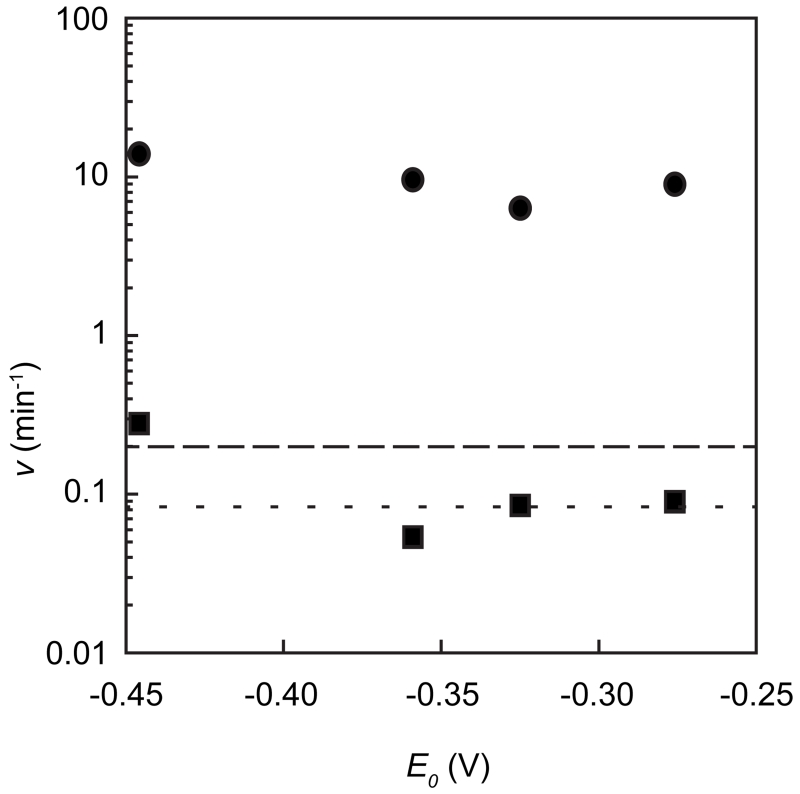
The data presented up to this point suggest that the differences in ability of Fld homologs to support turnover may result from differences in interactions between Fld and BsQueE. However, as we note above, there are differences between the published midpoint potentials of the Fld homologs used. Therefore, we examined if there is a correlation between the ability to support turnover and the midpoint potential using chemical mediators. Preliminary experiments with methyl viologen (Em= −0.446 V) indicated that BsQueE could be activated to the same levels (kCDG = 13.6 ± 3.8 min−1) as is achieved by saturating concentrations of BsFld with dithionite or Fpr/NADPH (Fig. 3). We carried out similar measurements with small molecule redox mediators with midpoint potentials ranging from −0.446 to −0.276 V. None of the mediators supported BsQueE turnover in the absence of dithionite. However, as shown in Fig. 4, at saturating concentrations of each mediator we observe activities that are comparable to those observed with BsFld homologs and dithionite. Therefore, the differences in midpoint potentials of Fld homologs are not the source of the differential activation of BsQueE by these proteins.
Figure 3.
BsQueE activity is enhanced by increasing methyl viologen concentration when electrons are supplied by dithionite. The data were fit using Eqn. 2 to determine kCDG. The activity of BsQueE was 0.2 min−1 in the absence of methyl viologen.
Figure 4.
Velocity of CDG formation (circles) or dAdo formation (squares) as a function of reduction potential of redox mediator used in the activity assay. Dithionite was the reductant used in all of the reactions. The redox mediators used were methyl viologen (Em= −0.446 V)20, benzyl viologen (Em= −0.359 V)20, neutral red (Em= −0.325 V)21, and lissamine blue BF (Em= −0.276 V, pH 7.5)22. The dashed lines represent the rates of formation for CDG (–––) and dAdo (---), 0.2 min−1 and 0.08 min−1 respectively, when BsQueE was activated by dithionite alone.
Uncoupled cleavage of SAM vs activity
All members of the radical SAM superfamily catalyze the reductive cleavage of SAM to afford a radical species, usually dAdo•, to initiate catalysis. However, it is well documented that these enzymes can reductively cleave SAM in a manner uncoupled to catalysis in the absence of substrate.27-31 While this activity is biologically undesirable, it has proved to be a very useful activity to characterize an enzyme as a member of the radical SAM superfamily in the absence of substantial quantities of substrate or in the event the substrate has yet to be determined.28-30 However, there is little direct comparison of the propensity for uncoupled cleavage relative to product formation using various reductants.
The differences in BsQueE activity that is achieved by dithionte alone relative to those observed in the presence of Fld may reflect a high proportion of abortive cleavage of SAM that masks the true activity. To address this, we measured the rate of formation for CDG and 5′-deoxyadenosine (dAdo) with dithionite alone or in the presence of the Fld homologs and with Fpr/NADPH to replace dithionite (Table 1). dAdo was readily detectable (0.08 min−1) when dithionite was used to deliver the reducing equivalents. In the presence of dithionite, the ratio of the rate of turnover to abortive cleavage is 2.5, which only increases slightly to 6.4 when EcFldA is added to mediate the reduction. In the presence of BsYkuN or BsYkuP, however, 10-fold more turnover to reductive cleavage was observed relative to that with EcFldA. The increase appears to arise largely from the ability of Fld homologs to support turnover, as the kdAdo values measured with EcFldA, BsYkuN, or BsYkuP with dithionite as reductant are within experimental error of one another. It is noteworthy that dAdo does not accumulate to detectable quantities when NADPH supplies the reducing equivalents through Fpr/Fld, indicating that the reductive cleavage of SAM to form dAdo• is tightly coupled to turnover.
The experiments measuring kCDG/kdAdo could not eliminate the possibility that differences in the midpoint potentials of Fld homologs is responsible. Therefore, we examined the ratio of turnover to abortive cleavage when redox mediators replaced the flavodoxin homologs (Fig. 4). Dithionite served as the reductant in these experiments. We note that in the presence of some redox mediators, quantifying [dAdo] was difficult because so little dAdo was being produced. In cases where we could measure detectable dAdo, the rate of formation was only slightly greater than that measured in the presence of dithionite alone. Overall, there appears to be no or perhaps a weak correlation between midpoint potential of the mediator and kCDG/kdAdo ratio. We conclude from these observations that the relative differences between the Fld systems may be, in fact, inherent to their interactions.
DISCUSSION
Reductive activation is essential for function of the >65,000 members of the radical SAM superfamily, which are distributed in 5136 species (Pfam PF04055 family).32 Of the three classes that comprise the superfamily, two use SAM as co-substrate whereas the Class I enzymes reform SAM on each turnover.33 Despite the central role that reductive cleavage plays in the function of all these enzymes, to date, there have been very few systematic studies on this reaction or the possible in vivo pathway for activation of these enzymes.
Flavodoxins are small FMN-dependent proteins found in bacteria and algae that mediate electron transfer events in vivo. A role for flavodoxins as non-specific mediators of reducing equivalents is suggested by the ability of flavodoxin from one species to interact with diverse targets. For example, the EcFldA interacts with flavodoxin reductase, cobalamin-dependent methionine synthase, and the radical SAM enzymes biotin synthase, pyruvate formate-lyase activating enzyme, and anaerobic ribonucleotide reductase.4,6-10 Moreover, EcFldA is commonly used in vitro to activate radical SAM enzymes from diverse bacterial sources.11-13,15,17,18
Many bacterial genomes encode two flavodoxin homologs. In E. coli only one of the homologs, EcFldA, is absolutely essential for cellular viability, suggesting that these proteins have non-overlapping functions in vivo.34 The B. subtilis genome also encodes two flavodoxin homologs (BsYkuN and BsYkuP), which in addition to its upstream redox partner, flavodoxin reductase, supports turnover of the P450 enzyme BioI.23 In this paper we have shown that the two B. subtilis flavodoxins, as well as FldA from E. coli, support turnover of the radical SAM enzyme QueE from B. subtilis. Interestingly, both BsYkuN and BsYkuP are more efficient activators of BsQueE than EcFldA, with BsYkuN and BsYkuP both supporting BsQueE activity at the same level under saturating conditions. However, the efficiency of the activation as measured by kCDG/KFLD indicates that BsYkuN is preferred by nearly 10-fold. The in vivo relevance of this observed preferential activation is unclear because the cellular concentrations and expression profiles of BsYkuN, BsYkuP, and BsQueE are currently unknown.
The dAdo• produced from reductive cleavage of SAM is sometimes quenched to form an abortive cleavage product in radical SAM enzymes. It has been suggested that higher proportions of dAdo from these abortive cleavage reactions are possible when dithionite is a reductant.31 Indeed, we do not detect dAdo when BsQueE is reduced with any Fld homolog using Fpr/NADPH to supply the reducing equivalents. Interestingly, with BsYkuN and BsYkuP, we observe substantially more CDG than dAdo even when dithionite is used. This arises almost entirely from the ~10-fold higher activity (kCDG) relative to that achieved by EcFldA.
The X-ray crystal structure of QueE from B. subtilis is not known, but the structure of the homolog from B. multivorans shows that the CX3CX2C motif characteristic of the radical SAM superfamily is punctuated by an 11-residue insertion between the first and second Cys residues in the conserved motif.18 The additional residues form a 310-helix at the surface of the protein further burying and shielding the [4Fe-4S] cluster from the solvent. In contrast to the B. subtilis QueE, dithionite appeared to more efficiently support turnover by the B. multivorans homolog relative to EcFldA.18 Whether robust activation of B. multivorans QueE by the cognate Fld homolog will be the same as that observed here with the B. subtilis QueE/Fld system remains to be established. Based on the observations presented here, we predict that protein sequence/surface specific interactions will play a significant role in Fld-dependent activation of radical SAM enzymes and that a better understanding of how the species-specific Fld/radical SAM partners interact will be essential in studies of the enzymes in the radical SAM superfamily.
Supplementary Material
Acknowledgments
Funding:
The work reported in this publication was supported by National Institutes of General Medical Sciences of the National Institutes of Health grant R01 GM72623 (awarded to VB). The content is solely the responsibility of the authors and does not necessarily represent the official views of the National Institutes of Health.
ABBREVIATIONS
- SAM
S-adenosyl-l-methionine
- dAdo•
5′-deoxyadenosyl radical
- dAdo
5′-deoxyadenosine
- NADPH
reduced β-nicotinamide adenine dinucleotide phosphate
- NADP+
β-nicotinamide adenine dinucleotide phosphate
- FMN
riboflavin 5′-monophosphate
- Fld
flavodoxin
- CPH4
6-carboxy-5,6,7,8-tetrahydropterin
- CDG
7-carboxy-7-deazaguanine
- DEAE
diethylaminoethyl
- E. coli
Escherichia coli
- FldA
E. coli flavodoxin
- Fpr
E. coli ferredoxin(flavodoxin):NADP+ oxidoreductase
- HEPES
4-(2-hydroxyethyl)-1-piperazineethanesulfonic acid
- HPLC
high-performance liquid chromatography
- IPTG
isopropyl β-d-1-thiogalactopyranoside
- PIPES
1,4-piperazinediethanesulfonic acid
- PMSF
phenylmethanesulfonyl fluoride
- QueE
CDG synthase
- SDS–PAGE
sodium dodecyl sulfate polyacrylamide gel electrophoresis
- TCA
trichloroacetic acid
- Tris
tris(hydroxymethyl)aminomethane
- LB
Lennox broth
Footnotes
AUTHOR CONTRIBUTIONS
A.P.Y. cloned and purified the E. coli FldA used in these experiments. N.A.B. and V.B designed and carried out the experiments, and wrote the manuscript.
Primers used to clone the three flavodoxin homologs used in this study from their respective genomic DNA. The material is available free of charge via the Internet at http://pubs.acs.org.
REFERENCES
- (1).Sofia HJ, Chen G, Hetzler BG, Reyes-Spindola JF, Miller NE. Radical SAM, a Novel Protein Superfamily Linking Unresolved Steps In Familiar Biosynthetic Pathways With Radical Mechanisms: Functional Characterization Using New Analysis And Information Visualization Methods. Nucleic Acids Res. 2001;29:1097–1106. doi: 10.1093/nar/29.5.1097. [DOI] [PMC free article] [PubMed] [Google Scholar]
- (2).Walsby CJ, Ortillo D, Broderick WE, Broderick JB, Hoffman BM. An Anchoring Role for FeS Clusters: Chelation of the Amino Acid Moiety of S-Adenosylmethionine to the Unique Iron Site of the [4Fe–4S] Cluster of Pyruvate Formate-Lyase Activating Enzyme. J. Am. Chem. Soc. 2002;124:11270–11271. doi: 10.1021/ja027078v. [DOI] [PubMed] [Google Scholar]
- (3).Chen D, Walsby C, Hoffman BM, Frey PA. Coordination and Mechanism of Reversible Cleavage of S-Adenosylmethionine by the [4Fe-4S] Center in Lysine 2,3-Aminomutase. J. Am. Chem. Soc. 2003;125:11788–11789. doi: 10.1021/ja036120z. [DOI] [PubMed] [Google Scholar]
- (4).Knappe J, Schacht J, Mockel W, Hopner T, Vetter H, Edenharder R. Pyruvate Formate-Lyase Reaction in Escherichia coli. The Enzymatic System Converting an Inactive Form of the Lyase into the Catalytically Active Enzyme. Eur J Biochem. 1969;11:316–327. doi: 10.1111/j.1432-1033.1969.tb00775.x. [DOI] [PubMed] [Google Scholar]
- (5).Chirpich TP, Zappia V, Costilow RN, Barker HA. Lysine 2, 3-Aminomutase. Purification And Properties Of A Pyridoxal Phosphate And S-Adenosylmethionine-Activated Enzyme. J. Biol. Chem. 1970;245:1778–1789. [PubMed] [Google Scholar]
- (6).Knappe J, Blaschkowski HP, Gröbner P, Schmitt T. Pyruvate Formate-Lyase Of Escherichia coli: The Acetyl-Enzyme Intermediate. Eur J Biochem. 1974;50:253–263. doi: 10.1111/j.1432-1033.1974.tb03894.x. [DOI] [PubMed] [Google Scholar]
- (7).Harder J, Eliasson R, Pontis E, Ballinger MD, Reichard P. Activation Of The Anaerobic Ribonucleotide Reductase From Escherichia coli By S-Adenosylmethionine. J. Biol. Chem. 1992;267:25548–25552. [PubMed] [Google Scholar]
- (8).Bianchi V, Reichard P, Eliasson R, Pontis E, Krook M, Jörnvall H, Haggård-Ljungquist E. Escherichia coli Ferredoxin NADP+ Reductase: Activation Of E. coli Anaerobic Ribonucleotide Reduction, Cloning Of The Gene (fpr), and Overexpression Of The Protein. J. Bacteriol. 1993;175:1590–1595. doi: 10.1128/jb.175.6.1590-1595.1993. [DOI] [PMC free article] [PubMed] [Google Scholar]
- (9).Bianchi V, Eliasson R, Fontecave M, Mulliez E, Hoover DM, Matthews RG, Reichard P. Flavodoxin Is Required for the Activation of the Anaerobic Ribonucleotide Reductase. Biochem. Biophys. Res. Commun. 1993;197:792–797. doi: 10.1006/bbrc.1993.2548. [DOI] [PubMed] [Google Scholar]
- (10).Ifuku O, Koga N, Haze S-I, Kishimoto J, Wachi Y. Flavodoxin is Required for Conversion of Dethiobiotin to Biotin in Escherichia coli. Eur J Biochem. 1994;224:173–178. doi: 10.1111/j.1432-1033.1994.tb20009.x. [DOI] [PubMed] [Google Scholar]
- (11).Layer G, Verfürth K, Mahlitz E, Jahn D. Oxygen-Independent Coproporphyrinogen-III Oxidase HemN from Escherichia coli. J. Biol. Chem. 2002;277:34136–34142. doi: 10.1074/jbc.M205247200. [DOI] [PubMed] [Google Scholar]
- (12).Cicchillo RM, Iwig DF, Jones AD, Nesbitt NM, Baleanu-Gogonea C, Souder MG, Tu L, Booker SJ. Lipoyl Synthase Requires Two Equivalents of S-Adenosyl-l-Methionine To Synthesize One Equivalent of Lipoic Acid. Biochemistry. 2004;43:6378–6386. doi: 10.1021/bi049528x. [DOI] [PubMed] [Google Scholar]
- (13).Brazeau BJ, Gort SJ, Jessen HJ, Andrew AJ, Liao HH. Enzymatic Activation Of Lysine 2,3-Aminomutase From Porphyromonas gingivalis. Applied and Environmental Microbiology. 2006;72:6402–6404. doi: 10.1128/AEM.01143-06. [DOI] [PMC free article] [PubMed] [Google Scholar]
- (14).Grove TL, Lee KH, St Clair J, Krebs C, Booker SJ. In vitro Characterization Of AtsB, A Radical SAM Formylglycine-Generating Enzyme That Contains Three [4Fe-4S] Clusters. Biochemistry. 2008;47:7523–7538. doi: 10.1021/bi8004297. [DOI] [PMC free article] [PubMed] [Google Scholar]
- (15).Grove TL, Ahlum JH, Qin RM, Lanz ND, Radle MI, Krebs C, Booker SJ. Further Characterization of Cys-Type and Ser-Type Anaerobic Sulfatase Maturating Enzymes Suggests a Commonality in the Mechanism of Catalysis. Biochemistry. 2013;52:2874–2887. doi: 10.1021/bi400136u. [DOI] [PMC free article] [PubMed] [Google Scholar]
- (16).McCarty RM, Somogyi A, Lin G, Jacobsen NE, Bandarian V. The Deazapurine Biosynthetic Pathway Revealed: in vitro Enzymatic Synthesis Of PreQ0 From Guanosine 5′-Triphosphate In Four Steps. Biochemistry. 2009;48:3847–3852. doi: 10.1021/bi900400e. [DOI] [PMC free article] [PubMed] [Google Scholar]
- (17).McCarty RM, Krebs C, Bandarian V. Spectroscopic, Steady-State Kinetic, and Mechanistic Characterization of the Radical SAM Enzyme QueE, Which Catalyzes a Complex Cyclization Reaction in the Biosynthesis of 7-Deazapurines. Biochemistry. 2013;52:188–198. doi: 10.1021/bi301156w. [DOI] [PMC free article] [PubMed] [Google Scholar]
- (18).Dowling DP, Bruender NA, Young AP, McCarty RM, Bandarian V, Drennan CL. Radical SAM Enzyme QueE Defines A New Minimal Core Fold And Metal-Dependent Mechanism. Nat. Chem. Biol. 2014;10:106–112. doi: 10.1038/nchembio.1426. [DOI] [PMC free article] [PubMed] [Google Scholar]
- (19).Vetter H, JR, Knappe J. Hoppe-Seyler’s Z. Physiol. Chem. 1971;352:433–446. doi: 10.1515/bchm2.1971.352.1.433. [DOI] [PubMed] [Google Scholar]
- (20).Michaelis L, Hill ES. The Viologen Indicators. The Journal of General Physiology. 1933;16:859–873. doi: 10.1085/jgp.16.6.859. [DOI] [PMC free article] [PubMed] [Google Scholar]
- (21).Clark WM, Perkins ME. Studies on Oxidation-Reduction. XVII Neutral Red. J. Am. Chem. Soc. 1932;54:1228–1248. [Google Scholar]
- (22).Horrobin S, Dennison SR, Holmes WS, Pritchard J, Thacker R. Oxidation-Reduction Indicators: Lissamine Blue BF. Trans. Faraday Soc. 1954;50:803–808. [Google Scholar]
- (23).Lawson RJ, Wachenfeldt von C, Haq I, Perkins J, Munro AW. Expression And Characterization Of The Two Flavodoxin Proteins Of Bacillus subtilis, YkuN And YkuP: Biophysical Properties And Interactions With Cytochrome P450 BioI. Biochemistry. 2004;43:12390–12409. doi: 10.1021/bi049131t. [DOI] [PubMed] [Google Scholar]
- (24).McIver L, Leadbeater C, Campopiano DJ, Baxter RL, Daff SN, Chapman SK, Munro AW. Characterisation Of Flavodoxin NADP+ Oxidoreductase And Flavodoxin; Key Components Of Electron Transfer In Escherichia coli. Eur J Biochem. 1998;257:577–585. doi: 10.1046/j.1432-1327.1998.2570577.x. [DOI] [PubMed] [Google Scholar]
- (25).Hall DA, Jordan-Starck TC, Loo RO, Ludwig ML, Matthews RG. Interaction of Flavodoxin with Cobalamin-Dependent Methionine Synthase. Biochemistry. 2000;39:10711–10719. doi: 10.1021/bi001096c. [DOI] [PubMed] [Google Scholar]
- (26).Hall DA, Vander Kooi CW, Stasik CN, Stevens SY, Zuiderweg ERP, Matthews RG. Mapping The Interactions Between Flavodoxin And Its Physiological Partners Flavodoxin Reductase And Cobalamin-Dependent Methionine Synthase. Proc. Natl. Acad. Sci. U.S.A. 2001;98:9521–9526. doi: 10.1073/pnas.171168898. [DOI] [PMC free article] [PubMed] [Google Scholar]
- (27).Moss ML, Frey PA. Activation Of Lysine 2,3-Aminomutase By S-Adenosylmethionine. J. Biol. Chem. 1990;265:18112–18115. [PubMed] [Google Scholar]
- (28).Duschene KS, Broderick JB. The Antiviral Protein Viperin Is A Radical SAM Enzyme. FEBS lett. 2010;584:1263–1267. doi: 10.1016/j.febslet.2010.02.041. [DOI] [PMC free article] [PubMed] [Google Scholar]
- (29).Estellon J, Ollagnier de Choudens S, Smadja M, Fontecave M, Vandenbrouck Y. An Integrative Computational Model For Large-Scale Identification Of Metalloproteins In Microbial Genomes: A Focus On Iron–Sulfur Cluster Proteins. Metallomics. 2014;6:1913–1930. doi: 10.1039/c4mt00156g. [DOI] [PubMed] [Google Scholar]
- (30).McGlynn SE, Boyd ES, Shepard EM, Lange RK, Gerlach R, Broderick JB, Peters JW. Identification and Characterization of a Novel Member of the Radical AdoMet Enzyme Superfamily and Implications for the Biosynthesis of the Hmd Hydrogenase Active Site Cofactor. J. Bacteriol. 2010;192:595–598. doi: 10.1128/JB.01125-09. [DOI] [PMC free article] [PubMed] [Google Scholar]
- (31).Grove TL, Benner JS, Radle MI, Ahlum JH, Landgraf BJ, Krebs C, Booker SJ. A Radically Different Mechanism for S-Adenosylmethionine-Dependent Methyltransferases. Science. 2011;332:604–607. doi: 10.1126/science.1200877. [DOI] [PubMed] [Google Scholar]
- (32).Finn RD, Bateman A, Clements J, Coggill P, Eberhardt RY, Eddy SR, Heger A, Hetherington K, Holm L, Mistry J, Sonnhammer ELL, Tate J, Punta M. Pfam: The Protein Families Database. Nucleic Acids Res. 2014;42:D222–D230. doi: 10.1093/nar/gkt1223. [DOI] [PMC free article] [PubMed] [Google Scholar]
- (33).Booker SJ. Anaerobic Functionalization Of Unactivated C–H Bonds. Curr. Opin. Chem. Biol. 2009;13:58–73. doi: 10.1016/j.cbpa.2009.02.036. [DOI] [PMC free article] [PubMed] [Google Scholar]
- (34).Gaudu P, Weiss B. Flavodoxin Mutants of Escherichia coli K-12. J. Bacteriol. 2000;182:1788–1793. doi: 10.1128/jb.182.7.1788-1793.2000. [DOI] [PMC free article] [PubMed] [Google Scholar]
Associated Data
This section collects any data citations, data availability statements, or supplementary materials included in this article.


