

Abstract
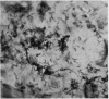
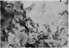
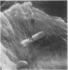
The skin surface biopsy technique has been used to investigate the erythrasma organism in situ in the stratum corneum in 11 patients. Staining by PAS and Gram stain showed the presence of a large number of organisms arranged haphazardly in some areas and in microcolonies in others. With the scanning electron microscope it was possible to see that smooth filamentous chains of microorganisms had penetrated horn cells and caused disturbance of the surface structure of these cells.
Enzyme histochemical tests showed that the erythrasma microorganism possessed a strong reactivity for NAD diaphorase and other mitochondrial enzymes. The reactivity was focal confirming a complex subcellular organization of organelles.
It is suggested that the erythrasma microorganism secretes a mucopolysaccharide sheath in some circumstances.
Full text
PDF


Images in this article
Selected References
These references are in PubMed. This may not be the complete list of references from this article.
- Dawber R. P., Marks R., Swift J. A. Scanning electron microscopy of the stratum corneum. Br J Dermatol. 1972 Mar;86(3):272–281. doi: 10.1111/j.1365-2133.1972.tb02228.x. [DOI] [PubMed] [Google Scholar]
- HOLT S. J. A new approach to the cytochemical localization of enzymes. Proc R Soc Lond B Biol Sci. 1954 Mar 25;142(907):160–169. doi: 10.1098/rspb.1954.0015. [DOI] [PubMed] [Google Scholar]
- Marks R., Dawber R. P. In situ microbiology of the stratum corneum. An application of skin surface biopsy. Arch Dermatol. 1972 Feb;105(2):216–221. [PubMed] [Google Scholar]
- Marks R., Dawber R. P. Skin surface biopsy: an improved technique for the examination of the horny layer. Br J Dermatol. 1971 Feb;84(2):117–123. doi: 10.1111/j.1365-2133.1971.tb06853.x. [DOI] [PubMed] [Google Scholar]
- Meinhof W. Zum histochemischen Nachweis von Enzymen des energieliefernden Stoffwechsels in Dermatophyten der Gattungen Microsporum, Epidermophyton und Keratinomyces. Arch Klin Exp Dermatol. 1968;232(3):279–294. [PubMed] [Google Scholar]
- Montes L. F., Black S. H., McBride M. E. Bacterial invasion of the stratum corneum in erythrasma. I. Ultrastructural evidence for a keratolytic action experted by Corynebacterium minutissimum. J Invest Dermatol. 1967 Nov;49(5):474–485. doi: 10.1038/jid.1967.168. [DOI] [PubMed] [Google Scholar]
- NACHLAS M. M., CRAWFORD D. T., SELIGMAN A. M. The histochemical demonstration of leucine aminopeptidase. J Histochem Cytochem. 1957 May;5(3):264–278. doi: 10.1177/5.3.264. [DOI] [PubMed] [Google Scholar]
- SARKANY I., TAPLIN D., BLANK H. The etiology and treatment of erythrasma. J Invest Dermatol. 1961 Oct;37:283–290. [PubMed] [Google Scholar]
- Somerville D. A. Erythrasma in normal young adults. J Med Microbiol. 1970 Feb;3(1):57–64. doi: 10.1099/00222615-3-1-57. [DOI] [PubMed] [Google Scholar]
- WACHSTEIN M., MEISEL E., NIEDZWIEDZ A. Histochemical demonstration of mitochondrial adenosine triphosphatase with the lead-adenosine triphosphate technique. J Histochem Cytochem. 1960 Sep;8:387–388. doi: 10.1177/8.5.387. [DOI] [PubMed] [Google Scholar]